
Can you provide ke explained lecture on CVS murmurs and valve disorders
Reading File
Now let me search the medical library for comprehensive content on CVS murmurs and valve disorders in parallel.
Finding Sources
Finding Sources
Finding Sources
Finding Sources
Reading File
Reading File
Finding Sources
Excellent results. Now let me pull the key textbook sections - Harrison's cardiac auscultation, Braunwald's valve disease, Tintinalli's murmur table, and Harriet Lane's innocent murmurs in parallel.
Reading File
Reading File
Reading File
Excellent content gathered. Let me now fetch more from Harrison's on specific valve disorders and the HOCM maneuver section, plus the Braunwald's auscultation section.
Reading File
Reading File
Now I have comprehensive content. Let me fetch the key diagrams and also get the Braunwald's content on individual valve disorders.
Loading Image
Loading Image
Finding Sources
I now have comprehensive, high-quality content from Harrison's, Braunwald's, Tintinalli's, and Fuster & Hurst's for a thorough lecture. Let me now compose the complete lecture.
CVS Murmurs and Valve Disorders - A Complete Lecture
Sources: Harrison's Principles of Internal Medicine 22E, Braunwald's Heart Disease, Tintinalli's Emergency Medicine, Fuster & Hurst's The Heart 15E
1. FOUNDATION: What is a Heart Murmur?
A heart murmur is a sound produced by turbulent blood flow within or near the heart. Murmurs are classified by:
- Timing: systolic, diastolic, or continuous
- Grade (Levine scale, 1-6)
- Quality: harsh, blowing, rumbling, musical
- Location and radiation
- Configuration: crescendo, decrescendo, crescendo-decrescendo (diamond-shaped), plateau (holosystolic)
Grading Scale (Tintinalli's)
| Grade | Description |
|---|---|
| 1 | Faint, may not be heard in all positions |
| 2 | Quiet, but heard immediately with stethoscope placement |
| 3 | Moderately loud |
| 4 | Loud |
| 5 | Heard with stethoscope partly off the chest wall |
| 6 | Heard when stethoscope is entirely off the chest wall |
Grade 4+ murmurs are associated with a palpable thrill.
2. THE CARDIAC CYCLE AND MURMUR TIMING
The diagram below from Harrison's shows the pressure gradients that generate each type of murmur, with simultaneous ECG and phonocardiographic recording:
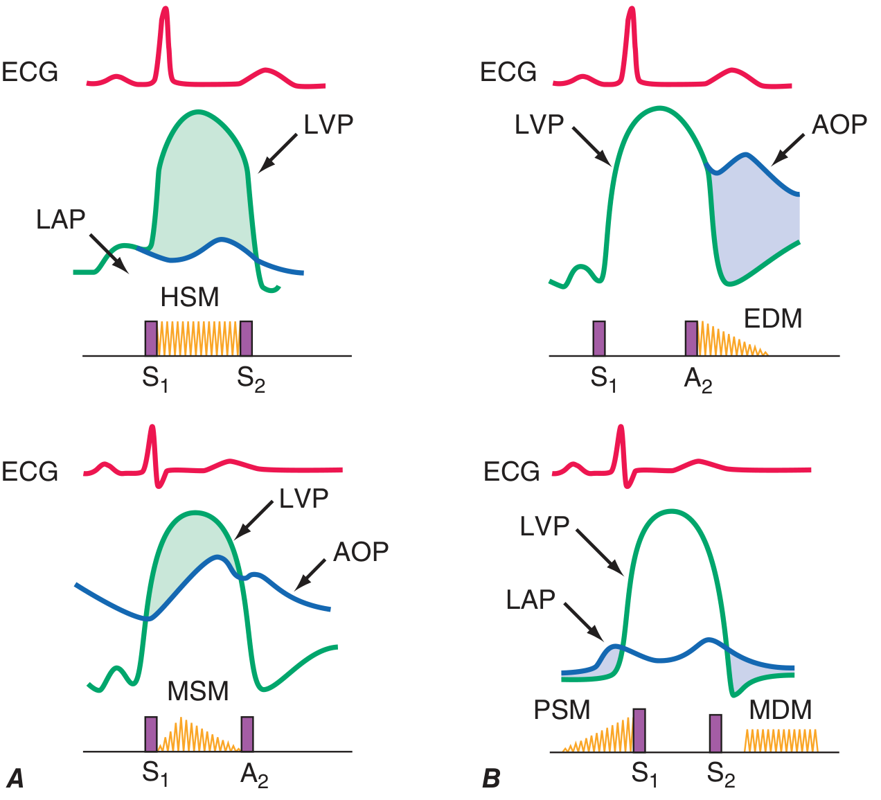
Key concept: A murmur exists when there is a pressure gradient across a valve or orifice. The direction of the gradient, and whether it exists during systole, diastole, or both, determines the murmur type.
- Holosystolic murmur (HSM): LV pressure > LA pressure throughout systole (MR, VSD, TR)
- Midsystolic murmur (MSM): LV > Aorta gradient during mid-systole (AS, HOCM)
- Early diastolic murmur (EDM): Aorta > LV during diastole (AR)
- Mid-diastolic murmur (MDM): LA > LV gradient in diastole (MS)
3. SYSTOLIC MURMURS
A. Midsystolic (Ejection) Murmurs
These begin after S1, peak in mid-systole, and end before S2 - giving a crescendo-decrescendo (diamond-shaped) pattern.
Causes include:
- Aortic stenosis (AS) - most common cause in adults
- Pulmonic stenosis
- HOCM
- Increased flow states: fever, anemia, thyrotoxicosis, pregnancy, ASD
- Aortic sclerosis (non-obstructive)
B. Holosystolic (Pansystolic) Murmurs
These span from S1 to S2 with a plateau configuration. They occur when there is a persistent pressure gradient from a high-pressure to a low-pressure chamber throughout systole.
Causes include:
- Mitral regurgitation (MR)
- Tricuspid regurgitation (TR)
- Ventricular septal defect (VSD)
C. Late Systolic Murmurs
- Mitral valve prolapse (MVP): mid-systolic click followed by a late systolic murmur crescendoing into S2
4. DIASTOLIC MURMURS
All diastolic murmurs are pathological (never innocent).
A. Early Diastolic (Decrescendo) Murmurs
Begin immediately after S2 and fade during diastole.
- Aortic regurgitation (AR): high-pitched, blowing, decrescendo murmur at the left sternal border (or right sternal border if aortic root dilated)
- Pulmonic regurgitation (PR): Graham Steell murmur - decrescendo, early to mid-diastolic, best heard at the 2nd left interspace, increases with inspiration; usually indicates pulmonary hypertension
B. Mid-Diastolic Murmurs (Rumbles)
Occur during passive ventricular filling.
- Mitral stenosis (MS): low-pitched, rumbling, at the apex; heard best in left lateral decubitus with bell
- Tricuspid stenosis (TS): lower left sternal border; increases with inspiration (Carvallo's sign)
- Austin Flint murmur: functional MS-like murmur in severe AR (regurgitant jet impinges on anterior mitral leaflet)
- Carey-Coombs murmur: short mid-diastolic murmur in acute rheumatic fever
C. Presystolic Murmurs
- MS in sinus rhythm: presystolic accentuation just before S1 due to atrial contraction
- TS: similar, augmented by inspiration
5. CONTINUOUS MURMURS
Begin in systole, peak near S2, and continue into diastole - implying a constant pressure gradient across both phases.
Causes:
- Patent ductus arteriosus (PDA): "machinery murmur" at upper left sternal border
- Ruptured sinus of Valsalva aneurysm: upper right sternal border, abrupt onset
- Coronary arteriovenous fistula
- Aortic coarctation: intercostal collaterals
- Cervical venous hum (innocent, compressible)
A continuous murmur spans S2, distinguishing it from a to-and-fro murmur (separate systolic + diastolic murmurs in mixed aortic valve disease, such as combined AS + AR).
6. INDIVIDUAL VALVE DISORDERS IN DETAIL
MITRAL STENOSIS (MS)
Mechanism: Obstruction to LV inflow - most often from rheumatic fever causing leaflet thickening, commissural fusion, and subvalvular scarring.
Murmur: Mid-diastolic low-pitched rumble at the apex (left lateral decubitus, bell of stethoscope). Presystolic accentuation in sinus rhythm (lost in AF).
Heart sounds: Loud snapping S1, opening snap (OS) after S2.
- Shorter A2-OS interval = more severe MS (higher LA pressure brings mitral leaflets to open position earlier)
Other signs: Tapping apical impulse (underfilled ventricle), small pulse volume, malar flush, AF.
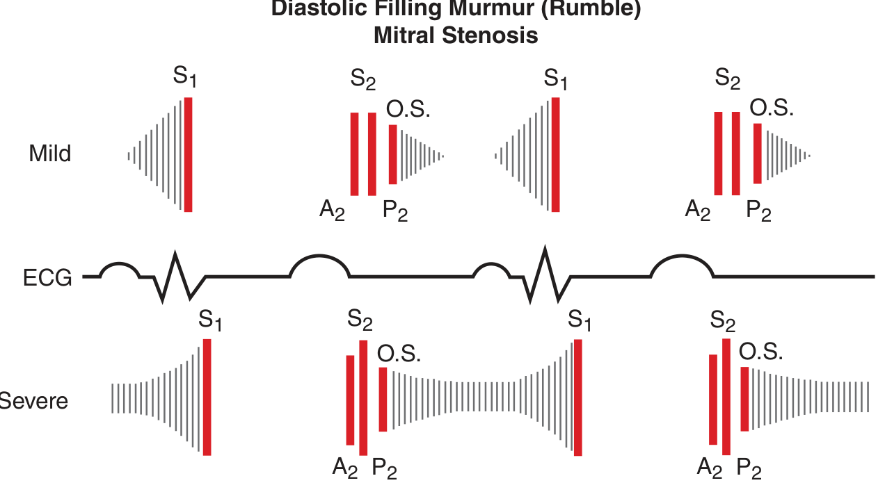
As severity increases, the A2-OS interval shortens and the rumble fills more of diastole. In severe MS, S2 splitting narrows and P2 becomes louder due to pulmonary hypertension.
MITRAL REGURGITATION (MR)
Mechanism: Failure of leaflet coaptation. Causes include:
- Primary (organic): Mitral valve prolapse, rheumatic disease, infective endocarditis, mitral annular calcification
- Secondary (functional): LV dilation, ischemic papillary muscle dysfunction
Murmur:
- Chronic: High-pitched, holosystolic (pansystolic) apical murmur radiating to axilla
- Acute: Harsh apical systolic murmur starting with S1, may end before S2 (because rapidly rising LA pressure equalizes with LV pressure before end-systole)
Heart sounds: S3 gallop (volume overload), soft S1 (incomplete leaflet coaptation), wide split S2.
Complications: LA enlargement → AF; LV volume overload → dilated cardiomyopathy; pulmonary hypertension (>50% with severe MR).
MITRAL VALVE PROLAPSE (MVP)
Mechanism: Myxomatous degeneration of valve leaflets causing billowing of one or both leaflets into the LA during systole.
Auscultation: Mid-systolic click + late systolic murmur (crescendo into S2).
Dynamic maneuvers (critical exam topic):
| Maneuver | Effect on Click | Reason |
|---|---|---|
| Standing/Valsalva (strain) | Moves CLOSER to S1 | Decreased preload; prolapse occurs earlier in smaller chamber |
| Squatting/Leg raise | Moves AWAY from S1 | Increased preload; prolapse delayed in larger chamber |
AORTIC STENOSIS (AS)
Mechanism: Obstruction to LV outflow - most common cause in adults is calcific degenerative disease; bicuspid aortic valve in younger patients; rheumatic disease.
Classic triad: Angina, syncope, heart failure (in order of appearance with worsening severity).
Murmur: Harsh crescendo-decrescendo midsystolic murmur, best heard at 2nd right interspace (aortic area), radiates to carotids.
- Gallavardin effect: The high-frequency components of the AS murmur may radiate to the apex, mimicking MR - a potential diagnostic trap.
Signs of severe AS:
- Parvus et tardus carotid pulse (small, slow-rising)
- Late-peaking, grade 3+ midsystolic murmur
- Soft or absent A2
- Sustained LV apical impulse
- S4 gallop (stiff, hypertrophied LV)
- Paradoxical splitting of S2 (delayed aortic valve closure)
Postpremature beat: The murmur of AS increases in intensity in the beat after a premature ventricular contraction (enhanced LV filling + post-extrasystolic potentiation) - whereas the MR murmur does not change significantly. This is a key bedside differentiating maneuver.
AORTIC REGURGITATION (AR)
Mechanism: Failure of aortic leaflet coaptation during diastole. Causes: bicuspid aortic valve, rheumatic disease, infective endocarditis, aortic root dilation (Marfan's, syphilis, hypertension, aortic dissection).
Murmur: High-pitched, blowing, decrescendo early diastolic murmur, best heard at the left sternal border (or right sternal border with aortic root dilation); patient sitting forward, breath held in expiration.
Austin Flint murmur: A mid-diastolic apical rumble in severe AR caused by the regurgitant jet impinging on the anterior mitral leaflet, producing functional mitral obstruction. No opening snap (distinguishes from MS).
Peripheral signs of severe chronic AR (wide pulse pressure, diastolic "runoff"):
- Corrigan's pulse (water-hammer): Rapid rise and collapse of carotid pulse
- Quincke's pulsations: Nail-bed capillary pulsations
- Hill's sign: Popliteal systolic BP exceeds brachial systolic BP by >20 mmHg
- Musset's sign: Head bobbing
- Traube's sign: Pistol-shot sounds over femoral artery
- Müller's sign: Uvular pulsations
- Wide pulse pressure: systolic elevated, diastolic very low
Acute AR (e.g., infective endocarditis, aortic dissection): Murmur is shorter and lower-pitched, peripheral signs absent. Premature mitral valve closure → soft S1. This is a surgical emergency.
TRICUSPID REGURGITATION (TR)
Mechanism: Usually functional (right ventricular dilation from pulmonary hypertension). Organic causes: rheumatic disease, carcinoid syndrome, IE (IV drug users), Ebstein's anomaly.
Murmur: Holosystolic murmur at the lower left sternal border; increases with inspiration (Carvallo's sign - positive in TR, negative in MR).
Signs: Prominent V waves in JVP, pulsatile hepatomegaly, peripheral edema.
PULMONIC STENOSIS (PS)
Mechanism: Usually congenital.
Murmur: Harsh crescendo-decrescendo midsystolic murmur at the 2nd left interspace; preceded by a pulmonic ejection sound (click) that is louder on expiration (unique among right-sided events).
Signs: RV heave, wide splitting of S2 (delayed P2), post-stenotic dilation of pulmonary artery.
7. DYNAMIC AUSCULTATION - BEDSIDE MANEUVERS
This is one of the most high-yield exam topics. The key principle: right-sided murmurs increase with inspiration; left-sided murmurs decrease.
| Maneuver | Effect | Which Murmurs Change |
|---|---|---|
| Inspiration | Increases right-sided murmurs | TR, TS, PS, PR (Carvallo's sign) |
| Valsalva (strain phase) | Decreases preload | Most murmurs ↓; MVP + HOCM ↑ |
| Rapid standing | Decreases preload | Most murmurs ↓; MVP + HOCM ↑ |
| Squatting | Increases preload + afterload | Most murmurs ↑; MVP click delays, HOCM ↓ |
| Hand grip | Increases afterload | MR, AR, VSD ↑; AS, HOCM ↓ |
| Post-PVC beat | Enhanced LV filling | AS ↑; MR unchanged |
Memory rule for MVP and HOCM: Any maneuver that reduces LV volume makes these murmurs worse (smaller LV = earlier prolapse in MVP, worse LVOT obstruction in HOCM). Any maneuver that increases LV volume ameliorates them.
8. INNOCENT MURMURS
Not all murmurs indicate disease. Innocent murmurs are:
- Always systolic (midsystolic, low-grade, ≤2/6)
- Short in duration
- Vibratory or musical quality (Still's murmur in children)
- No radiation
- No associated abnormal heart sounds, clicks, or thrills
- Change with position (often disappear when supine or standing)
- Present in children, adolescents, pregnancy, fever, anemia, thyrotoxicosis
Features that make a murmur NOT innocent (Harriet Lane criteria):
- Diastolic murmur
- Holosystolic or long murmur
- Grade 3+ pansystolic or harsh quality
- Abnormal heart sounds or clicks
- Abnormally strong or weak pulses
9. QUICK-REFERENCE SUMMARY TABLE
| Valve Disorder | Murmur | Timing | Best Heard | Key Signs |
|---|---|---|---|---|
| Mitral Stenosis | Low-pitched rumble | Mid-diastolic | Apex (LLD, bell) | Loud S1, opening snap, short A2-OS = severe |
| Mitral Regurgitation | High-pitched, blowing | Holosystolic | Apex → axilla | S3, soft S1, AF, wide split S2 |
| MVP | Click + late systolic | Late systolic | Apex | Mid-systolic click; click moves with position |
| Aortic Stenosis | Harsh, crescendo-decrescendo | Midsystolic | 2nd RSB → carotids | Parvus et tardus, soft A2, S4, LV heave |
| Aortic Regurgitation | High-pitched, blowing, decrescendo | Early diastolic | LSB (lean forward) | Wide pulse pressure, water-hammer pulse, Austin Flint |
| Tricuspid Regurgitation | Blowing holosystolic | Holosystolic | LLSB | Carvallo's sign (+), JVP V waves, pulsatile liver |
| Pulmonic Stenosis | Harsh, crescendo-decrescendo | Midsystolic | 2nd LSB | Ejection click (↑ expiration), wide S2, RV heave |
| Pulmonic Regurgitation | Blowing, decrescendo | Early diastolic | 2nd LSB | Graham Steell murmur, signs of pulmonary HTN |
| PDA | Machinery, continuous | Systolic + diastolic | Upper LSB | Bounding pulse, wide pulse pressure |
| VSD | Harsh, pansystolic | Holosystolic | LLSB | Thrill common; may disappear if defect closes |
| HOCM | Harsh | Midsystolic | LLSB | Worse with standing/Valsalva; better with squatting |
10. PATHOPHYSIOLOGY SUMMARY - PRESSURE AND VOLUME OVERLOAD
| Lesion | Type of Overload | LV Response | Compensation |
|---|---|---|---|
| AS | Pressure | Concentric hypertrophy | S4, prolonged systole |
| AR (chronic) | Volume | Eccentric hypertrophy + dilation | Wide pulse pressure |
| MR (chronic) | Volume | Eccentric hypertrophy | LA dilation, AF |
| MS | - | LV normal/small | LA dilation → AF → embolism |
| TR | Volume (RV) | RV dilation | Congestive hepatopathy |
Key Exam Mnemonics
"PASS" for when murmurs worsen with decreased preload (standing/Valsalva):
- P - Prolapse (MVP)
- A - HOCM (obstructive cardiomyopathy)
- S - Standing makes both worse
- S - Squatting makes both better
S2 splitting:
- Wide splitting → PS, RBBB, AS (paradoxical = delayed A2)
- Fixed splitting → ASD
- Paradoxical splitting → AS, LBBB, HOCM
Sources: Harrison's Principles of Internal Medicine 22E, Braunwald's Heart Disease 12E, Tintinalli's Emergency Medicine, Fuster & Hurst's The Heart 15E
This is a shared conversation. Sign in to Orris to start your own chat.