
Generate image Show the Circle of Willis (cerebral arterial circle) with all contributing arteries accurately branching from below.
Detailed anatomical diagram of the Circle of Willis (cerebral arterial circle) viewed from below (inferior view of the brain), showing all contributing arteries with accurate labels. Include: two Internal Carotid Arteries (ICA) entering from below, Anterior Cerebral Arteries (ACA) branching anteriorly from each ICA, Anterior Communicating Artery (ACoA) connecting the two ACAs at the midline, Middle Cerebral Arteries (MCA) branching laterally from each ICA, Posterior Communicating Arteries (PCoA) connecting ICA to Posterior Cerebral Arteries on each side, Basilar Artery ascending from below at the midline, two Vertebral Arteries merging to form the Basilar Artery inferiorly, Posterior Cerebral Arteries (PCA) branching from the top of the Basilar Artery. The circle is complete, symmetrical, and clearly annotated. Clean medical illustration style on a white background with color-coded vessels (arteries in red), bold label lines, and a legend.
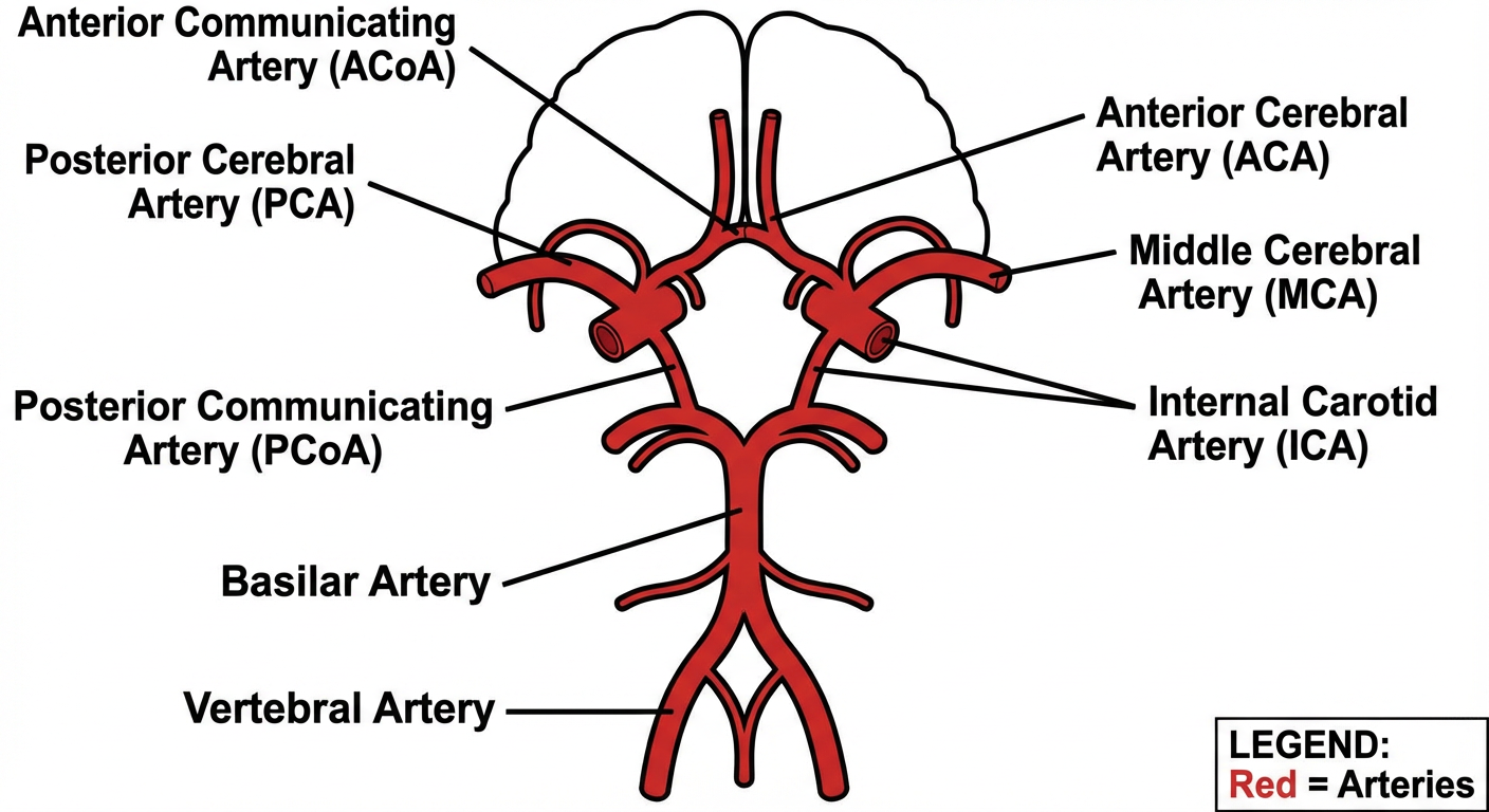
- ICA — Internal Carotid Artery (bilateral entry points)
- ACA — Anterior Cerebral Artery (bilateral, courses anteromedially)
- ACoA — Anterior Communicating Artery (bridges the two ACAs at the midline)
- MCA — Middle Cerebral Artery (bilateral, exits laterally into the Sylvian fissure)
- PCoA — Posterior Communicating Artery (bilateral, links ICA to PCA)
- Vertebral Arteries — ascend bilaterally and merge at the pontomedullary junction
- Basilar Artery — formed by the union of both vertebral arteries; travels rostrally along the pons
- PCA — Posterior Cerebral Artery (bilateral, arises at the basilar bifurcation)