
Internal capsule 5m short note
I'll start with a quick answer, then pull detailed content from the medical library.
The internal capsule is a compact band of white matter in the brain containing ascending and descending fiber tracts connecting the cortex with subcortical structures. It has five parts: anterior limb, genu, posterior limb, retrolenticular, and sublenticular portions. Lesions (commonly from hypertensive lacunar strokes) cause contralateral hemiplegia, hemisensory loss, and homonymous hemianopia depending on which portion is affected.
Searching sources...
Reading File
Now let me search the library for internal capsule content and a relevant image in parallel.
Finding Sources
Searching Images
internal capsule anatomy diagram parts limbs
Finding Sources
Good results. Let me now read the key sections from both Neuroanatomy and Localization in Clinical Neurology in parallel.
Reading File
Reading File
Reading File
Excellent content retrieved. Let me also fetch the internal capsule diagram and get the basal ganglia figure.
Loading Image
Reading File
I now have comprehensive content. Let me compile the complete short note.
Internal Capsule - Short Note (5 Marks)
Definition and Position
The internal capsule is a V-shaped (or arrowhead-shaped) compact band of white matter situated deep within each cerebral hemisphere. It contains all the ascending and descending projection fibers connecting the cerebral cortex with subcortical structures (thalamus, brainstem, spinal cord). On horizontal section it looks like two letter Vs with their points facing inward.
Relations:
- Medial: Head of caudate nucleus (anteriorly) and thalamus (posteriorly)
- Lateral: Lentiform nucleus (putamen + globus pallidus)
Parts / Subdivisions
The internal capsule has 5 parts:
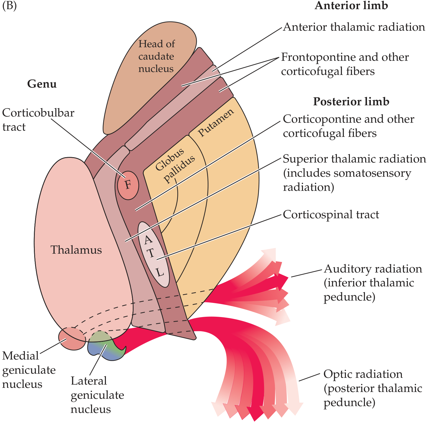
| Part | Location | Fibers Carried |
|---|---|---|
| Anterior limb | Between head of caudate and lentiform nucleus | Frontopontine fibers; anterior thalamic radiation (thalamocortical/corticothalamic connecting frontal lobe to thalamus); caudate-putaminal fibers |
| Genu ("knee") | At transition between anterior and posterior limbs, at the level of foramen of Monro | Corticobulbar (corticonuclear) fibers - motor fibers to cranial nerve nuclei; some corticopontine fibers |
| Posterior limb | Between lentiform nucleus and thalamus (lenticulothalamic segment, 3-4 cm long) | Corticospinal tract (somatotopically organized); superior thalamic radiation (somatosensory to parietal cortex); corticorubral, corticothalamic fibers |
| Retrolenticular | Behind the lentiform nucleus | Optic radiation (geniculocalcarine tract / radiatio optica of Gratiolet); corticotectal, corticonigral fibers |
| Sublenticular | Below the lentiform nucleus | Auditory radiation (inferior thalamic peduncle); temporopontine fibers |
Somatotopic Organization of the Posterior Limb
The corticospinal tract in the posterior limb is somatotopically organized from anterior to posterior:
- Face fibers - most anterior (genu/rostral posterior limb)
- Arm (shoulder > elbow > wrist > fingers)
- Trunk
- Leg (hip > knee > ankle > toes) - most posterior
- Bladder/rectum - most caudal
Because the fibers are so compact, even a small lesion here can produce weakness of the entire contralateral body (face + arm + leg).
Blood Supply
| Part | Artery | Source |
|---|---|---|
| Anterior limb | Recurrent artery of Heubner | Anterior cerebral artery (ACA) |
| Genu + inferior posterior limb | Anterior choroidal artery | Internal carotid artery (ICA) |
| Superior anterior + posterior limb | Lenticulostriate arteries | Middle cerebral artery (MCA) |
These are all end arteries - small perforating vessels with no collateral supply, making the internal capsule highly vulnerable to hypertensive lacunar strokes and hemorrhage.
Clinical Syndromes (Lesions of the Internal Capsule)
1. Posterior Limb Lesion
Most common (lacunar infarct or hypertensive hemorrhage)
- Contralateral hemiplegia - pure motor hemiplegia (face, arm, leg equally)
- Contralateral hemisensory loss - if superior thalamic radiation involved
- Combined = Sensorimotor stroke
2. Genu Lesion - "Capsular Genu Syndrome"
- Facial + lingual hemiparesis (corticobulbar involvement)
- Mild limb involvement
- Dysarthria, dysphagia
- Sometimes behavioral/cognitive changes (frontal-thalamic disconnection)
3. Retrolenticular Lesion
- Contralateral homonymous hemianopia (optic radiation interrupted)
- Visual agnosia
4. Complete Internal Capsule Lesion
Classic "capsular syndrome":
- Contralateral hemiplegia (UMN type: spasticity, hyperreflexia, upgoing plantars)
- Contralateral hemisensory loss (all modalities)
- Contralateral homonymous hemianopia = Contralateral Hemiplegia + Hemisensory loss + Hemianopia (the "three Hs")
5. Internal Capsular Hemorrhage
- Hypertensive hemorrhage is the most common cause
- Small hemorrhages in the genu or posterior limb may produce pure motor hemiplegia (PMH) or mixed sensorimotor syndrome
- Decorticate rigidity is seen on the hemiplegic side after hemorrhages/thromboses in the internal capsule (Ganong)
Key Points to Remember
- The internal capsule continues inferiorly as the cerebral peduncles (basis pedunculi) in the midbrain
- It is a major site for lacunar infarcts (due to end-artery occlusion, usually hypertensive)
- The entire corticospinal and corticobulbar projections are compacted here - hence small lesions produce large deficits
- Opposite of brainstem lesions: internal capsule lesions do NOT produce crossed findings (cranial nerve + contralateral body)
Sources: Neuroanatomy through Clinical Cases 3rd Ed.; Localization in Clinical Neurology 8e; Ganong's Review of Medical Physiology 26th Ed.
This is a shared conversation. Sign in to Orris to start your own chat.