
Evidence based treatment for Tarsal tunnel syndrome, DD, Imaging, Clinical assessment, Treatment and Role of non surgical pain intervention
"tarsal tunnel syndrome"[MeSH Terms] AND treatment
"tarsal tunnel syndrome" AND diagnosis AND imaging
PMID: 39811161
tarsal tunnel syndrome anatomy tibial nerve
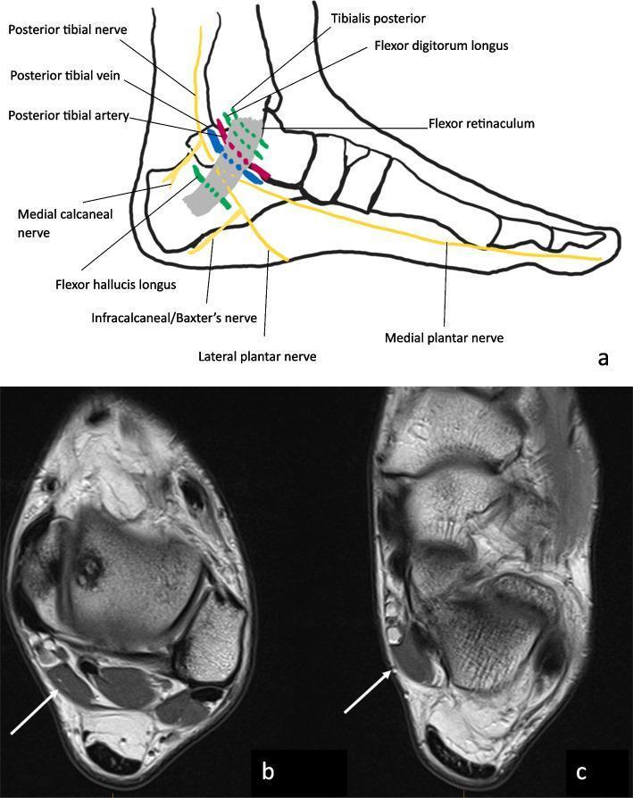
This educational composite focuses on the anatomy and pathology of the tarsal tunnel in the ankle. Image 'a' is a lateral anatomical diagram illustrating the contents of the tarsal tunnel beneath the flexor retinaculum. Key structures labeled include the posterior tibial nerve, artery, and vein, along with the tendons of the tibialis posterior, flexor digitorum longus, and flexor hallucis longus. It also depicts the branching of the medial and lateral plantar nerves and Baxter’s nerve (inferior calcaneal nerve). Images 'b' and 'c' are sequential axial proton density (PD) magnetic resonance images (MRI) of a 20-year-old male. White arrows point to an anatomical variation: an accessory flexor hallucis longus muscle located within the tarsal tunnel. This accessory structure occupies space within the tunnel, demonstrating a potential mechanical etiology for tarsal tunnel syndrome by compressing adjacent neurovascular structures. The MRI shows the muscle as a distinct, low-signal intensity mass relative to surrounding fat, characteristic of skeletal muscle tissue.
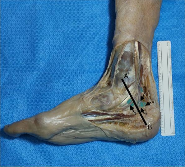
This clinical photograph shows a medial cadaveric dissection of the right distal leg, ankle, and foot, illustrating the anatomy of the tarsal tunnel and terminal branches of the tibial nerve. The Dellon–McKinnon malleolar-calcaneal line (DML) is superimposed as a black line, extending from point A' (center of the medial malleolus) to point B (medial process of the calcaneal tuberosity). The dissection reveals four specific nerve branches: 1. Medial plantar nerve (MPN), 2. Lateral plantar nerve (LPN), 3. Baxter’s nerve (the first branch of the LPN), and 4. Medial calcaneal nerve (MCN). The image demonstrates the spatial relationships between these nerves and the DML, which serves as a clinical landmark for identifying nerve entrapment sites in Tarsal Tunnel Syndrome. Bony landmarks such as the medial malleolus and calcaneus, along with the abductor hallucis muscle and various tendons, provide anatomical context for evaluating the branching patterns of the tibial nerve.
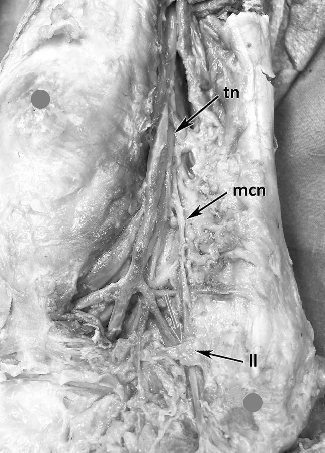
This clinical photograph displays a high-resolution cadaveric dissection of the medial ankle and hindfoot region, specifically highlighting the neurovascular architecture. The image focuses on the distal course and branching pattern of the tibial nerve (tn). A primary focus is the medial calcaneal branch (mcn/mcb), which is shown originating from the tibial nerve and coursing distally toward the posteromedial heel. A critical anatomical relationship is demonstrated where the medial calcaneal nerve enters an osteofibrous tube formed by the distal extension of the laciniate ligament (ll), indicated by a red arrow. This structural arrangement is educationally significant as it identifies a potential site for nerve entrapment. The dissection clearly illustrates the transition of the nerve from deeper tissue layers toward the surface. This visual serves as a topographical guide for understanding tarsal tunnel syndrome and the specific anatomy relevant to surgical decompression or ultrasound-guided regional anesthesia in the medial foot.
tarsal tunnel syndrome MRI ultrasound imaging findings
PMID: 35562562
PMID: 40368127
tarsal tunnel syndrome non-surgical pain intervention corticosteroid injection PRP ultrasound guided 2024 2025
Tarsal Tunnel Syndrome (TTS): Evidence-Based Clinical Review
1. Anatomy and Pathophysiology
- Medial calcaneal nerve (MCN) - purely sensory to the heel skin
- Medial plantar nerve (MPN) - mixed; innervates medial 3.5 digits and intrinsics
- Lateral plantar nerve (LPN) - mixed; innervates lateral 1.5 digits, Baxter's nerve (first branch of LPN) supplies the abductor digiti quinti
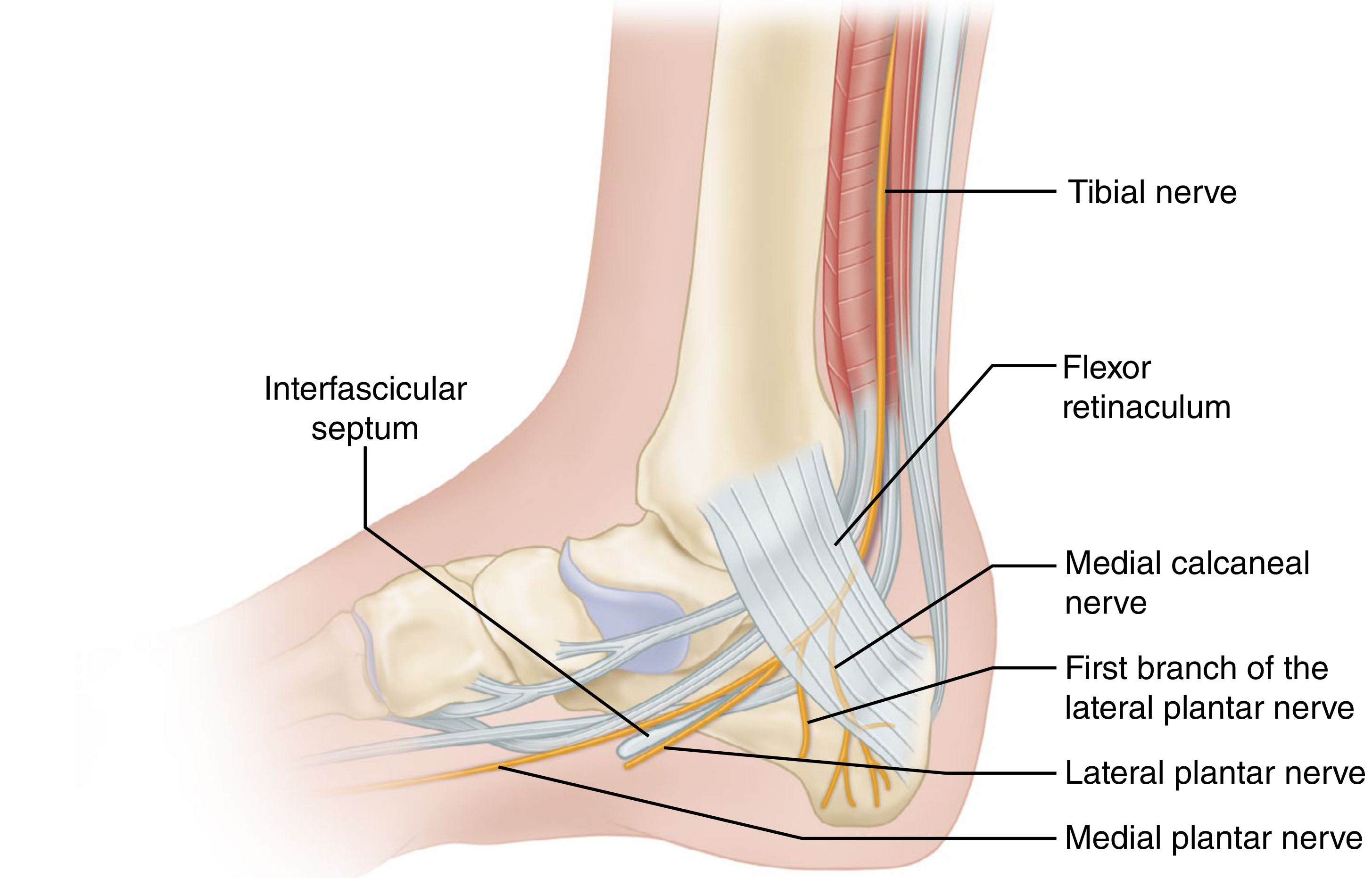
- Campbell's Operative Orthopaedics 15th Ed 2026, p.5024-5072
2. Etiology
| Category | Examples |
|---|---|
| Post-traumatic | Fracture fragments, calcaneal fracture malunion, post-cast fibrosis |
| Inflammatory/Synovial | Tenosynovitis of FHL/FDL, rheumatoid proliferative synovitis |
| Vascular | Posterior tibial varicosities (most common identifiable cause in some series) |
| Neoplastic | Ganglia, synovial cysts, neurilemmomas (schwannomas), lipomas |
| Osseous | Tarsal coalition, osteophytes, accessory ossicles |
| Anatomical variants | Accessory FDL/FHL tendons, accessory soleus, hypertrophy of abductor hallucis |
| Alignment/Biomechanical | Valgus hindfoot (chronic traction neuropathy), lateral-displacing calcaneal osteotomies |
| Systemic | Diabetes mellitus, hypothyroidism, rheumatoid arthritis, perineural fibrosis |
| Idiopathic | ~25% of cases - no identifiable cause |
- Rheumatology 2-Volume Set, Elsevier 2022, p.773; Campbell's 2026, p.5074-5081
3. Clinical Assessment
History
- Burning pain, paresthesias, or dysesthesias on the plantar foot, toes, or medial heel
- Symptoms may be nocturnal (waking patient from sleep), exertional, or at rest
- Retrograde radiation to the calf (Valleix phenomenon) in some patients
- Relief from walking (distinguishes TTS from plantar fasciitis where first-step pain is classic)
- Duration, onset (insidious vs. traumatic), aggravating/relieving factors, prior treatments
Physical Examination
| Finding | Significance |
|---|---|
| Tinel's sign over flexor retinaculum | Proximal entrapment; percussion triggers paresthesias distally |
| Point tenderness at medial heel soft spot (below abductor hallucis) | Distal branch entrapment |
| Sensory abnormalities | Check both plantar nerve distributions; dryness/scaliness over specific nerve territories |
| Motor findings | Atrophy of abductor hallucis (MPN) or abductor digiti minimi (LPN) - compare with contralateral side |
| Vasomotor changes | Temperature asymmetry, altered sweating pattern |
| Fusiform swelling posterior to medial malleolus | Suggests space-occupying lesion or varicosities |
Provocative Tests
- Ankle plantarflexed + foot inverted (increases tunnel pressure) + digital compression over tibial nerve
- Sensitivity 86%, Specificity 100% - highest diagnostic accuracy of any clinical test
-
Maximal passive ankle dorsiflexion + eversion + simultaneous MTP joint dorsiflexion
-
Hold for 5-10 seconds - reproduction of symptoms is positive
-
Comparable to Phalen's test for carpal tunnel syndrome
-
Campbell's Operative Orthopaedics 15th Ed 2026, p.5086-5145
4. Differential Diagnosis
| Condition | Key distinguishing features |
|---|---|
| Plantar fasciitis | First-step morning pain; point tenderness at plantar fascial insertion; resolves quickly; no neurogenic symptoms |
| Peripheral neuropathy (diabetic, systemic) | Bilateral, glove-and-stocking pattern; associated systemic disease |
| Lumbosacral radiculopathy (L4-S1) | Back pain, dermatomal pattern, positive SLR, spine imaging abnormality; electrodiagnostics show proximal lesion |
| Plantar heel pad atrophy | Diffuse plantar heel pain, age-related; no Tinel's, no paresthesias |
| Baxter's nerve entrapment (first branch LPN) | Lateral heel pain, entrapment distal to the laciniate ligament, particularly in runners |
| Abductor hallucis hypertrophy | Similar in runners; compression distal to tunnel; MRI distinguishes |
| Calcaneal stress fracture | Focal bone tenderness; positive squeeze test; MRI/bone scan confirms |
| Vascular insufficiency | Claudication pattern; absent pulses; ABI abnormal; imaging confirms |
| Arthritis of tarsal bones | Morning stiffness; joint swelling; radiographic changes |
| Morton's neuroma | Web space location; Mulder's click; 3rd-4th web space predominantly |
| CRPS Type I | Allodynia, trophic changes, disproportionate pain, vasomotor instability |
| Achilles tendinopathy | Posterior heel; tendon tenderness; no plantar paresthesias |
- Tintinalli's Emergency Medicine, p.293-301; Rheumatology 2022, p.773; Campbell's 2026
5. Investigations
Electrodiagnostic Studies (NCS/EMG)
- Nerve conduction studies are recommended - may show prolonged distal motor latency of plantar nerves, reduced sensory nerve action potential amplitude, or slowed conduction velocity
- EMG has insufficient evidence for routine recommendation
- Critically: normal electrodiagnostics do NOT exclude TTS and are not a contraindication to surgery - up to 10-30% of TTS patients have normal studies
- Useful to exclude systemic peripheral neuropathy (which would suggest systemic rather than localized injury)
- Prolonged distal motor latencies of plantar nerves occur in ~25% of RA patients
Imaging
- First-line to exclude osseous pathology: fractures, talocalcaneal coalition, osteophytes, accessory ossicles
- Weight-bearing views assess hindfoot alignment (valgus deformity)
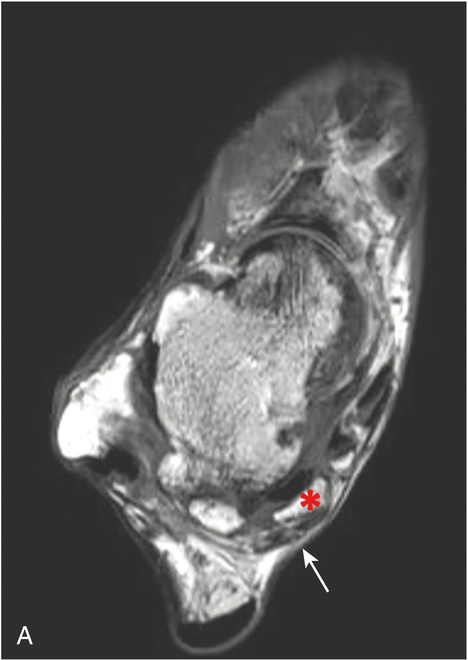
- Identifies the causative lesion in up to 88% of patients
- Demonstrates: ganglia, varicosities, accessory muscles, soft tissue tumors, post-traumatic heterotopic bone
- Essential for surgical planning (defines extent and location of compression)
- T1-weighted: anatomy, space-occupying lesions (lipomas, heterotopic bone)
- T2-weighted/STIR: edema, fluid-filled cysts, nerve signal change
- Shows nerve enlargement proximal to compression and flattening/signal change at the site
-
Dynamic assessment of nerve mobility and compressibility; identifies varicosities in real time
-
Lower cost, no radiation, available bedside
-
Operator-dependent; less comprehensive than MRI for soft tissue detail
-
Increasingly used for ultrasound-guided injection to improve accuracy and reduce risk to the neurovascular bundle
-
A 2025 paper (Reis Soares et al., PMID 41245551) specifically highlighted the role of USG in identifying vascular etiologies
-
Campbell's 2026, p.5147-5165; Rheumatology 2022, p.3217-3219
6. Treatment
Algorithm Overview
TTS Confirmed
    ↓
Conservative (6-12 weeks)
    ↓
Failure → Corticosteroid/Anesthetic Injection (non-surgical intervention)
    ↓
Continued failure OR identifiable space-occupying lesion → Surgical decompression
    ↓
Revision surgery if initial release fails (uncommon)
A. Conservative (Non-Surgical) Management
- Night splint or CAM walker boot: 6-12 weeks of ankle immobilization
- Avoidance of aggravating activities (prolonged standing, running)
- NSAIDs (oral): reduce perineural inflammation; short-term use
- Neuropathic agents: gabapentin (300-900 mg TID), pregabalin (75-150 mg BID), amitriptyline (10-75 mg nocte) - off-label but commonly used for the neuropathic component
- Topical agents: lidocaine patches, capsaicin cream
- Wide, cushioned, comfortable shoe
- For proximal TTS: standard longitudinal arch orthosis
- For distal TTS: orthosis with relief channel in the medial arch - a standard arch support may worsen distal symptoms by increasing pressure on the terminal branches
- Custom foot orthoses to correct hindfoot valgus biomechanics
- Tibial nerve mobilization/"neural flossing" techniques
- An RCT (2025, El-Nassag et al., PMID 40368127) showed that adding tibial nerve flossing (TNF) to conventional therapy (TENS + therapeutic US + gastrocnemius stretching) produced significantly greater improvement in pain, ankle ROM, and tibial nerve conduction velocity (p<0.05) vs. conventional therapy alone over 4 weeks
- Gastrocnemius/soleus stretching to reduce tension on the nerve
B. Non-Surgical Pain Interventions (Injection Therapies)
1. Corticosteroid + Local Anesthetic Injection (First-Line Injection Therapy)
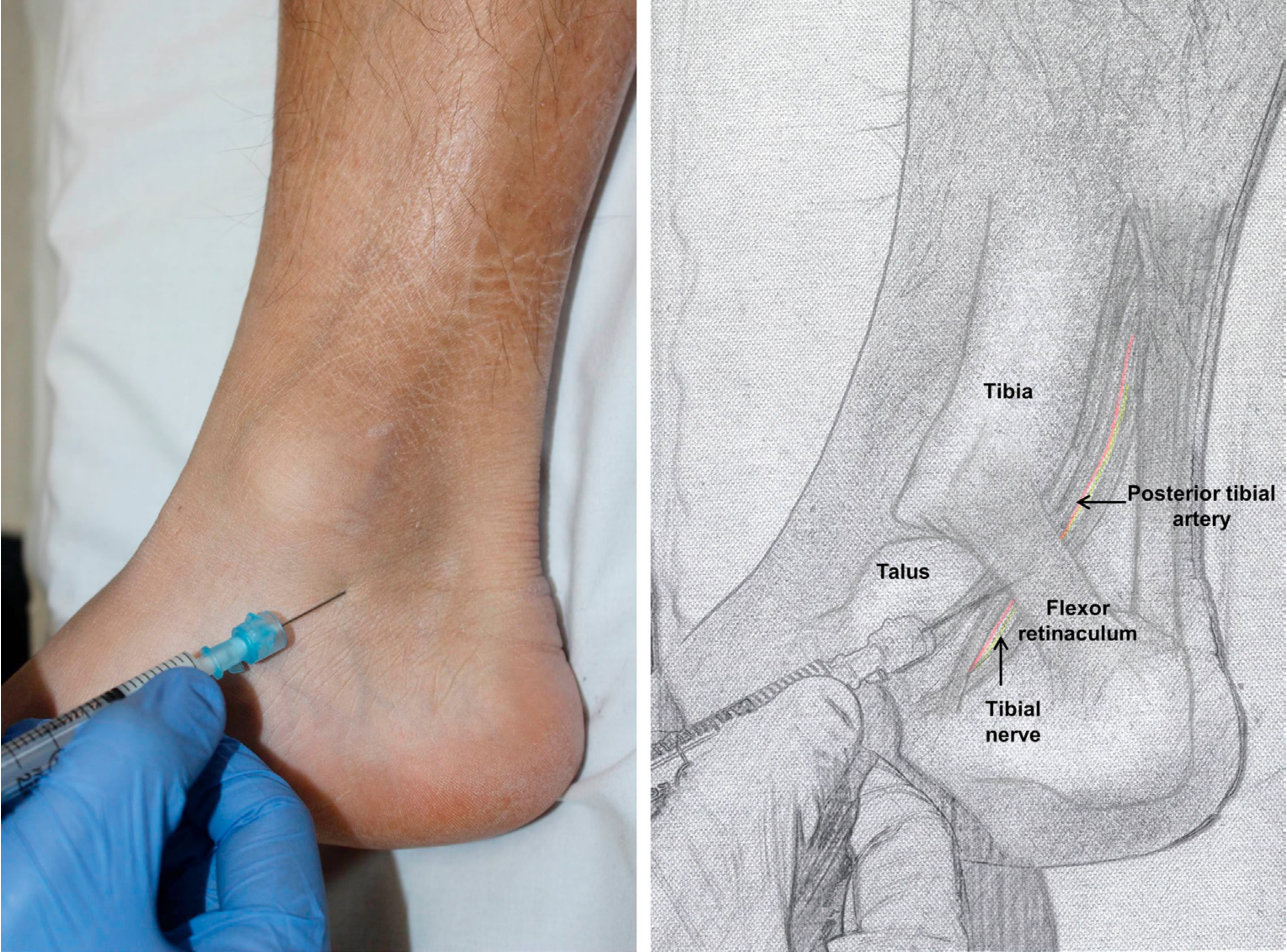
- Palpate medial malleolus and identify posterior tibial artery pulsation; mark artery to avoid
- Insert 25-gauge needle posterior to medial malleolus, anterior to the posterior tibial artery
- Direct needle parallel to skin, distally at 45 degrees along the line from big toe to heel
- Aspirate to confirm not intravascular, then inject slowly
- Injectate: hydrocortisone 25 mg/mL or methylprednisolone 10-20 mg ± 1-2 mL local anesthetic (e.g. 1% lidocaine)
- USG significantly reduces risk of inadvertent intravascular injection and tendon rupture
- Real-time visualization confirms injectate spread around the tibial nerve
- A retrospective cohort study published in Orthopedic Reviews found USG corticosteroid injection produced a mean pain reduction of ~3.6-3.9/10 (NRS) in both surgical and non-surgical groups; 63% of patients avoided surgery. Younger patients were more likely to fail conservative management
2. Hydrodissection / Nerve Release Injection Therapy
- Perineural injection with 5% dextrose (D5W) or dilute local anesthetic under ultrasound guidance
- Mechanically separates the nerve from surrounding adherent tissue/fascia
- Particularly useful in post-surgical TTS with perineural fibrosis
- Evidence is emerging; no TTS-specific RCTs, but supported by mechanism and carpal tunnel syndrome data
3. Platelet-Rich Plasma (PRP)
- Emerging option; theoretical benefit via neurotrophic and anti-inflammatory growth factors
- No TTS-specific controlled trial data available as of 2025
- Extrapolated from carpal tunnel syndrome evidence where PRP has shown benefit over corticosteroids for sustained relief at 6-12 months (Yang et al., PMID 38753671)
4. TENS (Transcutaneous Electrical Nerve Stimulation)
- Used as part of a multimodal conservative regimen
- Included in the RCT by El-Nassag et al. (2025) as standard care; beneficial for pain modulation
5. Therapeutic Ultrasound
- Phonophoresis with NSAIDs or corticosteroids directly to the tunnel
- Used adjunctively; evidence is empirical
- Firestein & Kelley's Textbook of Rheumatology, p.1050; Campbell's 2026, p.5182-5193
C. Surgical Treatment
- Failure of 6-12 weeks conservative + injection therapy
- Space-occupying lesion identified (ganglion, varicosity, tumor) - earlier surgery warranted
- Severe or progressive neurological deficit
- Identifiable MRI lesion (excision/decompression of specific cause)
- Symptom duration <1 year
- No systemic peripheral neuropathy
- Age 60-80 years
- Post-traumatic scarring within the tarsal canal
- Idiopathic TTS (no identifiable cause) - ~25% achieve little/no relief
- Protracted psychiatric illness
- Incision from 1 cm plantar to the navicular tuberosity, bisecting the area between medial malleolus and calcaneal tuberosity, ending 1 cm anterior to Achilles tendon
- Identify and ligate superficial veins connecting plantar and saphenous systems
- Incise flexor retinaculum along its entire length; extend 1-2 cm of deep fascia proximal to the upper retinacular edge
- Follow MPN and LPN beneath the abductor hallucis - both may pass through fascial slings requiring individual release
- Excise any space-occupying lesions; correct alignment disorders
- Use magnification, tourniquet, small scissors, and non-toothed forceps throughout
-
Causes of failure: incorrect diagnosis, inadequate release, poor technique, nerve scarring
-
Revision should address the specific cause; neurolysis with saphenous vein or collagen wrapping recommended for perineural scar adhesion
-
Outcomes of revision are unpredictable - counsel patients accordingly
-
Worsening symptoms immediately post-surgery may indicate iatrogenic nerve injury with subsequent neuroma formation
-
Campbell's Operative Orthopaedics 15th Ed 2026, p.5180-5290
7. Summary Evidence Table
| Intervention | Evidence Level | Notes |
|---|---|---|
| Conservative (splint, NSAIDs, orthotics) | Level IV | 6-12 weeks first-line; 63% success |
| Tibial nerve flossing (neural mobilization) | Level II (RCT 2025) | Effective adjunct to conventional PT |
| Corticosteroid injection (landmark) | Level III-IV | Short-medium term pain relief |
| USG corticosteroid injection | Level III | Preferred over landmark; safer |
| PRP injection | Level V | No TTS-specific RCT; extrapolated from CTS data |
| Hydrodissection/D5W | Level V | Emerging; mechanistically sound |
| Surgical decompression | Level IV | Best outcomes with identifiable lesion; ~75% success overall |
| Revision surgery | Level IV-V | Unpredictable; careful patient selection |
- Normal nerve conduction studies do NOT exclude TTS - clinical diagnosis must be primary
- MRI identifies causative lesion in up to 88% and is essential before surgery
- Distal TTS requires orthosis with a relief channel, not a standard arch support (which can worsen symptoms)
- Younger patients are more likely to fail conservative treatment and require surgery
- The Triple Compression Test (86% sensitivity, 100% specificity) is the most accurate clinical test
- Always consider "double crush" syndrome when proximal neurological pathology coexists