
What is myocardial infarction
myocardial infarction heart attack diagram pathophysiology
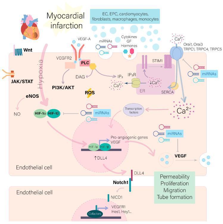
A pathophysiology diagram illustrating the intracellular signaling pathways in endothelial cells (ECs) activated following myocardial infarction (MI). The visual highlights the cellular response to ischemia and autocrine/paracrine signals from various heart cell populations (cardiomyocytes, fibroblasts, macrophages). Key pathways depicted include hypoxia-induced HIF-1α/HIF-1β stabilization, and activation of Wnt, JAK/STAT, PI3K/AKT, and eNOS/NO systems. The diagram emphasizes VEGF-A binding to VEGFR2, which triggers the PLC-IP3/DAG cascade, leading to Ca2+ release from the endoplasmic reticulum via IP3R and subsequent store-operated calcium entry (SOCE) through Orai and TRPC channels. These calcium signals and transcription factors drive the expression of pro-angiogenic genes like VEGF and DLL4. The model further illustrates the Notch signaling mechanism where DLL4 on a 'tip cell' activates Notch1/NICD in an adjacent 'stalk cell' (expressing Hes1/Hey1). The cumulative educational focus is on the hallmarks of angiogenesis: increased permeability, proliferation, migration, and tube formation essential for ischemic myocardial healing.
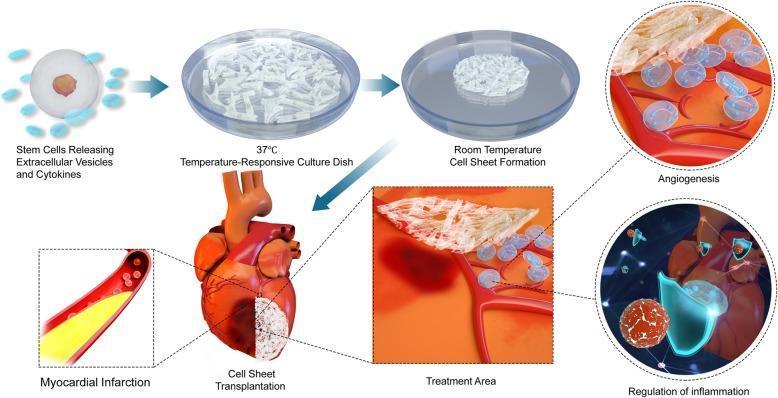
This pathophysiology diagram illustrates the process of cell sheet engineering for myocardial repair in ischemic heart disease. The multi-stage flowchart begins with stem cells releasing extracellular vesicles and cytokines. These cells are cultivated in a 37°C temperature-responsive culture dish, transitioning to room temperature to facilitate cell sheet formation. The resulting biological scaffold is then transplanted onto a heart with a white-shaded area representing myocardial infarction. Magnified 'Treatment Area' views detail the therapeutic mechanisms: Angiogenesis, showing the interaction between the fibrous cell sheet, transparent vesicles/cells, and the underlying vasculature; and Regulation of Inflammation, depicting immune cells and molecular signaling at the transplant site. This infographic summarizes stem cell-based regenerative therapy, focusing on paracrine signaling, tissue engineering, and vascularization strategies for treating damaged cardiac tissue.
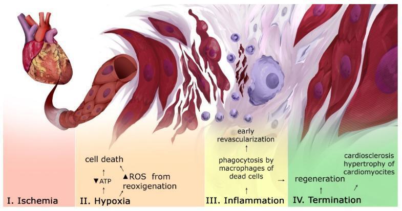
This pathophysiology diagram illustrates the chronological progression of tissue and cellular events following a myocardial infarction (MI), divided into four distinct stages. Stage I (Ischemia) shows an intact heart model. Stage II (Hypoxia) depicts the onset of cell death characterized by decreased ATP and increased reactive oxygen species (ROS) during reoxygenation, showing irregular, red-colored cardiomyocytes adjacent to a damaged capillary. Stage III (Inflammation) visualizes the early revascularization and immune response, including the phagocytosis of dead cell debris by macrophages (large irregular purple cells) and the infiltration of smaller leukocytes. Stage IV (Termination/Reparative phase) demonstrates myocardial regeneration and cardiosclerosis, where dead cells are replaced by dense white fibrous tissue (scarring) and surviving cardiomyocytes exhibit hypertrophy and more organized alignment. This educational infographic captures the transition from acute ischemic injury through the inflammatory cascade to chronic tissue remodeling and fibrosis, relevant for cardiovascular pathology and rehabilitative medicine study.
Myocardial Infarction (MI)
Definition
- Robbins & Kumar Basic Pathology, p. 353
Pathogenesis - How It Happens
- Plaque disruption - An atheromatous plaque is eroded or suddenly ruptured by endothelial injury, intraplaque hemorrhage, or mechanical forces. This exposes subendothelial collagen and necrotic plaque contents to circulating blood.
- Platelet activation - Platelets adhere, aggregate, and release thromboxane A₂, ADP, and serotonin, causing further platelet aggregation and vasospasm.
- Coagulation cascade - Tissue factor exposure activates the coagulation system, adding to the growing thrombus.
- Total occlusion - Within minutes, the thrombus can completely occlude the coronary artery lumen.
- Robbins & Kumar Basic Pathology, p. 353-354
What Happens to the Heart Muscle
- Within seconds: aerobic metabolism ceases; ATP drops; lactic acid accumulates.
- Within minutes: contractility is lost (reversible at this stage).
- After 20-40 minutes of sustained ischemia: irreversible damage and coagulative necrosis of myocytes begins.
- The earliest sign of necrosis is disruption of the sarcolemmal membrane, allowing intracellular enzymes (troponin, CK-MB) to leak into blood - the basis of biomarker testing.
- Has the highest oxygen consumption
- Is the last region to receive blood from epicardial vessels
- Is exposed to high intramural pressures during systole
- Guyton and Hall Medical Physiology, p. 271
- Robbins & Kumar Basic Pathology, p. 354-355
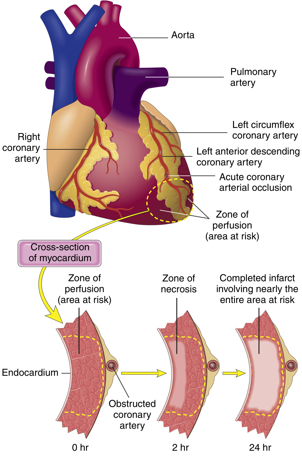
Coronary Artery Territory - Where Infarcts Occur
| Artery | % of MIs | Area infarcted |
|---|---|---|
| Left anterior descending (LAD) | 40-50% | Anterior LV wall, anterior 2/3 of septum, apex |
| Right coronary artery (RCA) | 30-40% | Right ventricle, inferior/posterior LV |
| Left circumflex (LCX) | 15-20% | Lateral left ventricle |
- Robbins & Kumar Basic Pathology, p. 355
Types of MI
| Type | Description |
|---|---|
| STEMI | ST-elevation MI - complete occlusion, full-thickness (transmural) infarct. Requires immediate reperfusion. |
| NSTEMI | Non-ST-elevation MI - partial occlusion, subendocardial infarct. Confirmed by elevated troponins without ST elevation. |
| Unstable Angina (UA) | Ischemia without biomarker elevation. Part of the ACS spectrum. |
- Frameworks for Internal Medicine, p. 33
Symptoms
- Chest pain/pressure - crushing, squeezing, or burning; often radiating to the left arm, jaw, or epigastrium
- Diaphoresis (heavy sweating)
- Nausea and vomiting
- Dyspnea (shortness of breath)
- Syncope or near-syncope
- Fatigue
Important: Women, diabetics, and post-operative patients often present atypically (without classic chest pain), more commonly with dyspnea, fatigue, or back pain.
- Frameworks for Internal Medicine, p. 32
Diagnosis
- ECG - obtained immediately. Key findings:
- STEMI: ST elevation in ≥2 contiguous leads; hyperacute (tall, broad-based, symmetrical) T-waves early on; later Q waves
- NSTEMI/UA: ST depression or T-wave inversions
- Cardiac biomarkers - Troponin I or T (most sensitive and specific); CK-MB. Rise and fall pattern is characteristic of MI.
- Coronary angiography - confirms occlusion and guides intervention
Causes of Death in Acute MI
- Decreased cardiac output (cardiogenic shock) - non-contracting myocardium; paradoxical systolic stretch worsens dysfunction
- Pulmonary edema - blood dams in the pulmonary vasculature
- Ventricular fibrillation - most common cause of sudden death; ischemia creates electrical instability. In 80-90% of out-of-hospital MI deaths, ventricular fibrillation is the terminal event.
- Cardiac rupture - rare but catastrophic; occurs in the first 1-2 weeks
- Guyton and Hall Medical Physiology, p. 271
Complications
- Arrhythmias (ventricular fibrillation, heart block)
- Heart failure / cardiogenic shock
- Mechanical complications: papillary muscle rupture (mitral regurgitation), ventricular septal defect, free wall rupture
- Pericarditis (Dressler syndrome)
- Ventricular aneurysm
- Mural thrombus with embolism
- Reperfusion injury - restoring blood flow can paradoxically worsen damage via reactive oxygen species and inflammatory mediators ("stunned myocardium")
Treatment
- Primary PCI (percutaneous coronary intervention) - preferred for STEMI if available within 90 minutes; involves balloon angioplasty and stenting
- Fibrinolytic therapy (thrombolytics) - used when PCI is not available within a timely window
- Antiplatelet agents - aspirin + P2Y12 inhibitor (clopidogrel, ticagrelor, prasugrel)
- Anticoagulants - heparin
- Beta-blockers - reduce myocardial oxygen demand, prevent arrhythmias
- ACE inhibitors - limit ventricular remodeling
- Statins - plaque stabilization and secondary prevention
- Nitrates - for pain and vasodilation
- Robbins & Kumar Basic Pathology
- Frameworks for Internal Medicine
Pathology Illustration (Stages After MI)

What should be disposed in different colors of bio degradable bin in hospital
hospital biomedical waste color coded bins categories disposal guidelines 2024
hospital biomedical waste color coded bins segregation chart
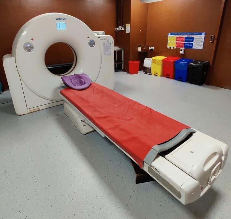
A clinical photograph of a modern Computed Tomography (CT) scanner set in a diagnostic imaging suite. The image features a white Siemens CT gantry with a large central bore and integrated digital display at the top. A motorized patient table extends from the gantry, equipped with a red protective cover and a contoured purple headrest. The clinical environment is further detailed by the presence of a color-coded waste segregation system consisting of red, yellow, blue, and black disposal bins along the back wall, beneath a medical facility poster. This visual provides educational context for diagnostic radiology equipment, the mechanics of cross-sectional imaging (slices), and the standard clinical setup required for trauma, oncology, and neurology diagnostics. The configuration illustrates the essential components used in human medical imaging for identifying internal pathologies such as fractures, tumors, or vascular abnormalities.
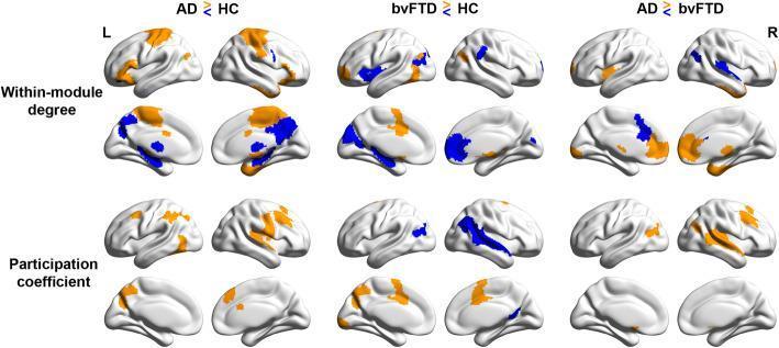
This diagnostic comparison chart utilizes brain surface plots derived from functional MRI (fMRI) data to visualize network-specific topological changes in Alzheimer’s disease (AD) and behavioral variant frontotemporal dementia (bvFTD) compared to healthy controls (HC). The image displays two primary graph theoretical metrics: Within-module degree (top rows), indicating intra-network connectedness, and Participation coefficient (bottom rows), representing inter-network distinctiveness (segregation). The cortical maps use a color-coded scale where orange denotes significant increases and blue denotes significant decreases (p < 0.05). In AD patients vs. HC, there is a prominent reduction in within-module degree in posterior regions like the precuneus and posterior cingulate cortex, alongside increased participation coefficients in the control and somatomotor networks, suggesting reduced network segregation. In contrast, bvFTD patients vs. HC exhibit significant connectivity decreases in frontal and insular regions within the salience/ventral attention network (SVAN). The head-to-head comparison (AD vs. bvFTD) further illustrates the divergent neurodegenerative patterns, emphasizing the posterior-dominant breakdown in AD versus the anterior/insular vulnerability in bvFTD.
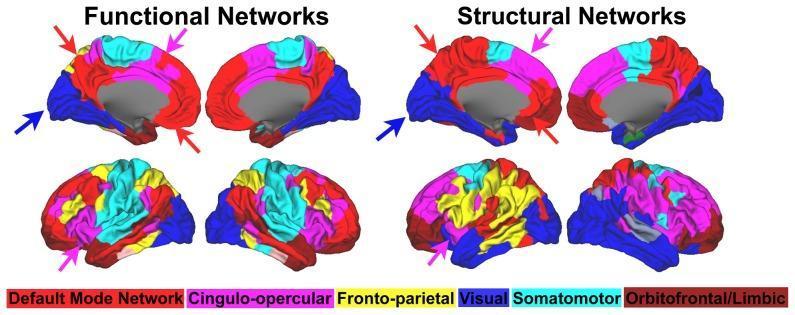
This Comparison Chart displays brain network community detection results from healthy adults, contrasting Functional Networks (left) and Structural Networks (right). The figure utilizes 3D cortical surface renderings in medial (top row) and lateral (bottom row) views. A color-coded visual key identifies six primary brain systems: Red (Default Mode Network), Pink (Cingulo-opercular), Yellow (Fronto-parietal), Dark Blue (Visual), Light Blue (Somatomotor), and Dark Red (Orbitofrontal/Limbic). Each cortical region is colored based on its assigned community membership. Arrows highlight specific anatomical regions, such as the posterior cingulate/precuneus and medial prefrontal cortex, where the spatial extent of communities like the Default Mode and Cingulo-opercular networks differs between the functional and structural modalities. The diagrams illustrate that while both network types share similar large-scale organizational principles, the functional connectivity patterns show higher system-specific segregation compared to the structural connectivity patterns. This visual is designed for advanced medical education in neuroanatomy and functional neuroimaging, specifically regarding the relationship between structural connectomics and resting-state fMRI networks.
https://www.diginerve.com/blogs/a-guide-to-different-types-o…
Hospital Biomedical Waste - Color-Coded Bin Guide
India - Bio-Medical Waste Management Rules 2016
🟡 YELLOW Bin / Yellow Non-Chlorinated Plastic Bag
| Sub-type | Examples |
|---|---|
| Human Anatomical Waste | Body parts, organs, limbs, tissues, fetuses |
| Animal Anatomical Waste | Animal body parts from veterinary/research settings |
| Discarded or Expired Medicine | Pharmaceuticals past expiry date |
| Microbiology & Lab Waste | Cultures, specimens, vaccine preparations, lab waste |
| Chemical Waste (Yellow-e) | Expired liquid chemicals, X-ray developer (spent hypo) stored in yellow container |
| Contaminated linen/bedding | Blood-soaked sheets, dressings, bandages with body fluids |
| Blood bags and blood products | Used/expired blood bags |
| Cytotoxic/Chemotherapy waste | All chemo drugs, cytotoxic/mutagenic agents |
🔴 RED Bin / Red Non-Chlorinated Plastic Bag (≥50 micron thickness)
| Examples |
|---|
| Syringes without needles (after needle removal) |
| IV tubes and sets, catheters |
| Soiled gloves, gowns, drapes |
| Urine bags, blood transfusion bags (empty) |
| Contaminated plastics |
| Specimen/sample collection containers (plastic) |
⬜ WHITE Bin / White Translucent Puncture-Proof Container
| Examples |
|---|
| Needles, syringes with fixed needles |
| Scalpels and surgical blades |
| Lancets |
| Broken glass with contamination |
| Metal implants / sharp metallic objects |
The CDC estimates ~385,000 needlestick injuries occur in hospital workers annually - proper white bin use is the primary prevention tool.
🔵 BLUE Bin / Blue-Marked Puncture-Proof Box
| Examples |
|---|
| Broken or discarded glass (non-contaminated or decontaminated) |
| Glass slides, vials, ampoules |
| Metallic implants removed from patients |
| Medicine vials (glass) |
Quick Reference Summary Table (India BMW Rules 2016)
| Color | Category | Key Contents | Treatment |
|---|---|---|---|
| 🟡 Yellow | Anatomical / Pharmaceutical / Chemo | Body parts, expired medicines, lab cultures, blood bags, chemo drugs | Incineration / Deep burial |
| 🔴 Red | Contaminated recyclable plastics | Syringes (no needle), IV sets, gloves, catheters, urine bags | Autoclave → Recycle |
| ⬜ White | Sharps | Needles, blades, scalpels, lancets, broken glass (contaminated) | Autoclave → Shred/Destroy |
| 🔵 Blue | Glassware / Metal implants | Glass vials, ampoules, slides, metallic implants | Disinfect → Secured landfill |
US/International (OSHA & WHO Standards)
| Color | Waste Type | Examples | Treatment |
|---|---|---|---|
| 🔴 Red | Infectious soft waste | Contaminated gloves, gowns, IV tubing, syringes (no needle) | Autoclave → landfill/recycle |
| 🟡 Yellow | Pathological + chemo waste | Tissues, body parts, blood bags, lab cultures, chemo/cytotoxic drugs | High-temp incineration |
| ⬜ White/Translucent | Sharps | Needles, scalpels, lancets | Autoclave → shred |
| 🔵 Blue | Non-hazardous pharma / glass | Non-RCRA expired drugs, glass | Landfill or incineration |
| ⬛ Black | RCRA hazardous pharma | Chemotherapy drugs meeting RCRA hazardous waste criteria | Regulated hazardous waste incineration |
What Should NOT Go in Biomedical Bins
- General paper/packaging waste - goes in regular general waste bins
- Food waste - general waste
- E-waste (batteries, circuit boards) - separate e-waste rules (E-waste Management Rules 2016)
- Radioactive waste - governed by Atomic Energy Act; separate handling entirely
- Liquid waste - sewage and liquid effluents treated in ETP (effluent treatment plant), not in color-coded bags
Key Rules to Remember
- Segregation happens at the point of generation - at the bedside, in the OT, in the lab
- Never mix categories - a yellow bag contaminated with sharps creates a serious safety hazard
- Bags should not be overfilled - fill only up to 3/4 capacity
- Label all bags with date, ward, and hospital name before transport
- Sharps never go in soft bags - always in rigid, puncture-proof white containers
- Chemotherapy waste always goes in yellow (incineration) - autoclaving does NOT destroy cytotoxic drugs
I have a interview for DMO in hospital only inpatient ward duties no emergency duty or casuality duties What questions doctor can ask me regarding my DMO job
DMO duty medical officer inpatient ward interview questions hospital India
DMO Interview Preparation - Inpatient Ward (No Emergency/Casualty)
1. Role Understanding & Responsibilities
Expected answer: Reviewing all admitted patients each shift, checking vitals charts, reviewing nursing notes, countersigning/updating medication orders, updating progress notes, calling consultants when required, declaring patients fit for procedures or discharge, documenting in medical records, and handover to the next DMO.
DMO manages day-to-day ward monitoring and implements treatment plans already made by the consultant. The consultant makes definitive treatment decisions; the DMO executes and watches for changes in patient condition.
Hand over all unstable or flagged patients verbally AND in writing - cover current vitals, recent changes, pending investigations, and any expected events. Use SBAR (Situation, Background, Assessment, Recommendation) format.
Patients who deteriorated overnight, those with abnormal vitals, post-operative patients in the first 24-48 hours, patients awaiting urgent reports, and those scheduled for procedures or discharge today.
2. Clinical Monitoring & Vitals
Go immediately, put the patient on oxygen (start with nasal prongs at 2-4 L/min, escalate to face mask/NRM if needed), check RR, HR, BP, auscultate chest, check if new onset or gradual, review current medications (sedatives?), call for ABG if available, and escalate to the consultant if no improvement.
Temperature, pulse, blood pressure, respiratory rate, SpO₂, urine output (in relevant patients), pain score, GCS (if neurological case), wound condition (if post-op), and input/output charts.
It's a scoring system based on 6-7 vital parameters. A rising score (typically ≥5) triggers escalation. It helps identify patients deteriorating before a crisis happens.
When there is: persistent hypoxia not responding to supplemental oxygen, hemodynamic instability (BP not responding to fluids), falling GCS/altered consciousness, worsening sepsis criteria, or any condition requiring invasive monitoring or ventilatory support.
3. Medication Management
Assess urgency - if it's a critical drug (e.g., insulin, anticoagulant, antiepileptic), escalate immediately to pharmacy and nursing in-charge. Document the issue. If unavailable, contact the consultant for an alternative. Never skip documentation.
Stop the suspected drug, assess severity (mild/anaphylaxis), give treatment accordingly (antihistamine, steroids, adrenaline for anaphylaxis), document in the patient's file, fill the ADR reporting form (Yellow Card / Pharmacovigilance), and inform the consultant.
Right Patient, Right Drug, Right Dose, Right Route, Right Time. (Some add: Right Documentation and Right Reason.)
Hold warfarin, inform consultant, monitor closely for signs of bleeding, check for interacting drugs/diet changes, and repeat INR. Vitamin K may be given per consultant advice.
4. Common Ward Scenarios
Call for help immediately, check for responsiveness and breathing, start BLS if no pulse/breathing, call for crash cart, attach defibrillator, follow ACLS protocol, inform consultant and senior, document time of events.
Wind, Water, Wound, Walking, Wonder drugs (the "5 Ws"):
- Day 1-2: Wind - atelectasis, pneumonia
- Day 3-5: Water - UTI, IV line infection
- Day 5-7: Wound - surgical site infection
- Day 7+: DVT/PE Examine accordingly, send cultures if indicated, review antibiotics with consultant.
If conscious and able to swallow - give 15g oral glucose (juice/glucose tablets). Recheck in 15 minutes. If unconscious - IV 25 mL of 50% dextrose (D50) or 150 mL of 10% dextrose, then start 10% dextrose infusion. Identify the cause (excess insulin? missed meal?), adjust insulin orders, and inform consultant.
Check hydration status and IV fluid input, check if catheter is blocked (flush/replace if catheterized), palpate for bladder distension, review medications (opioids cause urinary retention), check electrolytes/creatinine - if still no output after correcting factors, inform consultant, consider catheterization if not already done.
Listen respectfully to the family's concerns, explain the medical necessity of continued admission in simple language, involve the consultant in the conversation, document the discussion. If they insist on leaving against medical advice (LAMA), explain risks clearly, have them sign the LAMA form, and document thoroughly.
5. Documentation & Legal Aspects
Date and time, doctor's name and signature, patient's current complaints, examination findings, investigation results reviewed, assessment (diagnosis/changes), plan (medication changes, orders, referrals), and whether consultant was informed.
Confirm death (check for pulse, breathing, pupil response), note the time of death, inform the consultant, inform the family with empathy, complete the Death Certificate (if the cause is known and not suspicious), hand over the body to the mortuary as per hospital protocol, document everything, and inform the ward in-charge/nursing staff.
Leave Against Medical Advice. Document the patient's stated reason, risks explained to them, names of witnesses, and get the patient's signature on the LAMA form. A patient has the legal right to leave as long as they have decision-making capacity.
When the death is sudden/unexplained, within 24 hours of admission, post-operative death (especially within 24-48 hours), suspicious/medicolegal circumstances, injury-related death, or poisoning. In these cases, refer to the Medical Superintendent and/or police for medico-legal autopsy.
6. Infection Control & Hospital Protocols
Isolate the patient if indicated (e.g., MRSA, C. difficile), send appropriate cultures, review antibiotic policy with the consultant/microbiologist, report to the hospital infection control committee, implement contact precautions, and educate nursing staff on hand hygiene.
Gloves and gown at minimum. Add a mask if respiratory precautions apply. Remove PPE before leaving the patient's room and perform hand hygiene with soap and water or sanitizer.
Wash the wound immediately with soap and water (do not suck the wound), report to the occupational health/nodal officer, get baseline serology (HIV, HBV, HCV) for both source patient and injured staff, start PEP (Post-Exposure Prophylaxis) within 2 hours for HIV risk, document in the incident report.
7. Communication & Team Work
Use SBAR format:
- Situation: "Dr. X, patient in Bed 12, Mr. Y, is deteriorating."
- Background: "He was admitted 3 days ago with pneumonia, on IV antibiotics."
- Assessment: "His SpO₂ has dropped to 90% on 4L O₂, RR is 28, BP 90/60."
- Recommendation: "I think he may need ICU transfer. Can you please come and review?"
Remain calm and professional, focus on the patient's best interest, clarify your clinical reasoning, escalate to the consultant if the disagreement affects patient safety, and document the exchange.
8. Personal & Situational Questions
Focus on: interest in inpatient clinical medicine, desire to develop systematic patient monitoring skills, building clinical decision-making in a structured ward environment.
Prioritize by acuity using vitals flags. Delegate appropriate tasks to nursing staff. Use a structured checklist. Communicate early if the workload is unsafe. Document as you go rather than at the end of the shift.
Use a STAR answer (Situation, Task, Action, Result). Be specific, honest, and focus on what you learned.
Never act beyond your competence level. Call the consultant or senior on call. It is better to make one call and be corrected than to miss something serious. Document that you consulted.
Key Topics to Revise Before the Interview
| Topic | Why It Matters |
|---|---|
| Normal vitals and alarm limits | You'll be asked about these |
| ABG interpretation basics | Common ward skill |
| Insulin sliding scale management | Diabetic patients are very common |
| Blood transfusion reactions | Very common ward scenario |
| Post-op care basics (Day 1, 2, 3) | Especially surgical wards |
| DVT prophylaxis in admitted patients | Standard of care |
| Common ward drug dosages (heparin, dextrose, IV fluids) | Practical knowledge |
| SBAR communication | Key communication tool |
| Documentation standards | Legal requirement |
| Biomedical waste rules | Already covered above |
What i have to do if patient gets abdominal pain,hypertension,hypotension hyperthermia tachycardia in inpatient ward
DMO Ward Protocol: Managing 5 Acute Clinical Situations
Golden Rule for all 5: Assess → Stabilize → Investigate → Escalate to consultant using SBAR
1. 🔴 ABDOMINAL PAIN (New or Worsening in Inpatient)
Step 1 - Go Immediately & Do ABCDE First
- Is the patient hemodynamically stable? (BP, HR, SpO₂)
- Is the abdomen rigid/board-like? → Surgical emergency - call surgeon NOW
Step 2 - Take a Focused History (SOCRATES)
| Ask About | Why |
|---|---|
| Site and radiation | RUQ = liver/gallbladder; RIF = appendix; Epigastric = PUD/pancreatitis; Loin to groin = renal colic |
| Onset - sudden vs gradual | Sudden = perforation/rupture; Gradual = infection/obstruction |
| Character - colicky vs constant | Colicky = obstruction/stones; Constant = peritonitis/ischemia |
| Associated symptoms | Vomiting, fever, diarrhea, last bowel movement, urinary symptoms |
| Recent surgery | Post-op complications |
| Known conditions | Diabetes (DKA), IBD, peptic ulcer, gallstones |
Step 3 - Examine the Abdomen Systematically
- Inspect: Distension, visible peristalsis, scars
- Auscultate: Absent bowel sounds = ileus/peritonitis; High-pitched/tinkling = obstruction
- Percuss: Resonant = gas/obstruction; Dull = fluid/mass
- Palpate: Guarding (voluntary) vs Rigidity (involuntary = peritonitis), Rebound tenderness = peritoneal irritation
- Check PR/PV if indicated
- Check hernial orifices
Step 4 - Order Investigations
| Investigation | Looking For |
|---|---|
| CBC | Leucocytosis (infection), anaemia (bleeding) |
| LFT, Amylase/Lipase | Pancreatitis, hepatitis, cholecystitis |
| RFT, Electrolytes | Renal cause, DKA |
| Urine routine/microscopy | UTI, renal colic |
| Blood sugar | DKA |
| Serum β-hCG (females) | Ectopic pregnancy |
| Erect X-ray abdomen | Free gas under diaphragm (perforation), air-fluid levels (obstruction) |
| USG Abdomen | Gallstones, appendix, free fluid, organomegaly |
| CT Abdomen (with contrast) | If USG inconclusive; mesenteric ischemia, abscess |
| Lactate | If mesenteric ischemia suspected |
Step 5 - Immediate Management
- Keep patient NPO (nothing by mouth)
- Secure IV access - start IV fluids (Normal Saline or Ringer's Lactate)
- Pain relief - IV Paracetamol (1g) or Tramadol 50mg IV/IM after consultant advice (do not delay analgesia - this is an old myth)
- Antiemetic: Ondansetron 4mg IV if vomiting
- Insert Foley catheter if monitoring urine output needed
- If perforation/obstruction suspected: NGT insertion, mark time, call surgeon
- Serial abdominal exams - repeat every 1-2 hours and document
Escalate to Consultant When:
- Signs of peritonitis (rigidity, rebound tenderness, absent bowel sounds)
- Hemodynamic instability
- Fever >38.5°C with abdominal pain
- No improvement within 1-2 hours
- Post-op abdomen with sudden worsening
Textbook note: Management includes: ensure NPO, IV hydration, NGT decompression, serial exams, surgical/GI evaluation, pain control, antibiotics as indicated. - Harriet Lane Handbook
2. 🔴 HYPERTENSION (Sudden High BP in Ward)
First: Classify the Situation
| Category | BP Level | Features | Urgency |
|---|---|---|---|
| Incidental high BP | <180/110 | No symptoms, known hypertensive | Oral adjustment, follow-up |
| Hypertensive Urgency | ≥180/110 mmHg | No new end-organ damage | Oral medications, gradual reduction over 24-48 hrs |
| Hypertensive Emergency | Usually >180/110 mmHg | NEW end-organ damage | IV medications, ICU, immediate treatment |
Step 1 - Assess the Patient
- Check BP in both arms (difference >20 mmHg = aortic dissection?)
- Ask: Headache, visual blurring, chest pain, shortness of breath, confusion, hemiplegia?
- Check: Level of consciousness, papilledema (fundus), focal neurology, pedal edema, urine output
Step 2 - Look for End-Organ Damage (Emergency?)
| Organ | Sign | Action |
|---|---|---|
| Brain | Headache, confusion, seizures, focal deficit | CT head, call neurologist |
| Heart | Chest pain, pulmonary edema | ECG, troponin, call cardiologist |
| Kidney | Oliguria, rising creatinine | RFT, strict input/output |
| Eyes | Visual blurring, papilledema | Fundoscopy |
| Aorta | Tearing chest/back pain, BP difference between arms | CT aortogram, call surgeon |
Step 3 - Immediate Management
- Restart missed antihypertensive medications
- Add or increase oral antihypertensive per consultant
- Options: Oral Amlodipine 5mg, Losartan 50mg, or Clonidine
- Do NOT drop BP rapidly - reduce by no more than 25% over 24 hours (risk of stroke/MI from rapid reduction)
- Repeat BP every 30-60 minutes
- Shift to HDU/ICU - this is beyond routine ward management
- IV antihypertensives (Labetalol, Nicardipine, or Sodium Nitroprusside per consultant)
- Continuous BP monitoring (arterial line in ICU)
- Identify and treat the precipitating cause
Key point: "Rapid blood pressure reductions via either the oral or intravenous route run the short-term risk of precipitating myocardial, cerebral, or renal ischemia." - Goldman-Cecil Medicine
Step 4 - Document and Escalate
- Document time, BP readings, symptoms, drugs given
- Call consultant for any BP >180/110 with symptoms
3. 🔴 HYPOTENSION (Low BP in Ward)
Normal threshold: SBP < 90 mmHg or drop >40 mmHg from baseline
Step 1 - Go Immediately - ABCDE Assessment
- Is the patient conscious? Breathing? Pulse present?
- Check: HR, RR, SpO₂, temperature, urine output, capillary refill
Step 2 - Identify the Type of Shock
| Type | Clues | Common Causes in Ward |
|---|---|---|
| Hypovolemic | Tachycardia, cold/clammy, collapsed veins, low urine output | Bleeding (surgical), diarrhea/vomiting, poor intake, excessive diuretics |
| Septic | Fever or hypothermia, tachycardia, warm extremities initially | Hospital-acquired infection, post-op infection, UTI, pneumonia |
| Cardiogenic | Tachycardia, raised JVP, pulmonary edema, S3 gallop | MI, arrhythmia, heart failure decompensation |
| Distributive/Anaphylactic | Urticaria, bronchospasm, recent drug/blood transfusion | Drug reaction, blood transfusion reaction |
| Neurogenic | Bradycardia + hypotension, recent spinal procedure | Spinal anesthesia, high spinal cord injury |
Step 3 - Immediate Actions (First 5 Minutes)
- Call for help - alert nursing team and senior doctor
- Lay patient flat - raise legs 15-30° (Trendelenburg if tolerated)
- Secure 2 large-bore IV lines (16G or 18G)
- Oxygen - 15L/min via non-rebreather mask
- IV fluid challenge - 250-500 mL Normal Saline/Ringer's Lactate bolus FAST (unless cardiogenic shock - use cautiously)
- Stop any antihypertensives, diuretics, vasodilators currently running
- Monitor continuously - BP every 5 minutes, SpO₂, HR, urine output
Step 4 - Investigations
| Test | Purpose |
|---|---|
| CBC | Anaemia (bleeding), leucocytosis (sepsis) |
| Blood culture x 2 | If sepsis suspected (draw BEFORE antibiotics) |
| RFT, Electrolytes | Renal function, electrolyte imbalance |
| Blood glucose | Hypoglycemia can cause hypotension |
| Lactate | >2 mmol/L suggests tissue hypoperfusion |
| ECG | MI, arrhythmia causing cardiogenic shock |
| Troponin | MI |
| Urine output | Catheterize if not already - target >0.5 mL/kg/hour |
Step 5 - Targeted Treatment
- Hypovolemic: Aggressive IV fluids, transfuse if Hb <7-8 g/dL with active bleeding
- Septic: Blood cultures, start IV broad-spectrum antibiotics within 1 hour (per hospital antibiotic policy), fluid resuscitation, vasopressors if no response (ICU needed)
- Anaphylaxis: Adrenaline 0.5mg IM thigh immediately, IV fluids, IV hydrocortisone 200mg, IV chlorphenamine, call for senior help
When to Escalate Immediately:
- No response to 1L IV fluid bolus
- Lactate >2, worsening sensorium
- Signs of cardiogenic or septic shock
- Any unstable arrhythmia on ECG
4. 🔴 HYPERTHERMIA / HIGH FEVER (Temperature ≥38.5°C in Ward)
First: Distinguish Fever from True Hyperthermia
| Fever | True Hyperthermia | |
|---|---|---|
| Mechanism | Regulated - hypothalamus set-point raised by pyrogens | Unregulated - thermoregulation overwhelmed |
| Responds to antipyretics? | Yes (Paracetamol works) | No (antipyretics do not work) |
| Causes | Infection, malignancy, autoimmune, drugs | Heat stroke, malignant hyperthermia, NMS, thyroid storm |
"Antipyretics are not effective against heat illness and are not recommended to control environmental hyperthermia." - Rosen's Emergency Medicine
Step 1 - Assess Quickly
- Exact temperature (rectal is most accurate for high readings)
- Associated symptoms: Rigors, rash, neck stiffness, dysuria, cough, wound discharge
- Recent events: New drug, blood transfusion, IV line changes, surgical procedures
- Review drug chart: Recent antibiotics changed? New medications?
Step 2 - Common Causes by Post-Admission Day (5 Ws)
| Day | W | Cause |
|---|---|---|
| Day 1-2 | Wind | Atelectasis, aspiration pneumonia |
| Day 3-5 | Water | UTI, IV cannula infection |
| Day 5-7 | Wound | Surgical site infection, abscess |
| Day 7+ | Walking | DVT (low-grade fever) |
| Any day | Wonder drugs | Drug fever, transfusion reaction |
Step 3 - Investigations
| Test | Purpose |
|---|---|
| CBC with differential | Leucocytosis (bacterial), lymphocytosis (viral), eosinophilia (drug/parasitic) |
| Blood culture x 2 | Before giving antibiotics |
| Urine culture | UTI |
| Wound swab | Surgical site infection |
| Chest X-ray | Pneumonia, atelectasis |
| CRP, Procalcitonin | Bacterial infection marker |
| LFT | Hepatitis, cholangitis |
| Malarial antigen/smear | In endemic areas |
Step 4 - Management
- Remove/change IV cannula if insertion site red/swollen (commonest missed cause of hospital fever)
- Tepid sponging + fan
- IV/oral Paracetamol 1g every 6 hours (max 4g/day; reduce in liver disease)
- IV fluids to maintain hydration
- Start antibiotics only after cultures taken, and only per consultant guidance (avoid empiric antibiotics without indication)
- If drug fever suspected - withdraw the offending drug after consultant review
Escalate Immediately If:
- Temperature >40°C with altered sensorium (heat stroke or septic encephalopathy)
- Neck stiffness + fever (meningitis)
- Petechial/purpuric rash + fever (meningococcemia)
- Post-anesthetic patient with high fever + rigidity (malignant hyperthermia - rare, needs dantrolene)
- No antipyretic response + worsening vitals (escalate to ICU)
5. 🔴 TACHYCARDIA (HR >100 bpm in Ward)
Step 1 - Is the Patient Stable or Unstable?
| Unstable Signs | Action |
|---|---|
| Hypotension + tachycardia | Treat as shock (see above) |
| Chest pain + tachycardia | ECG immediately, call cardiology |
| Altered consciousness | Escalate to ICU immediately |
| SpO₂ falling | Oxygen + escalate |
Step 2 - Get an ECG Immediately (Always)
| ECG Pattern | Likely Diagnosis |
|---|---|
| Narrow complex, regular, rate 150 bpm | SVT or Atrial Flutter (2:1) |
| Narrow complex, irregularly irregular | Atrial Fibrillation |
| Wide complex, regular | Ventricular Tachycardia (VT) - EMERGENCY |
| Sinus tachycardia (P before every QRS) | Reactive - find the cause |
Step 3 - If Sinus Tachycardia (P wave before every QRS), Find the Cause
| Cause | Clue |
|---|---|
| Pain | Check pain score, wound, catheter |
| Fever | Check temperature (HR rises 10 bpm per °C) |
| Hypovolemia | Low BP, dry mucous membranes, poor skin turgor |
| Anaemia | Pallor, recent blood loss, low Hb |
| Hypoxia | Low SpO₂ |
| Anxiety/agitation | Confused post-op patient |
| Pulmonary embolism | Sudden dyspnea, pleuritic chest pain, post-op day 3-7 |
| Thyroid storm | Hyperthyroid history, tremor, sweating |
| Drugs | Salbutamol, atropine, theophylline, IV dopamine |
| Sepsis | Fever, hypotension, leucocytosis |
Step 4 - Immediate Investigations
- ECG (12-lead)
- SpO₂, ABG if hypoxic
- CBC, RFT, Thyroid profile
- Troponin (if chest pain)
- D-dimer / CTPA (if PE suspected)
- Blood culture (if sepsis suspected)
Step 5 - Management
- Oxygen if SpO₂ <95%
- Treat underlying cause (fluids for hypovolemia, paracetamol for fever, antibiotics for sepsis)
- SVT with haemodynamic stability: Valsalva maneuver → IV Adenosine 6mg rapid push (under consultant supervision)
- AF (new onset): Rate control (IV Metoprolol or Diltiazem) under consultant guidance
- VT: DO NOT treat alone - escalate, call code/senior immediately
- Beta-blockers only under consultant guidance - do not give blindly if hypotension is present
Always Escalate For:
- VT or VF on ECG
- HR >150 with hypotension
- No clear reversible cause identified
- New-onset AF in a non-AF patient
Summary Quick Reference Card
| Condition | First Action | Key Investigation | Don't Forget |
|---|---|---|---|
| Abdominal Pain | Keep NPO + IV access + examine abdomen | Erect X-ray, USG, Amylase | Rigid abdomen = call surgeon immediately |
| Hypertension | Classify urgency vs emergency | ECG, RFT, fundoscopy | Never drop BP too fast |
| Hypotension | Lay flat + IV fluids + oxygen | Lactate, blood culture, ECG | 2 large-bore IVs first |
| Hyperthermia | Remove/change IV line + Paracetamol + cultures | Blood/urine culture, CXR | Draw cultures BEFORE antibiotics |
| Tachycardia | ECG immediately | 12-lead ECG, SpO₂, CBC | Wide complex VT = call senior NOW |
Always document: Time of call, vitals at time of assessment, actions taken, consultant informed (time + name), and patient's response to treatment.
GCS scoring
Glasgow Coma Scale GCS scoring chart eye verbal motor
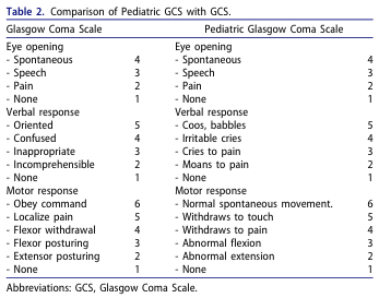
Table 2. Comparison of Pediatric GCS with GCS. <table><thead><tr><th>Glasgow Coma Scale</th><th></th><th>Pediatric Glasgow Coma Scale</th><th></th></tr></thead><tbody><tr><td>Eye opening</td><td></td><td>Eye opening</td><td></td></tr><tr><td>- Spontaneous</td><td>4</td><td>- Spontaneous</td><td>4</td></tr><tr><td>- Speech</td><td>3</td><td>- Speech</td><td>3</td></tr><tr><td>- Pain</td><td>2</td><td>- Pain</td><td>2</td></tr><tr><td>- None</td><td>1</td><td>- None</td><td>1</td></tr><tr><td>Verbal response</td><td></td><td>Verbal response</td><td></td></tr><tr><td>- Oriented</td><td>5</td><td>- Coos, babbles</td><td>5</td></tr><tr><td>- Confused</td><td>4</td><td>- Irritable cries</td><td>4</td></tr><tr><td>- Inappropriate</td><td>3</td><td>- Cries to pain</td><td>3</td></tr><tr><td>- Incomprehensible</td><td>2</td><td>- Moans to pain</td><td>2</td></tr><tr><td>- None</td><td>1</td><td>- None</td><td>1</td></tr><tr><td>Motor response</td><td></td><td>Motor response</td><td></td></tr><tr><td>- Obey command</td><td>6</td><td>- Normal spontaneous movement.</td><td>6</td></tr><tr><td>- Localize pain</td><td>5</td><td>- Withdraws to touch</td><td>5</td></tr><tr><td>- Flexor withdrawal</td><td>4</td><td>- Withdraws to pain</td><td>4</td></tr><tr><td>- Flexor posturing</td><td>3</td><td>- Abnormal flexion</td><td>3</td></tr><tr><td>- Extensor posturing</td><td>2</td><td>- Abnormal extension</td><td>2</td></tr><tr><td>- None</td><td>1</td><td>- None</td><td>1</td></tr></tbody></table> Abbreviations: GCS, Glasgow Coma Scale.
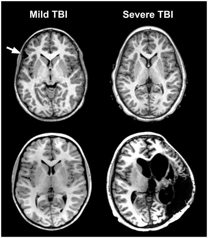
This comparison chart displays four axial T1-weighted magnetic resonance imaging (MRI) slices of the brain, categorized by Traumatic Brain Injury (TBI) severity to illustrate the disparity between clinical Glasgow Coma Scale (GCS) scores and structural pathology. The left column shows two cases classified as 'Mild TBI'. The top left slice exhibits focal encephalomalacia in the frontal lobe (indicated by a white arrow) and subtle asymmetry of the anterior horn of the lateral ventricle, suggesting regional parenchymal volume loss. The bottom left slice shows no visible gross abnormalities. The right column displays 'Severe TBI' cases. The top right slice appears structurally normal despite a low GCS score, while the bottom right slice demonstrates massive structural damage, including profound parenchymal loss, severe brain distortion, and large fluid-filled cystic cavities or significantly enlarged ventricles. This visual comparison highlights the heterogeneity of neuropathological outcomes in TBI, emphasizing that clinical severity indices may not fully reflect underlying structural integrity or long-term neuroanatomical changes.
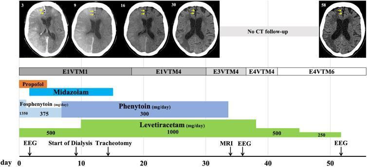
This infographic combines a diagnostic imaging timeline with a clinical management chart for a patient with cerebral arterial gas embolism (CAE). The top section features five axial head CT scans at days 3, 9, 16, 30, and 58. Yellow arrows highlight persistent, small, dark hypodense air pockets in the right frontal lobe/cingulate gyrus, demonstrating prolonged air retention. The lower section includes a timeline (Day 0 to 58) tracking the Glasgow Coma Scale (GCS) scores, which improve from E1VTM1 to E4VTM6. A pharmacological Gantt chart depicts the administration and dosage of sedatives (Propofol, Midazolam) and anticonvulsants (Fosphenytoin, Phenytoin, Levetiracetam). Annotated events include the start of dialysis, tracheotomy, and scheduling of EEG and MRI. This visual resource illustrates the clinical progression, long-term radiographic findings of intracranial air, and intensive seizure management in post-traumatic neurocritical care.
Glasgow Coma Scale (GCS) - Complete Guide
"The GCS provides a practical indicator of gross neurologic status by assessing motor function, verbal responses, and the patient's ability to open his or her eyes voluntarily or in response to external commands and stimuli." - Harrison's Principles of Internal Medicine, 22e
The 3 Components
👁️ E - Eye Opening (Max 4)
| Score | Response | How to Elicit |
|---|---|---|
| 4 | Spontaneous | Eyes already open before any stimulus - blinking normally |
| 3 | To sound/speech | Opens eyes when you speak or shout the patient's name |
| 2 | To pressure/pain | Opens eyes only when pain stimulus applied (nail bed pressure, sternal rub) |
| 1 | None | No eye opening even to painful stimulus |
| NT | Non-testable | Eyes swollen shut, facial trauma |
🗣️ V - Verbal Response (Max 5)
| Score | Response | What It Means |
|---|---|---|
| 5 | Oriented | Correctly states name, place, and date - fully alert |
| 4 | Confused | Speaks in sentences, can hold conversation, but disoriented |
| 3 | Words | Only single intelligible words (e.g., "help", "no", "pain") |
| 2 | Sounds | Only moaning, groaning - no recognizable words |
| 1 | None | Complete silence even to pain |
| NT | Non-testable | Intubated patient - write "VT" (verbal, tube) |
🤚 M - Motor Response (Max 6)
| Score | Response | Clinical Appearance |
|---|---|---|
| 6 | Obeys 2-part command | "Hold up 2 fingers" - follows instructions |
| 5 | Localizes to pain | Moves hand UP toward the painful stimulus purposefully |
| 4 | Normal flexion (withdrawal) | Pulls limb AWAY from pain - simple reflex withdrawal |
| 3 | Abnormal flexion (Decorticate) | Arm flexes at elbow + wrist, legs extend - indicates cortical damage |
| 2 | Extension (Decerebrate) | Arms AND legs extend and internally rotate - indicates brainstem damage |
| 1 | None | No movement at all - flaccid |
Visual Memory Aid for Motor Scores
M6 = Does what you SAY (obeys commands)
M5 = Reaches toward PAIN (localizes)
M4 = Pulls AWAY from pain (withdraws)
M3 = Arm BENDS abnormally (decorticate) ← bad
M2 = Arm STRAIGHTENS (decerebrate) ← worse
M1 = NOTHING (flaccid) ← worst
Decorticate vs Decerebrate Posturing
| Posture | Score | Arm Position | Leg Position | Level of Lesion |
|---|---|---|---|---|
| Decorticate | M3 | Flexed at elbow, fist clenched | Extended | Above midbrain (cortex/internal capsule) |
| Decerebrate | M2 | Extended + internally rotated | Extended | Midbrain/upper pons (brainstem) |
Decerebrate (M2) is worse than Decorticate (M3) - it indicates more caudal (lower) brain damage.
Total GCS Score = E + V + M
| Total Score | Severity | Clinical Interpretation |
|---|---|---|
| 15 | Normal | Fully alert and oriented |
| 13-14 | Mild impairment | Minor head injury / confusion |
| 9-12 | Moderate impairment | Moderate brain injury |
| ≤8 | Severe impairment | Coma - patient cannot protect airway |
| 3 | Deepest coma | No response in any component |
Key Clinical Rule
GCS ≤ 8 = Intubate A patient with GCS ≤8 cannot protect their own airway. This is the threshold for considering intubation/securing airway in most clinical guidelines.
How to Document GCS Properly
GCS 11 = E3 V3 M5
❌ Incorrect: GCS 11 (alone - doesn't tell you which components are affected)"Individual patients are best described by the three components of the coma scale (eye, verbal, motor, e.g., E3V4M6); the derived total coma score is less informative and should only be used to characterize groups of patients." - Harrison's Principles of Internal Medicine, 22e (2025)
How to Apply Pain Stimulus Correctly
| Type | Technique | Use For |
|---|---|---|
| Nail bed pressure | Press pen firmly on fingernail | Peripheral - for motor localization |
| Sternal rub | Knuckles rubbed on sternum | Central stimulus |
| Trapezius pinch | Pinch trapezius muscle | Central stimulus |
| Supraorbital pressure | Press above eyebrow on orbital rim | Central - careful, avoid in facial fractures |
Always use the best response from the best limb for the motor score.
GCS in Special Situations (Non-Testable - NT)
| Situation | Component Affected | Document As |
|---|---|---|
| Eyes swollen shut / orbital fracture | Eye (E) | E-NT |
| Intubated / tracheostomy | Verbal (V) | V-T or V-NT |
| Paralyzed limbs / spinal cord injury | Motor (M) | M-NT |
| Sedated patient | All components | Document time and sedation given |
Paediatric GCS (for children <5 years)
| Score | Verbal (Child) |
|---|---|
| 5 | Coos, babbles (infant) / Normal words (child) |
| 4 | Irritable crying |
| 3 | Cries to pain |
| 2 | Moans to pain |
| 1 | None |
- Pediatric GCS comparison, Harriet Lane Handbook
GCS Severity Classification Summary
| GCS | TBI Grade | Action in Ward |
|---|---|---|
| 15 | Normal | Routine monitoring |
| 13-14 | Mild | Neuro observations every 1-2 hrs, watch for deterioration |
| 9-12 | Moderate | Hourly neuro obs, inform consultant, CT head |
| ≤8 | Severe | Airway at risk - inform consultant immediately, prepare for ICU/intubation |
| ≤8 + falling | Deteriorating | Emergency - call senior NOW |
Quick Memory Trick
"456 - Eyes, Verbal, Motor"
- Eyes max = 4
- Verbal max = 5
- Motor max = 6 Total max = 15