
Hypotension and hypertension
Reading File
The topic is broad, so let me clarify what you need before diving in.
Asking for Preferences
What would you like to know about hypotension and hypertension? · What is your context?
I'll provide a comprehensive overview of both conditions from the medical library.
Searching sources...
Finding Sources
Finding Sources
Reading File
Reading File
Finding Sources
Finding Sources
Finding Sources
Finding Sources
Reading File
Finding Sources
Reading File
Finding Sources
Finding Sources
Reading File
I now have enough material from the medical library to give a thorough, structured overview of both conditions. Here is a comprehensive comparison:
Hypotension and Hypertension
HYPERTENSION
Definition and Classification
Hypertension is persistently elevated blood pressure (BP). The AHA/ACC guidelines define five categories (per JNC-based classifications):
| Category | Systolic (mmHg) | Diastolic (mmHg) |
|---|---|---|
| Normal | < 120 | < 80 |
| Elevated | 120-129 | < 80 |
| Stage 1 Hypertension | 130-139 | 80-89 |
| Stage 2 Hypertension | 140-159 | 90-99 |
| Stage 3 Hypertension | ≥ 160 | ≥ 100 |
These categories carry progressively increasing cardiovascular (CV) risk, independent of other risk factors. The Brenner and Rector Kidney textbook notes that death from heart disease and stroke increases linearly from BP levels as low as 115/75 mmHg - meaning there is no true "safe floor" of BP in terms of cardiovascular risk.
Types
1. Primary (Essential) Hypertension (~90-95% of cases)
- No identifiable single cause
- Linked to genetics, diet (sodium), obesity, physical inactivity, stress, and aging
2. Secondary Hypertension (~5-10% of cases)
- Renovascular disease (renal artery stenosis)
- Chronic kidney disease (CKD)
- Primary aldosteronism
- Pheochromocytoma
- Obstructive sleep apnea
- Thyroid disorders
- Drug-induced (NSAIDs, oral contraceptives, stimulants)
ICD classification also includes: hypertensive heart disease (402), hypertensive CKD (403), and combined hypertensive heart and kidney disease (404).
Pathophysiology
BP = Cardiac Output x Systemic Vascular Resistance. Hypertension results from:
- Increased sympathetic nervous system activity
- Renin-Angiotensin-Aldosterone System (RAAS) overactivation - angiotensin II causes vasoconstriction and sodium/water retention
- Endothelial dysfunction - reduced nitric oxide, increased endothelin
- Increased sodium retention by the kidneys (pressure-natriuresis curve shift)
- Structural arterial changes (arteriosclerosis, reduced compliance)
Risk Factors
- Age (risk doubles each decade after 55)
- Hypertension is a 2.5-fold risk factor for PAD in men and a 3.9-fold risk in women (Kannel and McGee, 1985)
- Obesity, diabetes, dyslipidemia, smoking
- Family history
Target Organ Damage (Complications)
| Organ | Complication |
|---|---|
| Heart | Left ventricular hypertrophy, heart failure, coronary artery disease |
| Brain | Stroke (ischemic and hemorrhagic), hypertensive encephalopathy |
| Kidneys | Hypertensive nephrosclerosis, CKD progression |
| Eyes | Hypertensive retinopathy |
| Arteries | Accelerated atherosclerosis, aortic dissection |
Per Comprehensive Clinical Nephrology, the association of BP with CV and kidney complications is continuous over the entire BP range - risk starts rising even before the hypertension threshold.
Treatment
Lifestyle Modifications (all stages):
- DASH diet (low sodium, high potassium/fruits/vegetables)
- Weight loss, regular aerobic exercise
- Limit alcohol, smoking cessation
Pharmacotherapy:
First-line agents (from Brenner and Rector's The Kidney):
- Thiazide diuretics (e.g., chlorthalidone, hydrochlorothiazide)
- ACE inhibitors (e.g., lisinopril, ramipril) - preferred in CKD, diabetes, heart failure
- Angiotensin Receptor Blockers (ARBs) (e.g., losartan) - ACE inhibitor intolerant patients
- Calcium channel blockers (e.g., amlodipine) - particularly effective in elderly and Black patients
- Beta-blockers - preferred in heart failure with reduced EF, post-MI
The landmark ALLHAT trial showed no significant difference in major CV outcomes between thiazide diuretics, CCBs, and ACE inhibitors for primary hypertension.
In CKD, treatment should begin when BP > 130/80 mmHg, using RAAS blocking agents (ACE inhibitors or ARBs) which provide kidney and cardiovascular benefits beyond BP reduction alone.
HYPOTENSION
Definition
Hypotension is generally defined as a systolic BP < 90 mmHg or a mean arterial pressure (MAP) < 65 mmHg, though clinical context always matters. The ICD classification includes:
- 458.0 - Orthostatic (postural) hypotension
- 458.1 - Chronic hypotension (permanent, idiopathic)
- 458.2 - Iatrogenic hypotension (due to medical treatment)
- 458.8/458.9 - Other/unspecified
Types and Causes
1. Orthostatic (Postural) Hypotension
A drop in SBP ≥ 20 mmHg or DBP ≥ 10 mmHg within 3 minutes of standing. Causes:
- Volume depletion (dehydration, hemorrhage, diuretics)
- Autonomic failure: Shy-Drager syndrome (multiple system atrophy), Riley-Day syndrome, diabetic autonomic neuropathy, Guillain-Barré, amyloidosis, AIDS, alcoholic neuropathy
- Drugs: antihypertensives, diuretics, nitrates, CCBs, alpha-blockers, L-dopa, tricyclic antidepressants, MAOIs, sildenafil, opiates, phenothiazines
- (Bradley and Daroff's Neurology in Clinical Practice)
2. Vasovagal (Neurally-mediated) Syncope
- Most common cause of transient hypotension leading to syncope
- Affects 20-25% of young people
- Triggered by emotional stimuli, pain, prolonged standing, heat, venipuncture
- Features: pallor, diaphoresis, nausea, bradycardia
3. Distributive Shock (Septic/Anaphylactic/Neurogenic)
- Septic shock: hypotension + vasopressor requirement + lactate > 2 mmol/L (Sepsis-3 definition, per Rosen's Emergency Medicine)
- Anaphylaxis: histamine-mediated massive vasodilation
- Neurogenic shock: spinal cord injury/trauma, spinal anesthesia - loss of sympathetic vasomotor tone
4. Hypovolemic Shock
- Hemorrhage (trauma, GI bleed, ruptured aortic aneurysm)
- Volume depletion (burns, vomiting, diarrhea, pancreatitis)
- Signs of hypoperfusion: tachycardia, dusky/mottled skin, delayed capillary refill, altered mental state, low urine output, elevated lactate, low central venous O2 saturation
5. Cardiogenic Shock
- Massive MI, severe heart failure, arrhythmias, cardiac tamponade, tension pneumothorax
- Cardiac failure causing systemic hypoperfusion with lactic acidosis and organ dysfunction
6. Iatrogenic Hypotension
- Vasodilators (nitrates, hydralazine) - especially in RV infarction, hypertrophic obstructive cardiomyopathy, aortic stenosis
- Over-diuresis
Clinical Features
Symptoms relate to organ hypoperfusion:
- Dizziness, lightheadedness, blurred vision (cerebral hypoperfusion)
- Weakness, fatigue, pallor, diaphoresis
- Oliguria (renal hypoperfusion)
- Chest pain (cardiac ischemia)
- Altered consciousness, syncope (severe)
Management
Orthostatic Hypotension:
- Increase fluid/salt intake; compression stockings
- Avoid precipitating drugs
- Fludrocortisone (mineralocorticoid - increases sodium retention)
- Midodrine (alpha-1 agonist - increases vascular tone)
- Droxidopa (norepinephrine prodrug) - approved for neurogenic orthostatic hypotension
Hemorrhagic Shock (from Rosen's Emergency Medicine):
- Ensure adequate ventilation/oxygenation
- Immediate hemorrhage control (direct pressure, REBOA)
- Isotonic crystalloid infusion (10-20 mL/kg)
- Packed red blood cell (PRBC) transfusion if perfusion is poor and hemorrhage control is delayed
- Balanced transfusion (PRBCs : FFP : platelets) for massive hemorrhage
- Treat coincident dysrhythmias
Septic Shock:
- IV fluid resuscitation
- Vasopressors (norepinephrine is first-line) if hypotension persists despite adequate fluid resuscitation
- Antibiotics within 1 hour
- Source control
Cardiogenic Shock:
- Treat underlying cause (revascularization in STEMI)
- Vasopressors/inotropes (dobutamine, norepinephrine)
- Mechanical circulatory support (IABP, Impella) in refractory cases
Quick Comparison Table
| Feature | Hypertension | Hypotension |
|---|---|---|
| BP | > 130/80 mmHg (Stage 1) | < 90/60 mmHg |
| Primary risk | Chronic: silent end-organ damage | Acute: tissue hypoperfusion/shock |
| Most common type | Essential (primary) | Orthostatic |
| Key mechanism | Increased SVR / RAAS activation | Decreased CO / low SVR / volume loss |
| Chronic complications | Stroke, MI, CKD, retinopathy | Recurrent falls, syncope, end-organ ischemia |
| First-line treatment | Lifestyle + thiazide/ACE/ARB/CCB/BB | Treat cause + fluids +/- vasopressors |
Sources: Brenner and Rector's The Kidney | Rosen's Emergency Medicine | Bradley and Daroff's Neurology in Clinical Practice | Comprehensive Clinical Nephrology, 7th ed. | Pfenninger and Fowler's Procedures for Primary Care
Hypothyroidism and hyperthyroidism
Searching sources...
Finding Sources
Finding Sources
Finding Sources
Finding Sources
Reading File
Reading File
Reading File
Finding Sources
Reading File
Finding Sources
Reading File
Finding Sources
Reading File
Finding Sources
Reading File
Loading Image
I now have comprehensive material from the library. Here is the full structured overview:
Hypothyroidism and Hyperthyroidism
The Thyroid Axis (Background)
The hypothalamus releases TRH (thyrotropin-releasing hormone) → stimulates the pituitary to release TSH (thyroid-stimulating hormone) → stimulates the thyroid gland to produce T4 (thyroxine) and T3 (triiodothyronine). T4 is converted to the more active T3 in peripheral tissues. Both exert negative feedback on the hypothalamus and pituitary.
- Primary thyroid disease: problem is in the gland itself
- Secondary: problem in the pituitary (low TSH)
- Tertiary: problem in the hypothalamus (low TRH)
HYPERTHYROIDISM
Definition
Hyperthyroidism is an excess of thyroid hormones in the body. The terms hyperthyroidism and thyrotoxicosis are often used interchangeably - thyroid storm is its life-threatening extreme manifestation.
Causes
Primary Hyperthyroidism (excess production by the thyroid gland):
| Cause | Notes |
|---|---|
| Graves' disease | Most common - 85% of all cases; TSH-receptor antibodies drive uncontrolled hormone synthesis; occurs 10x more in women |
| Toxic multinodular goiter | Second most common |
| Toxic (hyperfunctioning) adenoma | Single overactive nodule |
| Thyroiditis (subacute/de Quervain) | Painful; hormone released from inflamed follicles |
| Hashimoto's thyroiditis (early) | Initial "hashitoxicosis" phase before eventual hypothyroidism |
| Iodine-induced hyperthyroidism | Wolff-Chaikoff effect failure |
| Factitious thyrotoxicosis | Exogenous thyroxine intake |
| Struma ovarii | Ectopic thyroid in ovarian teratoma |
| Neonatal thyrotoxicosis | From maternal Graves' disease (transplacental TSH receptor antibodies) |
Secondary Hyperthyroidism (rare):
- TSH-secreting pituitary adenoma (PitNET)
Pathophysiology
Excess thyroid hormone causes:
- Hypermetabolic state - increased basal metabolic rate, heat production, weight loss despite increased appetite
- Beta-adrenergic overstimulation - tachycardia, tremor, anxiety, diarrhea, sweating
- Cardiac effects - increased contractility and output → tachycardia, palpitations, cardiomegaly, atrial fibrillation; in severe/chronic cases: high-output heart failure, thyrotoxic cardiomyopathy
- Bone resorption - osteoporosis and fracture risk (thyroid hormone stimulates osteoclasts)
- Sympathetic nervous system overactivity - nervous system features below
Clinical Features (Robbins Pathology)
| System | Features |
|---|---|
| General | Weight loss despite increased appetite, heat intolerance, sweating, fever |
| Cardiovascular | Tachycardia, palpitations, wide pulse pressure, systolic hypertension, atrial fibrillation, heart failure |
| Nervous system | Tremor, anxiety, emotional lability, insomnia, inability to concentrate |
| Musculoskeletal | Proximal muscle weakness (thyroid myopathy), decreased muscle mass |
| Skin | Soft, warm, flushed, moist skin; fine hair; onycholysis |
| Eyes | Wide staring gaze, lid lag (sympathetic overstimulation of superior tarsal muscle) |
| GI | Diarrhea, hyperdefecation, fat malabsorption |
| Reproductive | Oligomenorrhea/amenorrhea; reduced fertility |
| Thyroid | Goiter (diffuse in Graves'; not always present) |
Graves' disease specific:
- Exophthalmos (proptosis) - due to accumulation of loose connective tissue and GAGs behind the orbit (NOT just sympathetic activation); severity does not parallel thyroid dysfunction
- Pretibial myxedema - non-pitting, waxy skin thickening over the shins
- Thyroid acropachy - clubbing + periosteal new bone formation (rare)
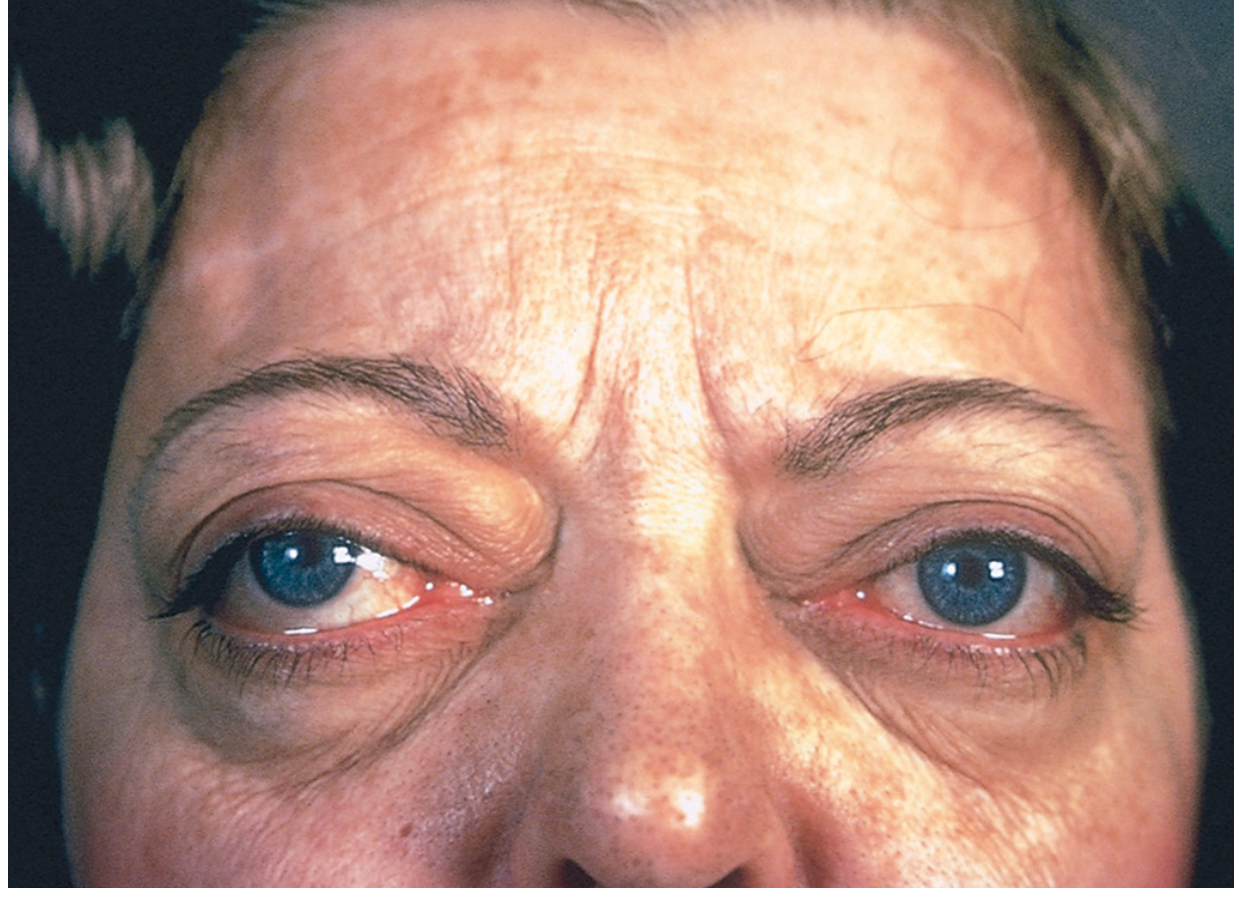
Apathetic hyperthyroidism - occurs in elderly patients; the typical hyperadrenergic features are blunted; diagnosis often found incidentally on workup for unexplained weight loss or worsening cardiac disease.
Diagnosis
| Test | Finding in Primary Hyperthyroidism |
|---|---|
| TSH | Low (suppressed) - most sensitive single test; decreased even in subclinical disease |
| Free T4 | Elevated |
| Free T3 | Elevated (T3 toxicosis if only T3 is elevated) |
| TSI / TRAb antibodies | Positive in Graves' disease |
| Radioactive iodine uptake (RAIU) | Diffusely increased (Graves'), focal uptake (toxic adenoma), decreased (thyroiditis) |
- In secondary hyperthyroidism (pituitary adenoma): TSH is normal or elevated (paradoxical)
- "T3 toxicosis": free T4 may be normal - must measure T3 directly
Treatment
Step 1 - Symptom control:
- Beta-blockers (propranolol 60-80 mg every 4h, or atenolol) - control tachycardia and adrenergic symptoms; propranolol also inhibits peripheral T4→T3 conversion
Step 2 - Reduce thyroid hormone synthesis (thionamides):
- Methimazole (preferred; once-daily dosing) - first-line for most patients
- Propylthiouracil (PTU) - preferred in pregnancy (1st trimester), thyroid storm (also blocks T4→T3 conversion), and methimazole allergy; avoid in children
Step 3 - Definitive therapy:
- Radioactive iodine (RAI / I-131) - most common definitive treatment; causes ablation of thyroid tissue; contraindicated in pregnancy
- Surgery (thyroidectomy) - for large goiters, compressive symptoms, malignancy suspicion, pregnancy, or patient preference
- Both RAI and surgery often result in hypothyroidism requiring lifelong levothyroxine
For Graves' ophthalmopathy:
- Mild: artificial tears, smoking cessation, selenium 100 mcg bid
- Moderate-severe: IV methylprednisolone (500 mg weekly x6, then 250 mg weekly x6)
- Non-responsive: orbital decompression surgery; teprotumumab (anti-IGF-1R monoclonal antibody)
Thyroid Storm (Thyrotoxic Crisis)
A life-threatening emergency - mortality 4-25% even with treatment (Harrison's Principles of Internal Medicine, 22e).
Precipitants: infection, surgery, trauma, stroke, DKA, abrupt cessation of antithyroid drugs, radioiodine in untreated hyperthyroid patient, childbirth
Features: fever, severe tachycardia, delirium/agitation, seizures, coma, vomiting, diarrhea, jaundice, cardiac arrhythmias
Management (Harrison's 22e):
- PTU 500-1000 mg loading dose, then 250 mg every 4h (drug of choice - also blocks T4→T3 conversion)
- One hour later: Stable iodide (SSKI 5 drops every 6h) - Wolff-Chaikoff effect (delay prevents iodine from being used to make new hormone)
- Propranolol 60-80 mg PO every 4h (or 2 mg IV every 4h) - controls heart rate; also inhibits T4→T3; use esmolol IV if titration needed
- Glucocorticoids - hydrocortisone 300 mg IV bolus, then 100 mg every 8h (reduces hormone release, inhibits conversion, covers adrenal insufficiency)
- Supportive: cooling, IV fluids, oxygen, cholestyramine (sequesters thyroid hormones in gut), antibiotics if infection, ICU monitoring
HYPOTHYROIDISM
Definition
A condition caused by any structural or functional derangement that reduces thyroid hormone production. Prevalence: overt hypothyroidism ~0.3%; subclinical hypothyroidism >4%. Nearly 10x more common in females than males; prevalence increases with age.
Causes
(Robbins Pathologic Basis of Disease)
Primary Hypothyroidism (most cases):
| Cause | Notes |
|---|---|
| Hashimoto's thyroiditis | Most common cause in iodine-sufficient countries; autoimmune destruction |
| Iodine deficiency | Most common cause worldwide (endemic areas) |
| Post-ablative | After radioiodine therapy or thyroidectomy for hyperthyroidism/cancer |
| Drugs | Lithium, amiodarone, iodides, p-aminosalicylic acid |
| Congenital | Thyroid agenesis/hypoplasia, dyshormonogenetic goiter (defects in T3/T4 synthesis steps) |
| External irradiation | Head/neck radiation |
Secondary/Central Hypothyroidism (rare):
- Pituitary failure (low TSH) - e.g., pituitary adenoma, Sheehan syndrome
- Hypothalamic failure (low TRH) - e.g., craniopharyngioma
Hashimoto's Thyroiditis (Autoimmune Hypothyroidism)
The most common cause in iodine-sufficient areas. Pathogenesis involves breakdown of self-tolerance to thyroid autoantigens, leading to:
- CD8+ cytotoxic T-cell destruction of thyrocytes
- Cytokine-mediated injury (IFN-γ)
- Autoantibodies: anti-thyroid peroxidase (anti-TPO) and anti-thyroglobulin antibodies
Histology (Robbins): Dense lymphocytic infiltrate with germinal center formation; follicular atrophy; oncocyte (Hürthle cell) metaplasia of follicular epithelium - characteristic finding
"Hashitoxicosis": early transient phase of hyperthyroidism as follicle disruption releases stored hormone, before autoimmune destruction causes permanent hypothyroidism.
Hashimoto patients are at increased risk for other autoimmune diseases: type 1 diabetes, SLE, myasthenia gravis, Sjögren syndrome.
Pathophysiology
Insufficient thyroid hormone leads to:
- Reduced basal metabolic rate - generalized "slowing" of all body functions
- Reduced sympathetic tone - opposite of hyperthyroid picture
- Accumulation of hyaluronic acid and GAGs in tissues - myxedema (non-pitting edema)
- Hyponatremia - impaired free-water excretion
- Dyslipidemia - reduced LDL receptor expression → elevated LDL and total cholesterol
Clinical Features
(Kaplan & Sadock's Psychiatry, Harrison's, Robbins)
| System | Features |
|---|---|
| General | Fatigue, lethargy, cold intolerance, weight gain |
| Cardiovascular | Bradycardia, reduced cardiac output, pericardial effusion, hypertension (diastolic), dyslipidemia |
| Nervous system | Cognitive slowing ("brain fog"), depression, somnolence, psychosis ("myxedema madness") |
| Musculoskeletal | Stiffness and cramping, proximal weakness, carpal tunnel syndrome |
| Skin | Dry, coarse skin; non-pitting periorbital edema; coarse/sparse hair; prolonged relaxation phase of deep tendon reflexes (pathognomonic) |
| GI | Constipation, reduced gut motility |
| Voice/tongue | Coarsening and deepening of voice; macroglossia (thickened tongue) |
| Reproductive | Menorrhagia, anovulation, reduced fertility, hyperprolactinemia |
| Metabolic | Elevated LDL/cholesterol, hypoglycemia tendency, hyponatremia |
Key signs: prolonged DTR relaxation phase, periorbital puffiness, macroglossia, bradycardia, dry skin, coarse hair.
Diagnosis
| Test | Finding in Primary Hypothyroidism |
|---|---|
| TSH | Elevated (most sensitive; elevated even in subclinical disease) |
| Free T4 | Decreased |
| Anti-TPO antibodies | Positive in Hashimoto's |
| Cholesterol/LDL | Elevated |
| CBC | Normocytic or macrocytic anemia |
- Subclinical hypothyroidism: Normal free T4, mildly elevated TSH. Management controversial; consider treatment if anti-TPO positive (will likely progress to overt hypothyroidism)
- In secondary hypothyroidism (pituitary): TSH is low or inappropriately normal despite low free T4
Treatment
Levothyroxine (LT4) - synthetic T4; the standard of care.
- Full replacement dose: 1.6 mcg/kg/day orally
- Elderly or those with coronary artery disease: start at 12.5-25 mcg/day and increase by 12.5-25 mcg every 2-3 months to avoid precipitating angina or MI
- Monitor TSH 6-8 weeks after each dose change; target TSH within normal range
- In pregnancy: requirements increase by ~30-50%; TSH should be kept 0.1-2.5 mIU/L
Myxedema Coma
The end-stage, life-threatening emergency of untreated hypothyroidism. Mortality 20-40% despite intensive treatment (Harrison's 22e).
Who: Almost always elderly; often precipitated by an acute stress.
Precipitants: Sedatives/anesthetics/antidepressants, pneumonia, CHF, MI, GI bleeding, stroke, sepsis, cold exposure
Features: Reduced consciousness (± seizures), severe hypothermia (can reach 23°C/74°F), bradycardia, hypoventilation (hypoxia + hypercapnia), hypoglycemia, dilutional hyponatremia, hypotension
Emergency Management (Harrison's 22e):
- LT4 IV bolus 200-400 mcg as loading dose, then 1.6 mcg/kg/day IV (25% less than oral dose due to better IV bioavailability)
- + Liothyronine (T3) 5-20 mcg IV loading dose, then 2.5-10 mcg every 8h (T4→T3 conversion is impaired in myxedema - use lower doses in elderly/cardiac patients)
- Hydrocortisone 50 mg IV every 6h (adrenal reserve is impaired in severe hypothyroidism)
- Ventilatory support - usually needed for first 48h; monitor ABGs
- Warming - external warming ONLY if temp <30°C (risk of cardiovascular collapse otherwise); use space blankets
- Avoid hypotonic fluids - worsen hyponatremia; use hypertonic saline only for severe hyponatremia
- Broad-spectrum antibiotics pending exclusion of infection
- Minimize sedatives - metabolism impaired; use reduced doses
- ICU admission
Side-by-Side Comparison
| Feature | Hypothyroidism | Hyperthyroidism |
|---|---|---|
| TSH | High | Low |
| T3/T4 | Low | High |
| Most common cause | Hashimoto's (iodine-sufficient areas) | Graves' disease (85%) |
| Weight | Gain | Loss (despite increased appetite) |
| Metabolic rate | Decreased | Increased |
| Heart rate | Bradycardia | Tachycardia |
| BP | Diastolic hypertension | Systolic hypertension / wide pulse pressure |
| Temperature | Cold intolerance, hypothermia | Heat intolerance, sweating |
| Skin | Dry, coarse, cool; myxedema | Warm, moist, flushed |
| Bowels | Constipation | Diarrhea/hyperdefecation |
| Reflexes | Prolonged relaxation phase | Brisk/hyperreflexia |
| Mood/Cognition | Depression, slow cognition | Anxiety, emotional lability |
| Eyes | Periorbital puffiness | Exophthalmos (Graves'), lid lag |
| Hair | Coarse, sparse | Fine, diffuse hair loss |
| Menstrual | Menorrhagia | Oligomenorrhea/amenorrhea |
| Cholesterol | Elevated | Low/normal |
| Emergency | Myxedema coma (mortality 20-40%) | Thyroid storm (mortality 4-25%) |
| Treatment | Levothyroxine (LT4) | Beta-blocker + thionamide ± RAI/surgery |
Sources: Robbins, Cotran & Kumar - Pathologic Basis of Disease | Harrison's Principles of Internal Medicine, 22e | Tintinalli's Emergency Medicine | Kaplan & Sadock's Comprehensive Textbook of Psychiatry | Katzung's Basic and Clinical Pharmacology, 16e
This is a shared conversation. Sign in to Orris to start your own chat.