
Tell me from some textbook what is external obligue aponeurosis made of?
External Oblique Aponeurosis - What It Is Made Of
Definition (from Mulholland & Greenfield's Surgery, 7e)
Structural Composition
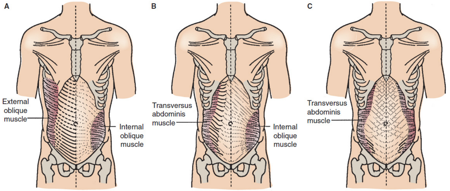
- It is the tendinous/aponeurotic continuation of the external abdominal oblique muscle, where muscle fibers transition into a broad flat sheet of fibrous tissue.
- The fascicles run in an oblique direction (downward and medially, mirroring the fiber direction of the muscle above).
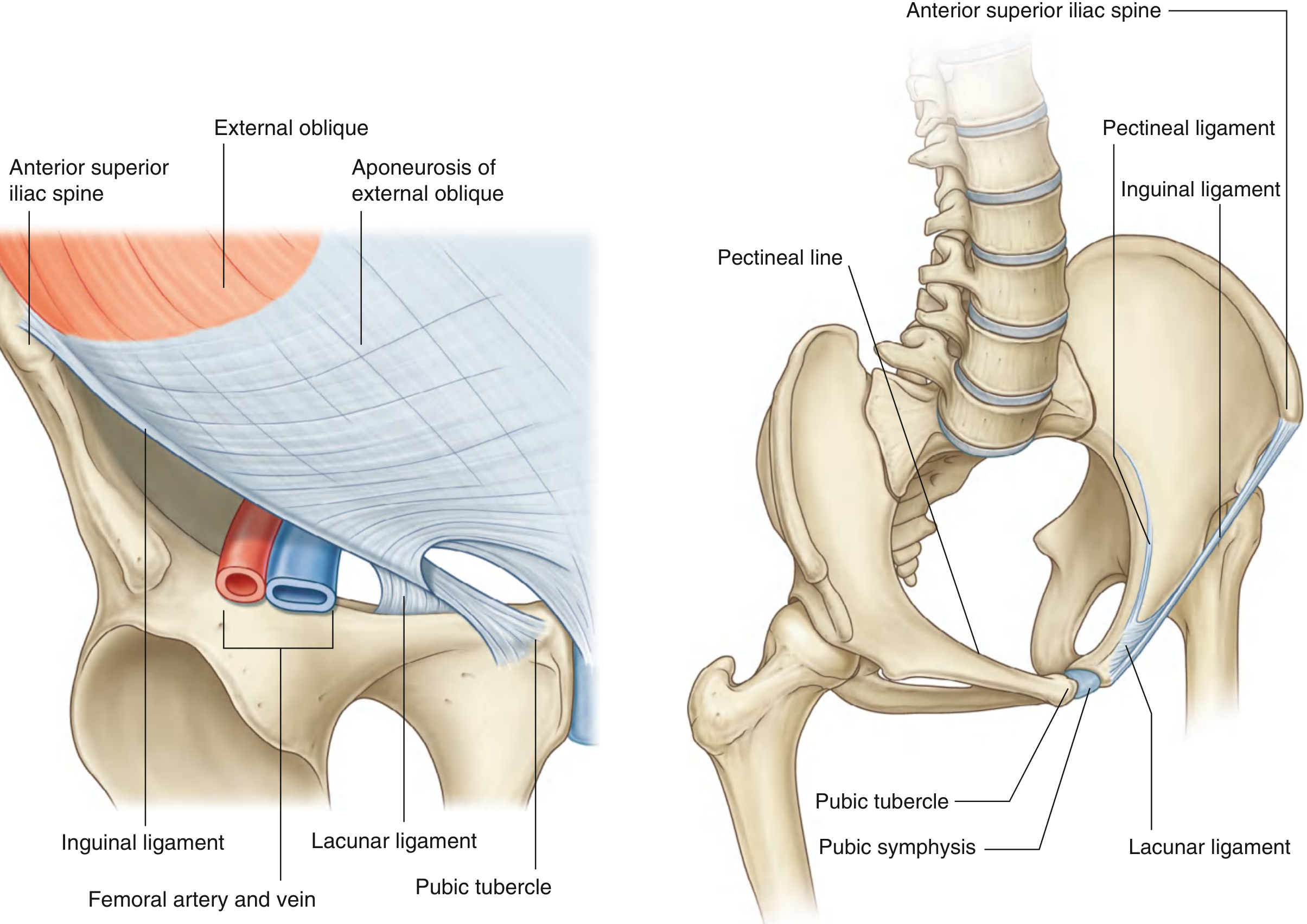
- The lower aponeurotic fibers are anteroinferiorly oriented and fold upon themselves to form the inguinal ligament.
- At the midline, its fibers decussate and interweave with those of the internal oblique and transversus abdominis aponeuroses to form the linea alba.
- Some fibers even cross the midline to reinforce the anterior rectus sheath of the opposite side.
Key Structures Formed From the Aponeurosis
| Structure | How It's Formed |
|---|---|
| Inguinal ligament | Inferior edge of the aponeurosis folds beneath itself; densely interwoven fibers |
| Superficial (external) inguinal ring | Medial fibers split into a medial crus and a lateral crus |
| Anterior wall of inguinal canal | The aponeurosis forms the entire anterior wall |
| Anterior rectus sheath | Aponeurosis contributes to the anterior leaf |
| Linea alba | Midline decussation of all three aponeuroses |
| Lacunar ligament | Fan-shaped medial extension toward the pectineal line |
Summary in Plain Terms
- Mulholland and Greenfield's Surgery: Scientific Principles and Practice, 7e, Ch. 72 (External Abdominal Oblique Muscle and Associated Ligaments, pp. 3634-3635)
- THIEME Atlas of Anatomy: General Anatomy and Musculoskeletal System, Section 5.7 (Anterior Trunk Wall: The Inguinal Canal)
Great I was ready to ask me what is inguinal ligament made of
Inguinal Ligament - What It Is Made Of
The Short Answer
From the Textbooks
"The lower border of the external oblique aponeurosis forms the inguinal ligament on each side. This thickened reinforced free edge of the external oblique aponeurosis passes between the anterior superior iliac spine laterally and the pubic tubercle medially. It folds under itself forming a trough, which plays an important role in the formation of the inguinal canal."
"The inguinal ligament is formed by obliquely oriented anteroinferior aponeurotic fibers of the external abdominal oblique. The ligament is formed when the aponeurosis folds beneath itself."
"The inferior edge of the EO aponeurosis forms the inguinal (Poupart) ligament... This structure turns posteriorly to form the shelving edge of the inguinal ligament."
Key Points Summarized
| Feature | Detail |
|---|---|
| Tissue type | Dense fibrous (collagenous) connective tissue - aponeurotic fibers |
| Origin | Anteroinferior fibers of the external oblique aponeurosis |
| How it forms | The aponeurosis folds (rolls) under itself at its lower border |
| Attachments | Laterally: anterior superior iliac spine (ASIS); Medially: pubic tubercle |
| Shape | Forms a trough/gutter facing upward and posteriorly ("shelving edge") |
| Extensions | Medial fibers continue as the lacunar ligament (to pecten pubis), then as Cooper's (pectineal) ligament |
- Gray's Anatomy for Students, p. 340 (Associated ligaments)
- Mulholland and Greenfield's Surgery, 7e, p. 3634-3635 (External Abdominal Oblique Muscle and Associated Ligaments)
- Sabiston Textbook of Surgery, p. 1680 (Anterior Anatomy)
- THIEME Atlas of Anatomy: General Anatomy and Musculoskeletal System, Section 5.7
Can u some how elaborate with 3D diagram
A detailed 3D anatomical diagram showing the formation of the inguinal ligament from the external oblique aponeurosis. The image should show: 1. The external oblique muscle (upper left, with diagonal muscle fibers running downward and medially, shown in red-pink) 2. The external oblique aponeurosis (flat, sheet-like fibrous continuation of the muscle, shown in light blue/white with a cross-hatched fiber pattern) 3. The inferior border of the aponeurosis rolling/folding underneath itself to form the inguinal ligament (shown as a thick rounded cord/band in yellow-gold) 4. The inguinal ligament stretching from the anterior superior iliac spine (ASIS) on the left to the pubic tubercle on the right, with both bony landmarks labeled 5. A cut-away cross-section showing the trough/shelving edge shape of the folded ligament 6. The lacunar ligament as a fan-shaped medial extension from the pubic tubercle end 7. Clear arrows and labels: "External oblique muscle", "External oblique aponeurosis", "Inferior border folds under itself", "Inguinal ligament (Poupart's ligament)", "ASIS", "Pubic tubercle", "Lacunar ligament", "Shelving edge (trough)" 8. The pelvis and hip bones shown in bone-white for anatomical context Medical illustration style, clean white background, educational 3D rendering with depth and shading