

Screenshot_2026-08-14-02-36-28-641_com.google.android.apps.docs.jpg
Answer all questions according to mbbs first year
Reading File
This is a comprehensive AIIMS Rajkot 1st MBBS Physiology Paper II exam. Given the budget constraints, I'll answer all questions thoroughly from my medical knowledge at the MBBS first year level. Let me search the library for key topics in parallel.
Finding Sources
Finding Sources
Finding Sources
Finding Sources
Finding Sources
Finding Sources
Finding Sources
Searching Images
ECG waves cardiac action potential phases diagram
Searching Images
blood coagulation intrinsic extrinsic pathway flowchart
Here are complete, exam-ready answers for all questions in the AIIMS Rajkot 1st MBBS Physiology Paper II (2024 Batch):
AIIMS Rajkot - 1st MBBS Physiology Paper II - Complete Answers
SECTION A
Q.1 - Clinical Case: 55-year-old male with chest pain
a) Most Probable Diagnosis (1 mark)
Acute Myocardial Infarction (AMI) - Inferior wall MI (or Anterior MI depending on leads, but the clinical picture is classic for AMI).
Justification:
- Squeezing, central chest pain radiating to the left arm
- Nausea, sweating, diaphoresis
- Risk factors: smoking, hypertension, sedentary lifestyle
- BP 160/100, HR 90, anxious and diaphoretic
This is a STEMI (ST-Elevation Myocardial Infarction) until proven otherwise.
b) ECG Waves and Phases of Cardiac Action Potential (5 marks)
The Normal ECG has the following waves:
| Wave/Interval | Represents | Duration |
|---|---|---|
| P wave | Atrial depolarization | 0.08-0.1 sec |
| PR interval | AV conduction time | 0.12-0.20 sec |
| QRS complex | Ventricular depolarization | 0.06-0.10 sec |
| ST segment | Plateau of ventricular AP | Isoelectric normally |
| T wave | Ventricular repolarization | - |
| QT interval | Total ventricular electrical activity | 0.35-0.44 sec |
| U wave | Repolarization of Purkinje fibers (sometimes seen) | - |
Cardiac Muscle Action Potential - 5 Phases:
+20 mV ___
        /   \
       /     \___________  Phase 2 (plateau)
      /                   \
Phase 0                    \  Phase 3
(rapid depol)               \
                             \___  Phase 4 (resting)
-90 mV __________________________|
Phase 0 - Rapid Depolarization
- Rapid opening of fast Na⁺ channels (voltage-gated)
- Na⁺ rushes into the cell
- Membrane potential rises from -90 mV to +20 mV
- ECG correlation: QRS complex
Phase 1 - Initial Rapid Repolarization
- Inactivation of fast Na⁺ channels
- Brief opening of transient outward K⁺ (Ito) channels
- Slight repolarization (notch)
- ECG correlation: J point
Phase 2 - Plateau Phase (unique to cardiac muscle)
- Slow L-type Ca²⁺ channels open (Ca²⁺ influx)
- Slow K⁺ efflux balances Ca²⁺ influx
- Membrane potential maintained near 0 mV
- Functionally important: triggers Ca²⁺-induced Ca²⁺ release from SR → contraction
- ECG correlation: ST segment (isoelectric in normal; elevated in STEMI)
Phase 3 - Rapid Repolarization
- L-type Ca²⁺ channels close
- Rapid K⁺ efflux (IKr, IKs channels open)
- Membrane returns toward resting potential
- ECG correlation: T wave
Phase 4 - Resting Membrane Potential
- Na⁺/K⁺-ATPase pump restores ionic gradients
- Resting potential = -90 mV (maintained by K⁺ leak channels)
- ECG correlation: isoelectric baseline (between T and next P wave)
Key diagram to draw: Draw a ventricular AP (phases 0-4) and below it draw an ECG strip, aligning each phase with its corresponding ECG component. Show the plateau (Phase 2) corresponding to the ST segment.
c) Inferior Wall MI - ECG Findings and Leads (2+2 = 4 marks)
ECG Findings in Inferior Wall MI:
(i) Acute/Evolving changes:
- ST elevation (convex upward / tombstone shaped) in inferior leads
- Reciprocal ST depression in anterior leads (I, aVL)
- Hyperacute T waves (tall, peaked) - earliest change
(ii) Evolving changes (hours to days):
- Q waves (pathological: >0.04 sec wide, >1/4 of R wave amplitude) - indicate transmural necrosis
- T wave inversion
- ST elevation gradually resolves
(iii) Old/Established MI:
- Persistent Q waves
- T wave may normalize
Leads in which changes are prominent:
The inferior wall of the heart is supplied by the Right Coronary Artery (RCA) (in 80% - right dominant) or Left Circumflex Artery.
The inferior wall faces the inferior/diaphragmatic surface, recorded by:
| Lead | Records |
|---|---|
| Lead II | Inferior wall (positive electrode on left leg) |
| Lead III | Inferior wall (positive electrode on left leg, reference on right arm) |
| aVF | Inferior wall (augmented unipolar - left foot) |
So: ST elevation in leads II, III, and aVF = Inferior wall MI.
Reciprocal changes (ST depression): in leads I and aVL.
(If RCA is occluded proximally): Also look for ST elevation in V4R (right-sided lead) suggesting Right Ventricular MI associated with inferior MI.
Q.2 - Short Notes (5 × 4 = 20 marks)
A. Role of RAAS in Blood Pressure Regulation (4 marks)
The Renin-Angiotensin-Aldosterone System (RAAS):
Triggers for Renin Release (from Juxtaglomerular cells of kidney):
- Decreased renal perfusion pressure (baroreceptor mechanism)
- Decreased Na⁺ delivery to macula densa
- Sympathetic stimulation (via β₁ receptors)
The Cascade:
Angiotensinogen (liver)
        ↓ [Renin - from JG cells]
Angiotensin I (inactive decapeptide)
        ↓ [ACE - Angiotensin Converting Enzyme, mainly in lungs]
Angiotensin II (active octapeptide)
        ↓
    ┌───────────────────────────────────┐
    ↓                                   ↓
Vasoconstriction              Adrenal Cortex (Zona Glomerulosa)
(↑ TPR → ↑ BP)                        ↓ [Aldosterone release]
    ↓                         Na⁺ reabsorption in DCT/collecting duct
Also: ADH release             H₂O retention → ↑ Blood Volume → ↑ BP
Thirst stimulation
Sympathetic activation
Actions of Angiotensin II:
- Arteriolar vasoconstriction - most potent vasoconstrictor in the body
- Stimulates aldosterone - Na⁺ and H₂O retention (↑ blood volume)
- Stimulates ADH (vasopressin) release - water retention
- Stimulates thirst center in hypothalamus
- Cardiac hypertrophy and renal mesangial contraction
- Efferent arteriole constriction - maintains GFR during hypotension
Net Result: RAAS increases BP by:
- Short-term: vasoconstriction (↑ TPR)
- Long-term: Na⁺/water retention (↑ blood volume → ↑ cardiac output)
Clinical Relevance: ACE inhibitors (e.g., Enalapril) and ARBs (e.g., Losartan) block this system → antihypertensive effect.
B. Flowchart of Intrinsic and Extrinsic Pathways of Blood Coagulation (4 marks)
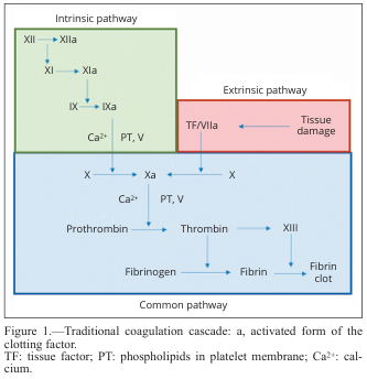
EXTRINSIC PATHWAY (faster - seconds):
Tissue Damage → Release of Tissue Factor (TF / Factor III)
                         ↓
TF + Factor VII + Ca²⁺ → Factor VIIa-TF complex
                         ↓
                  Activates Factor X → Xa
INTRINSIC PATHWAY (slower - minutes, activated by contact):
Contact with collagen/glass → Factor XII activated → XIIa
                         ↓
        XIIa → XI → XIa
                         ↓
        XIa + Ca²⁺ → IX → IXa
                         ↓
        IXa + Factor VIIIa + Ca²⁺ + PF3 → Factor X activated → Xa
COMMON PATHWAY:
Factor Xa + Factor Va + Ca²⁺ + Phospholipid (Prothrombinase complex)
                         ↓
        Prothrombin (Factor II) → Thrombin (IIa)
                         ↓
        Fibrinogen (Factor I) → Fibrin (loose/soluble)
                         ↓
        Factor XIII + Ca²⁺ → Cross-linked Fibrin (stable clot)
Key Points:
- Extrinsic pathway assessed by PT (Prothrombin Time)
- Intrinsic pathway assessed by aPTT (activated Partial Thromboplastin Time)
- Vitamin K-dependent factors: II, VII, IX, X (and Protein C, S)
C. Counter-Current Multiplier in Kidney (4 marks)
The counter-current mechanism in the kidney is responsible for production of concentrated urine (up to 1200 mOsm/L in humans).
Location: Loop of Henle (especially long loops of juxtamedullary nephrons)
Components:
- Descending limb of Loop of Henle - permeable to water, impermeable to solutes
- Ascending limb of Loop of Henle - impermeable to water, actively transports NaCl out (thick ascending limb = TAL)
Mechanism:
Cortex (300 mOsm)
        ↓ fluid flows DOWN descending limb
Medulla
        ↓
At each level:
- TAL pumps NaCl out (active transport via Na⁺-K⁺-2Cl⁻ cotransporter / NKCC2)
- Tubular fluid in descending limb equilibrates with interstitium by losing water
- As fluid descends → becomes more concentrated
- At bend → ~1200 mOsm
- As fluid ascends in TAL → NaCl pumped out without water → fluid becomes dilute
- At cortex → tubular fluid ~100 mOsm (hypotonic)
The "Multiplier" Effect:
- At any horizontal level, there is a small gradient (~200 mOsm) between ascending and descending limbs
- The counter-current flow multiplies this small horizontal gradient into a large vertical medullary osmotic gradient (300-1200 mOsm from cortex to papilla)
Role of Vasa Recta:
- Acts as counter-current exchanger (not multiplier)
- Preserves medullary gradient by passive exchange of solutes and water
- Prevents "washout" of the gradient
Collecting Duct:
- In the presence of ADH, collecting duct becomes permeable to water
- Water moves from tubule into hypertonic medullary interstitium (osmosis)
- Concentrated urine (up to 1200 mOsm) is produced
Without ADH: Collecting duct remains impermeable → dilute urine (~50-100 mOsm)
Urea Recycling: Urea from inner medullary collecting duct (IMCD, under ADH) contributes ~500 mOsm to medullary gradient, along with NaCl (~500 mOsm). This is maintained in the papilla.
D. Define Shock - Distributive Types of Shock (4 marks)
Definition of Shock:
Shock is a state of acute circulatory failure resulting in inadequate tissue perfusion and cellular oxygen delivery, leading to cellular dysfunction and if untreated, organ failure and death. The hallmark is a mismatch between oxygen delivery (DO₂) and oxygen consumption (VO₂).
Classification of Shock:
| Type | Mechanism | CO | SVR |
|---|---|---|---|
| Hypovolemic | ↓ Blood volume | ↓ | ↑ |
| Cardiogenic | Pump failure | ↓ | ↑ |
| Distributive | Abnormal vasodilation | ↑ or N | ↓↓ |
| Obstructive | Outflow obstruction | ↓ | ↑ |
Distributive Types of Shock:
Characterized by: Massive vasodilation → ↓ SVR → maldistribution of blood flow → tissue hypoperfusion despite normal or ↑ cardiac output. Also called "warm shock" due to vasodilation.
(1) Septic Shock (most common distributive shock)
- Cause: Gram-negative bacteria (endotoxin/LPS), Gram-positive bacteria, fungi
- Mechanism: LPS → macrophage activation → IL-1, IL-6, TNF-α → iNOS activation → massive NO production → vasodilation
- Features: fever, warm flushed skin, hypotension, ↑ HR, leukocytosis
- Phases: Early (warm/hyperdynamic) → Late (cold/hypodynamic)
(2) Anaphylactic Shock
- Cause: Type I hypersensitivity reaction (bee sting, drug allergy, food allergy)
- Mechanism: IgE → mast cell degranulation → massive histamine release → vasodilation + ↑ vascular permeability + bronchospasm
- Features: urticaria, angioedema, bronchospasm, hypotension
- Treatment: Adrenaline (epinephrine) IM
(3) Neurogenic Shock
- Cause: Spinal cord injury (above T6), severe pain, emotional shock, spinal anesthesia
- Mechanism: Loss of sympathetic tone → uncontrolled vasodilation + bradycardia (loss of cardiac sympathetics)
- Features: hypotension + bradycardia + warm dry skin (unusual triad)
- Distinguishing: bradycardia (not tachycardia as in other shocks)
(4) Toxic Shock Syndrome (sometimes classified separately)
- Cause: Staphylococcal/Streptococcal exotoxins acting as superantigens
- Leads to massive T-cell activation → cytokine storm → distributive shock
Q.3 - Short Answer Questions (4 × 2 = 8 marks)
A. Role of Neutrophils in Immune Response (2 marks)
Neutrophils (Polymorphonuclear leukocytes / PMNs) are the first line of cellular defense and form the largest component of white blood cells (60-70%).
Functions:
-
Chemotaxis: Respond to chemotactic signals (C5a, IL-8, LTB4, fMLP) and migrate to site of infection/inflammation
-
Phagocytosis:
- Recognize bacteria via opsonins (IgG, C3b) through Fc receptors and complement receptors
- Engulf pathogens into phagosomes
- Phagosome fuses with granules → phagolysosome
-
Killing Mechanisms:
- Oxygen-dependent (Respiratory burst): NADPH oxidase → superoxide (O₂⁻) → H₂O₂ → HOCl (hypochlorous acid via myeloperoxidase) → kills bacteria
- Oxygen-independent: Lysozyme (degrades bacterial cell wall), defensins, lactoferrin, elastase, cathepsins
-
Degranulation:
- Primary (azurophilic) granules: myeloperoxidase, lysozyme, defensins
- Secondary (specific) granules: lactoferrin, collagenase, B12-binding protein
-
NET formation (Neutrophil Extracellular Traps):
- Release chromatin + antimicrobial proteins to trap and kill bacteria extracellularly
-
Cytokine Release: IL-1β, TNF-α, IL-6, IL-12 → amplify inflammatory response
B. Pathophysiological Basis of Erythroblastosis Foetalis (2 marks)
Also called Hemolytic Disease of the Newborn (HDN).
Most Common Cause: Rh incompatibility (Rh-negative mother + Rh-positive fetus)
Pathophysiology:
1st Pregnancy:
Rh- mother + Rh+ fetus
→ At delivery (or abortion/trauma): Rh+ fetal RBCs enter maternal circulation
→ Maternal immune system recognizes Rh antigen (D antigen) as foreign
→ Primary immune response → IgM antibodies (don't cross placenta) - No harm to 1st baby

Sensitization occurs (mother becomes anti-D IgG positive)

2nd Pregnancy (Rh+ fetus again):
→ Maternal anti-D IgG antibodies cross placenta (IgG crosses, IgM does not)
→ Anti-D IgG coats fetal Rh+ RBCs
→ Fetal macrophages (spleen) recognize coated RBCs → phagocytosis → HEMOLYSIS

Consequences:
→ Severe hemolytic anemia in fetus
→ Compensatory extramedullary hematopoiesis (liver, spleen, bone marrow)
→ Nucleated RBCs (erythroblasts) in peripheral blood → "Erythroblastosis"
→ Hydrops fetalis (severe cases): generalized edema, ascites, pleural effusion
→ Hyperbilirubinemia → Kernicterus (bilirubin deposition in brain) in neonate
Prevention: Anti-D immunoglobulin (RhoGAM) given to Rh- mother at 28 weeks and within 72 hours of delivery
ABO incompatibility: Can cause mild HDN in 1st pregnancy (naturally occurring IgG anti-A/anti-B), usually milder.
C. Cushing's Reflex (2 marks)
Also called the Vasopressor response or CNS ischemic response.
Definition: An emergency protective cardiovascular response triggered when cerebral perfusion pressure (CPP) falls dangerously low, usually due to raised intracranial pressure (ICP).
Trigger: When ICP rises to near the level of Mean Arterial Pressure (MAP) → CPP (= MAP - ICP) falls → brain ischemia → neurons of vasomotor center in medulla oblongata become ischemic and hypercapnic
Response (Classic Cushing's Triad - clinical presentation):
- ↑ Blood pressure (marked systolic hypertension) - ischemic vasomotor center fires maximally → intense sympathetic discharge → vasoconstriction and ↑ cardiac output
- Bradycardia - baroreceptor response to extreme hypertension → vagal activation → bradycardia
- Irregular/slow respirations (Cheyne-Stokes or agonal breathing) - due to brainstem compression
Mechanism:
↑ ICP → ↓ CPP → Brain ischemia → Ischemic vasomotor center fires
→ Massive sympathetic discharge → ↑ BP
→ Baroreceptors detect ↑ BP → Vagus nerve → Bradycardia
Clinical Significance:
- Cushing's triad (hypertension + bradycardia + irregular breathing) is a late and ominous sign of severely raised ICP (herniation imminent)
- Seen in: head injury, intracranial hemorrhage, brain tumors
- Requires emergency intervention (mannitol, head elevation, neurosurgery)
D. Wenckebach Phenomenon in Heart Block (2 marks)
Wenckebach phenomenon = Mobitz Type I Second-Degree AV Block
Definition: A progressive prolongation of the PR interval on each successive beat until a P wave is not conducted (blocked), followed by a dropped QRS. The cycle then repeats.
ECG Features:
- PR interval progressively lengthens beat by beat
- Until one P wave is not followed by a QRS (dropped beat)
- After the dropped beat, PR interval resets to its shortest value
- Cycle repeats (e.g., 3:2 or 4:3 ratio of P:QRS)
Mechanism:
- AV node conducts each successive impulse with increasing difficulty (fatigue/decremental conduction)
- Eventually fails to conduct → dropped beat
- During the pause (dropped beat), AV node recovers
- The greatest increment in PR prolongation occurs between the 1st and 2nd beat; subsequent increments are smaller → PP interval shortens before the dropped beat, giving a characteristic "grouped beating" pattern
Site of block: AV node (supra-Hisian) - has a narrow QRS
Common Causes:
- Inferior MI (RCA supplies AV node)
- Increased vagal tone (athletes, during sleep)
- Digitalis toxicity
- Myocarditis
- Drug effects (β-blockers, calcium channel blockers)
Clinical: Usually benign and transient. May not require pacing. Differentiate from Mobitz II (sudden drop without prior PR prolongation) which is more dangerous and often requires pacemaker.
SECTION B
Q.4 - Neural Regulation of Respiration + Cheyne-Stokes + Kussmaul (6+3 = 9 marks)
Neural Regulation of Respiration (6 marks)
Respiratory Centers (located in brainstem):
A. Medullary Centers (Primary):
(1) Dorsal Respiratory Group (DRG):
- Location: Nucleus tractus solitarius (NTS), dorsal medulla
- Function: Generates the basic rhythm of inspiration (ramp signal)
- Contains mainly inspiratory neurons (I neurons)
- Receives afferents from peripheral chemoreceptors (CN IX, X) and lung stretch receptors
- Sends impulses to phrenic nerve (C3,4,5) → diaphragm
(2) Ventral Respiratory Group (VRG):
- Location: Nucleus ambiguus + nucleus retroambiguus, ventral medulla
- Contains both inspiratory and expiratory neurons
- Quiet breathing: VRG is relatively inactive (expiration is passive)
- Active breathing/forced expiration: VRG expiratory neurons fire → activate accessory muscles (internal intercostals, abdominals)
- Pre-Botzinger complex (pacemaker of respiratory rhythm) is within the VRG
B. Pontine Centers (Modulatory):
(3) Pneumotaxic Center (Pontine Respiratory Group):
- Location: Nucleus parabrachialis, upper pons
- Function: Inhibits inspiration → limits inspiratory volume → controls respiratory rate
- Sends inhibitory signals to DRG → turns off inspiration (inspiratory "off-switch")
- When active: rapid, shallow breathing
- When inhibited: slow, deep breathing (apneusis if absent + vagus cut)
(4) Apneustic Center:
- Location: Lower pons
- Function: Prolongs inspiration (sends continuous excitatory drive to DRG)
- Normally inhibited by pneumotaxic center and Hering-Breuer reflex
- If unopposed: produces apneusis (prolonged inspiratory gasps)
Hering-Breuer Reflex:
- Lung stretch receptors (in bronchial smooth muscle) → activated when lungs inflate sufficiently
- Signals via Vagus nerve to DRG → inhibits inspiration (prevents over-inflation)
- Important in newborns and during tidal volumes >1L in adults
Chemical Regulation (works alongside neural):
Central Chemoreceptors:
- Location: Ventral surface of medulla (near respiratory centers)
- Sensitive to: CO₂ / [H⁺] in CSF (NOT directly to O₂)
- Mechanism: CO₂ crosses BBB → combines with H₂O → H₂CO₃ → H⁺ + HCO₃⁻ → ↑[H⁺] in CSF → stimulates chemoreceptors → ↑ ventilation
- Main regulator of normal breathing
Peripheral Chemoreceptors:
- Location: Carotid bodies (CN IX) and Aortic bodies (CN X)
- Sensitive to: ↓ PaO₂ (mainly, when <60 mmHg), ↑ PaCO₂, ↓ pH
- Respond to hypoxia, hypercapnia, acidosis
- Important in: hypoxic drive (e.g., COPD patients)
Cheyne-Stokes Breathing (1.5 marks)
Definition: An abnormal breathing pattern characterized by a cyclic waxing and waning of tidal volume, with periods of apnea (cessation of breathing) alternating with periods of hyperpnea (increased depth of breathing).
Pattern:
Apnea → gradual ↑ tidal volume (crescendo) → peak → gradual ↓ (decrescendo) → Apnea again
[Each cycle: 30 seconds to 3 minutes]
Mechanism:
- During apnea: PaCO₂ rises, PaO₂ falls
- When CO₂ rises enough to stimulate respiratory centers → breathing resumes and increases (hyperpnea)
- Hyperventilation causes PaCO₂ to fall below normal → apnea again
- The oscillation continues due to delayed feedback (long circulation time from lungs to brain)
Causes:
- Heart failure (most common in adults) - ↑ circulation time (lung to brain) = delayed CO₂ feedback
- Severe brain damage (stroke, head injury, brain tumors)
- Normal in premature infants and some normal adults during sleep at high altitude
- Uremia, narcotic overdose
Kussmaul's Breathing (1.5 marks)
Definition: A pattern of deep, rapid, labored breathing that is regular in rhythm (not cyclic like Cheyne-Stokes). Also called "air hunger."
Pattern: Deep + fast + regular = "hunger for air"
Mechanism:
- Occurs in severe metabolic acidosis
- ↓ blood pH → stimulates peripheral and central chemoreceptors → hyperventilation
- Goal: to blow off CO₂ (compensatory respiratory alkalosis)
- PaCO₂ falls as compensation for metabolic acidosis (Henderson-Hasselbalch relationship)
Causes:
- Diabetic Ketoacidosis (DKA) - most classic cause (acetone breath + Kussmaul breathing)
- Lactic acidosis
- Renal failure (uremic acidosis)
- Methanol/salicylate poisoning
Distinction from Cheyne-Stokes:
| Feature | Cheyne-Stokes | Kussmaul |
|---|---|---|
| Pattern | Cyclic, waxing-waning + apnea | Regular, deep, rapid |
| Rhythm | Irregular | Regular |
| Cause | Heart failure, brain lesions | Metabolic acidosis |
| CO₂ | Low (post-hyperventilation) | Low (compensatory) |
Q.5 - Short Notes (4 × 5 = 20 marks)
A. HCl Secretion in Stomach (5 marks)
Site: Parietal cells (Oxyntic cells) of gastric glands in the body and fundus of the stomach
Normal gastric acid: pH 1-2, concentration ~150 mEq/L HCl
Mechanism of HCl Secretion (The Proton Pump mechanism):
Step-by-step:
- CO₂ + H₂O → H₂CO₃ (catalyzed by carbonic anhydrase inside parietal cell)
- H₂CO₃ → H⁺ + HCO₃⁻
- H⁺ is pumped into gastric lumen by H⁺/K⁺-ATPase (Proton Pump) on the apical membrane (exchange: H⁺ out, K⁺ in)
- Cl⁻ is secreted into the lumen through Cl⁻ channels → combines with H⁺ → HCl
- HCO₃⁻ exchanges for Cl⁻ on the basolateral side (via Cl⁻/HCO₃⁻ antiporter) → HCO₃⁻ enters blood ("alkaline tide")
Stimulation of HCl secretion:
| Stimulus | Mechanism |
|---|---|
| Acetylcholine (vagus nerve) | Muscarinic (M₃) receptors → ↑ IP₃/Ca²⁺ → activates proton pump |
| Histamine (from ECL cells) | H₂ receptors → ↑ cAMP → PKA → activates proton pump |
| Gastrin (from G cells, antrum) | CCK-B receptors on parietal cells → ↑ Ca²⁺ → stimulates acid |
Phases of Gastric Secretion:
- Cephalic phase (30%): Thought, smell, taste of food → vagal stimulation
- Gastric phase (60%): Food in stomach → gastrin release, distension
- Intestinal phase (10%): Chyme in duodenum (initial stimulation, then inhibition)
Inhibition:
- Low pH (<2) in antrum → inhibits gastrin release (local feedback)
- Secretin, GIP, VIP, somatostatin → inhibit acid secretion
- Prostaglandins (E₂) → protect mucosa
Clinical: Proton pump inhibitors (PPIs like Omeprazole) block H⁺/K⁺-ATPase → treat peptic ulcer, GERD. H₂-blockers (Ranitidine) block histamine receptors.
B. Phases of Deglutition (Swallowing) (5 marks)
Definition: Deglutition is the act of swallowing, which moves a bolus of food/liquid from the mouth to the stomach.
Three Phases:
Phase 1: Oral (Voluntary) Phase
- Fully voluntary and conscious
- Tongue pushes bolus posteriorly against the hard palate and then to the oropharynx
- Lips sealed, teeth occluded
- Duration: ~1 second
- Triggers the swallowing reflex
Phase 2: Pharyngeal (Involuntary) Phase
- Triggered by touch receptors in posterior pharynx → CN IX → swallowing center in medulla (nucleus tractus solitarius + nucleus ambiguus)
- Entirely reflex and involuntary
- Protective mechanisms occur simultaneously:
- Soft palate elevates → closes nasopharynx (prevents nasal regurgitation)
- Larynx elevates and moves anteriorly
- Epiglottis folds over laryngeal inlet (seals airway)
- True and false vocal cords adduct (glottis closes)
- Respiration is inhibited (deglutition apnea)
- Upper esophageal sphincter (UES) relaxes
- Pharyngeal peristaltic waves propel bolus into esophagus
- Duration: 1-2 seconds
- Cranial nerves involved: V (trigeminal), IX (glossopharyngeal), X (vagus), XII (hypoglossal)
Phase 3: Esophageal (Involuntary) Phase
- Entirely involuntary
- Primary peristalsis: Continuation of pharyngeal peristaltic wave, propels bolus down esophagus
- Secondary peristalsis: If bolus is not cleared, distension triggers additional peristaltic waves (mediated locally + via vagus)
- Upper 1/3 of esophagus: striated muscle (voluntary)
- Lower 2/3: smooth muscle (involuntary, under Auerbach's plexus)
- Lower Esophageal Sphincter (LES) relaxes ahead of bolus → allows entry into stomach
- LES then contracts to prevent reflux
- Duration: 8-20 seconds (liquids faster, solids slower)
- Gravity assists but is not essential (can swallow upside down)
Swallowing Center:
- Located in medulla oblongata (NTS + nucleus ambiguus)
- Coordinates the sequential contraction/relaxation pattern
- Receives afferents from pharyngeal touch receptors via CN IX
C. Tubuloglomerular Feedback (5 marks)
Definition: Tubuloglomerular feedback (TGF) is an intrinsic autoregulatory mechanism by which the kidney adjusts GFR based on the NaCl concentration sensed at the macula densa in the early distal tubule.
Anatomical Basis - Juxtaglomerular Apparatus (JGA):
- Macula densa: Specialized cells in the wall of the thick ascending limb (TAL) of the loop of Henle, at the point where it abuts its own glomerulus
- Juxtaglomerular (JG) cells: Modified smooth muscle cells in the afferent arteriole wall; produce renin
- Extraglomerular mesangial cells (Lacis cells): Connective tissue cells, may transmit signals
Mechanism:
↑ GFR → ↑ tubular flow → ↑ NaCl delivery to macula densa
                ↓
Macula densa cells sense ↑ [NaCl] via NKCC2 transporter
                ↓
Release of adenosine + TXA₂ (vasoconstrictors)
                ↓
Afferent arteriole CONSTRICTS → ↓ GFR (feedback correction)
↓ GFR → ↓ tubular flow → ↓ NaCl delivery to macula densa
                ↓
Macula densa releases prostaglandins (PGI₂, PGE₂) → vasodilator
                ↓
Afferent arteriole DILATES → ↑ GFR (feedback correction)
                ↓
Also: ↓ NaCl → stimulates renin release → RAAS activation
Functional Role:
- Autoregulation of GFR: Maintains constant GFR despite changes in systemic blood pressure (works alongside myogenic mechanism)
- Matches filtration rate to tubular reabsorptive capacity: Prevents delivery of excess NaCl to distal nephron
- Links RAAS to tubular NaCl sensing: Low NaCl at macula densa → renin release → Ang II → aldosterone → Na⁺ reabsorption
Summary:
- TGF is a negative feedback loop
- Sensor: Macula densa (NaCl sensor)
- Effector: Afferent arteriole (changes GFR)
- Mediators: Adenosine (constriction), PGs (dilation), ATP, NO
D. Role of DCT and Collecting Tubule in Urine Formation (5 marks)
Distal Convoluted Tubule (DCT):
The DCT is the final fine-tuning segment before the collecting duct. It handles about 5-10% of filtered Na⁺.
Na⁺ reabsorption:
- Via NCC (Na⁺-Cl⁻ cotransporter) on apical membrane (electroneutral)
- Impermeable to water (diluting segment, like TAL)
- Creates dilute tubular fluid
- Regulated by: Thiazide diuretics block NCC (used for hypertension)
Early DCT:
- Reabsorbs Na⁺ and Cl⁻ (NCC)
- Reabsorbs Ca²⁺ (via TRPV5 channel, regulated by PTH and Calcitriol)
- Reabsorbs Mg²⁺ (via TRPM6)
Late DCT (connects to collecting duct):
- Contains principal cells and intercalated cells (like collecting duct)
- Aldosterone begins to act here
Collecting Duct (CD):
Most critical segment for final regulation of urine concentration and composition.
Contains two cell types:
1. Principal Cells (majority):
- Apical: ENaC (epithelial Na⁺ channel) for Na⁺ reabsorption
- Basolateral: Na⁺/K⁺-ATPase
- K⁺ is secreted via ROMK channels (apical)
- Regulated by Aldosterone: ↑ Na⁺ reabsorption + ↑ K⁺ secretion (↑ ENaC and Na⁺/K⁺-ATPase expression)
- Water permeability regulated by ADH (vasopressin):
- ADH → V2 receptor → ↑ cAMP → PKA → insertion of Aquaporin-2 (AQP2) into apical membrane
- Water follows osmotic gradient into hypertonic medullary interstitium → concentrated urine
2. Intercalated (Type A and B) Cells:
- Type A (alpha) intercalated cells: Secrete H⁺ (via H⁺-ATPase) and reabsorb HCO₃⁻ → acidify urine → important in metabolic acidosis
- Type B (beta) intercalated cells: Secrete HCO₃⁻ → important in metabolic alkalosis
Summary of DCT + Collecting Duct:
| Function | Hormone | Effect |
|---|---|---|
| Na⁺ reabsorption | Aldosterone | ↑ ENaC, Na⁺/K⁺-ATPase |
| K⁺ secretion | Aldosterone | ↑ ROMK |
| Water reabsorption | ADH | ↑ AQP2 insertion |
| H⁺ secretion | (Acid-base regulation) | Acidifies urine |
| Ca²⁺ reabsorption | PTH | ↑ TRPV5 in early DCT |
Net contribution: DCT and collecting duct together determine the final composition and volume of urine, responding to hormonal signals (aldosterone, ADH, PTH) to maintain body homeostasis.
Q.6 - Short Answer Questions (4 × 2 = 8 marks)
A. Caisson Disease (2 marks)
Also called Decompression Sickness or "The Bends."
Background: Affects divers, caisson workers (underwater construction), and those in hyperbaric environments who ascend too rapidly.
Mechanism:
- At high pressure, gases (especially N₂ - nitrogen) dissolve in blood and tissues in greater amounts (Henry's Law: amount of gas dissolved ∝ partial pressure)
- If pressure is reduced too rapidly (rapid ascent), N₂ comes out of solution as bubbles in blood and tissues before it can be exhaled
- Gas bubbles form in: joints (especially knees, shoulders), muscles, fat, spinal cord, inner ear, blood vessels, lungs
Clinical Features:
- Type I (mild): Joint pain ("the bends"), skin rash, itching
- Type II (serious):
- Neurological: paresthesias, paralysis, spinal cord infarction
- Pulmonary: "the chokes" - chest pain, cough, dyspnea (pulmonary emboli)
- Inner ear: vertigo, hearing loss (staggers)
- Cardiovascular: arterial gas embolism
Prevention: Controlled slow ascent with decompression stops (allows N₂ to diffuse out slowly via lungs)
Treatment: Hyperbaric oxygen therapy (100% O₂ at 2.8 atm) - recompression reduces bubble size + replaces N₂ with O₂ (rapidly metabolized)
B. Migrating Motor Complex (MMC) (2 marks)
Definition: The Migrating Motor Complex (MMC) is a cyclic pattern of organized electrical and contractile activity that sweeps through the gastrointestinal tract (stomach to ileum) during the interdigestive (fasting) period.
Cycle Duration: Approximately 90-120 minutes per cycle
4 Phases (Phases I-IV):
| Phase | Duration | Activity |
|---|---|---|
| Phase I | 40-60 min | Quiescence - no contractions |
| Phase II | 20-30 min | Irregular contractile activity |
| Phase III | 5-15 min | Regular, intense contractions (the "activity front") - the "housekeeper" wave |
| Phase IV | Few min | Transition back to Phase I |
Phase III is the most important: Migrates from stomach to terminal ileum, sweeping undigested material, bacteria, and desquamated cells toward the colon - called the "intestinal housekeeper"
Regulation:
- Primarily regulated by Motilin (hormone from M cells of duodenum/jejunum)
- Motilin peaks just before Phase III activity front
- Also regulated by intrinsic enteric nervous system
- Abolished by eating (fed pattern replaces the MMC pattern)
Functions:
- Clears undigested residues from small intestine
- Prevents bacterial overgrowth (sweeps bacteria toward colon)
- Maintains colonic flora in proper location
Clinical: Absence of MMC → small intestinal bacterial overgrowth (SIBO). Erythromycin is a motilin agonist (prokinetic drug).
C. Effect of Moderate Exercise on Heart Rate and Blood Pressure (2 marks)
During moderate exercise, the cardiovascular system undergoes the following changes:
Heart Rate (HR):
- Increases (tachycardia) - from resting ~70 bpm to 120-150 bpm (moderate exercise)
- Mechanisms:
- Neural (primary): ↑ sympathetic outflow (via cardiac accelerator nerves) → ↑ SA node firing rate
- Withdrawal of vagal tone → further ↑ HR
- Bainbridge reflex (atrial reflex): ↑ venous return → atrial stretch → reflex tachycardia
- Circulating catecholamines: Epinephrine/norepinephrine from adrenal medulla → β₁ receptor → ↑ HR
Blood Pressure:
- Systolic BP increases (120 mmHg → 160-180 mmHg during moderate exercise)
- Due to ↑ cardiac output (↑ HR × ↑ stroke volume)
- Diastolic BP remains relatively unchanged or slightly decreases (↑ or ↓ by <10 mmHg)
- Due to vasodilation in exercising muscles (↓ TPR balances ↑ CO)
- Pulse pressure increases (↑ SBP with stable DBP = widened pulse pressure)
- Mean Arterial Pressure (MAP): Slightly increases
- Total Peripheral Resistance (TPR): Decreases (vasodilation in muscles due to local metabolites: CO₂, H⁺, lactate, adenosine, K⁺, NO)
Summary:
| Parameter | Change |
|---|---|
| Heart Rate | ↑↑ |
| Stroke Volume | ↑ |
| Cardiac Output | ↑↑ (up to 4-5x) |
| Systolic BP | ↑ |
| Diastolic BP | No change or slight ↓ |
| TPR | ↓ (muscle vasodilation) |
| Skin blood flow | ↑ (thermoregulation) |
| Splanchnic/renal flow | ↓ (redistribution) |
D. Heat Stroke (2 marks)
Definition: A life-threatening emergency characterized by core body temperature >40°C (104°F) associated with central nervous system dysfunction (altered consciousness, seizures, coma), due to failure of thermoregulatory mechanisms.
Types:
- Classic Heat Stroke: Occurs in elderly, very young, or those with chronic illness during heat waves; due to environmental hyperthermia overwhelming heat dissipation
- Exertional Heat Stroke: Occurs in young, physically active individuals during intense exercise in hot/humid conditions; heat production exceeds dissipation
Pathophysiology:
- Core temperature rises above the set point
- Hypothalamus initially triggers sweating, cutaneous vasodilation, ↑ cardiac output
- When compensatory mechanisms are overwhelmed → temperature continues to rise
- At >41°C: cellular proteins begin to denature, mitochondrial dysfunction
- Endothelial damage → inflammatory cascade (cytokines, IL-1, TNF-α)
- Multiorgan failure: CNS (cerebral edema, hemorrhage), liver, kidneys (ATN), heart, coagulopathy (DIC)
- Sweating is characteristically absent in classic heat stroke (anhidrosis - sweat glands fail)
- Exertional type may still have sweating
Clinical Features:
- Hot, dry, flushed skin (classic type: anhidrotic)
- Core temp >40°C
- CNS dysfunction: confusion, delirium, seizures, coma (key distinguishing feature from heat exhaustion)
- Hypotension, tachycardia
- Nausea, vomiting
Distinction from Heat Exhaustion: Heat exhaustion - core temp <40°C, CNS intact, profuse sweating, weakness (less severe)
Treatment: Rapid cooling is the priority
- Remove from heat, undress
- Ice packs to neck, axillae, groin (areas of major vessels)
- Evaporative cooling (spraying water + fanning) is most effective
- Cold IV fluids
- Target: cool to <39°C within 30 minutes
---All answers are now complete. Here is a quick reference summary of what was covered:
Quick Reference Summary
Paper Structure Covered:
- Section A (38 marks): Q1 + Q2 + Q3
- Section B (37 marks): Q4 + Q5 + Q6
Key Points to Remember for the Exam:
Q1 - AMI:
- Diagnosis = Acute MI (history + risk factors)
- Phase 0 (QRS) = fast Na⁺; Phase 2 (ST segment) = Ca²⁺ plateau; Phase 3 (T wave) = K⁺ efflux
- Inferior MI = ST elevation in II, III, aVF (RCA territory)
Q2 - Short Notes:
- RAAS = Renin → Ang I → ACE → Ang II → vasoconstriction + aldosterone
- Coagulation: Extrinsic (TF + VII) vs Intrinsic (XII→XI→IX→VIII) → common pathway (X→V→prothrombin→thrombin→fibrin)
- Counter-current multiplier = TAL actively pumps NaCl, creates 300-1200 mOsm corticopapillary gradient
- Distributive shock: Septic, Anaphylactic, Neurogenic (all cause ↓ SVR despite ↑/normal CO)
Q3 - Short Answers:
- Neutrophils: Chemotaxis → Phagocytosis → Respiratory burst (NADPH oxidase → O₂⁻ → HOCl via MPO)
- Erythroblastosis: Rh- mother sensitized in 1st pregnancy → anti-D IgG crosses placenta in 2nd pregnancy → fetal hemolysis
- Cushing's Reflex: ↑ ICP → brain ischemia → ↑ BP + bradycardia + irregular breathing (triad = ominous sign)
- Wenckebach = Mobitz I = progressive PR prolongation → dropped beat → reset
Q4 - Respiration:
- DRG = inspiration rhythm; Pneumotaxic center = limits inspiration; Apneustic center = prolongs inspiration
- Cheyne-Stokes = cyclic apnea-hyperpnea, seen in heart failure (long circulation time)
- Kussmaul = deep, rapid, regular breathing = compensation for metabolic acidosis (DKA)
Q5 - Short Notes:
- HCl: H⁺/K⁺-ATPase (proton pump) on parietal cells; stimulated by ACh, histamine, gastrin
- Deglutition: Oral (voluntary) → Pharyngeal (reflex, glottis closes) → Esophageal (peristalsis, LES relaxes)
- TGF: Macula densa senses NaCl → adenosine → afferent arteriole constriction → ↓ GFR (negative feedback)
- DCT/CD: Na⁺ via ENaC (aldosterone), Water via AQP2 (ADH), H⁺ via intercalated cells
Q6 - Short Answers:
- Caisson disease = rapid ascent → N₂ bubbles → joint pain, neurological deficits; treat with hyperbaric O₂
- MMC: 90-120 min cycles (fasting); Phase III = "housekeeper wave"; regulated by Motilin
- Moderate exercise: ↑ HR, ↑ SBP, DBP unchanged, ↑ CO, ↓ TPR (muscle vasodilation)
- Heat stroke: Core temp >40°C + CNS dysfunction; classic = anhidrotic; treat with rapid cooling
Good luck with your examinations!
This is a shared conversation. Sign in to Orris to start your own chat.