
How to manage hypernatremia in pediatrics
hypernatremia management children
Management of Hypernatremia in Pediatrics
Definition and Overview
Diagnostic Approach
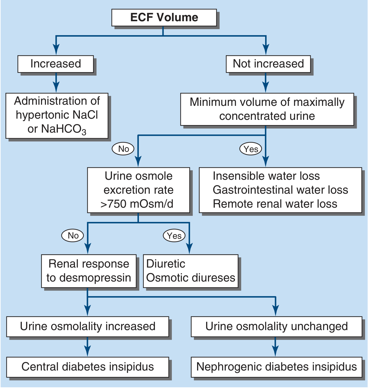
| Type | Cause | Example |
|---|---|---|
| Hypovolemic | Water loss > Na loss | Diarrhea, vomiting, burns, osmotic diuresis |
| Euvolemic | Pure water loss | Diabetes insipidus (central or nephrogenic), insensible losses |
| Hypervolemic | Excess Na+ gain | Hypertonic saline/NaHCO3 administration, hyperaldosteronism |
Clinical Features
- Irritability, high-pitched cry (infants)
- Mental status changes, lethargy
- Muscular weakness, hyper- or hyporeflexia
- Tremors, myoclonus, asterixis, chorea
- Nuchal rigidity, increased peripheral tone
- Seizures, intracerebral hemorrhage
- Death (in severe untreated cases)
Management: Step-by-Step Approach
Step 1 - Assess Volume Status (Priority #1)
- Give isotonic NS 20 mL/kg IV boluses, reassessing after each bolus until stable
- Do NOT give hypotonic fluids while the patient is in shock
- Once euvolemia is achieved, then address the free water deficit
Step 2 - Calculate Free Water Deficit
Free water deficit (mL) = 4 mL × body weight (kg) × [desired change in serum Na (mEq/L)]
Water deficit (L) = TBW × ([Na+]/140 - 1) where TBW = 0.6 × weight (kg) in children
- Free water deficit = 4 mL × 10 kg × (165-145) = 800 mL
Step 3 - Determine Rate of Correction (Critical in Pediatrics)
| Duration | Target Correction Rate |
|---|---|
| Acute hypernatremia (<48h) | No faster than 1 mEq/L/hr, no more than 10-12 mEq/L/day |
| Chronic hypernatremia (>48h) | More conservative: 5-8 mEq/L/day |
| General pediatric rule | Never exceed 0.5 mEq/L/hr - case series show seizures at faster rates |
| Maximum in first 24h | No more than 15 mEq/L in the first 24 hours |
Animal studies and pediatric case series specifically suggest that correction >0.5 mEq/L/hr can provoke seizures due to cerebral edema from the osmolyte-loaded brain cells drawing in water. - Comprehensive Clinical Nephrology, 7th Edition
Step 4 - Choose Replacement Fluid
| Situation | Fluid Choice |
|---|---|
| Hemodynamic instability | Normal saline (0.9% NaCl) boluses first |
| Mild-moderate hypernatremia, volume stable | 0.45% NaCl (half-normal saline) |
| Moderate-severe once stable | D5W or 0.2% NaCl (quarter NS) |
| Oral/NG route available | Oral water or dilute feeds - preferred when possible |
- The preferred route is oral or nasogastric water/feeds when the child is not vomiting and is hemodynamically stable
- IV D5W or quarter NS are alternatives
- For hypovolemia, 0.45% NS corrects both volume and partially addresses the free water deficit simultaneously
- Subtract bolus fluids already given from the calculated deficit
- Give half the remaining deficit over the first 8 hours
- Give the remaining half over the next 16 hours
- Add maintenance fluids and estimated ongoing losses on top of this
Step 5 - Treat the Underlying Cause
- Treat with DDAVP (desmopressin) - a vasopressin analog
- Intranasal: 5-30 mcg once or twice daily
- Oral: 0.05-0.8 mg/day in divided doses
- Low-sodium diet + thiazide diuretics (induces mild volume depletion, enhancing proximal water reabsorption)
- NSAIDs (indomethacin) can be added in select cases
- Remove offending drugs (lithium, ifosfamide) if applicable
- Discontinue hypertonic infusions
- Diuretics to eliminate excess sodium (furosemide)
- Dialysis if sodium cannot be reduced without causing volume overload, or if Na+ >180 mEq/L
- Oral rehydration solution (ORS) is preferred when tolerated
- Use low-osmolarity ORS (the 2024 systematic review, PMID 39641334 confirms low-osmolarity ORS is effective for childhood diarrhea-related dehydration)
Step 6 - Monitor Closely
- Check serum Na+ every 1-2 hours initially
- Adjust infusion rate based on trending results
- Monitor urine output - risk of acute tubular necrosis in severe cases
- A single calculation is never enough - the dynamic balance of ongoing losses requires frequent reassessment
- Watch for signs of cerebral edema during correction: worsening headache, vomiting, seizures, altered consciousness
Special Considerations in Neonates/Infants
- Neonatal hypernatremic dehydration is frequently linked to breastfeeding failure/inadequate intake. A 2024 systematic review (PMID 39111773) identified maternal risk factors including primiparous mothers, flat/inverted nipples, and delayed lactogenesis.
- Use TBW = 0.7 × weight (kg) in neonates (higher body water content)
- Oral rehydration is preferred over IV when possible
- Formula preparation errors (concentrated formula) are a preventable cause
Maintenance Fluid Choice After Correction
Summary Algorithm
Hypernatremia (Na+ >145 mEq/L)
         |
    Hemodynamically unstable?
    YES → NS boluses 20 mL/kg until stable
         |
    Now stable → Calculate free water deficit
    (4 mL × kg × desired ΔNa+)
         |
    Choose fluid (½NS, ¼NS, D5W, or oral)
         |
    Correct at ≤0.5 mEq/L/hr
    (max 10-12 mEq/L/day, 15 mEq/L in first 24h)
         |
    Monitor Na+ q1-2h, adjust rate
         |
    Identify & treat underlying cause
    (DI → DDAVP or thiazide; excess Na+ → stop source/diurese)
Dengue Fever management in pediatrics
dengue fever management children
Dengue Fever Management in Pediatrics
Overview
WHO Classification (2009)
| Category | Definition |
|---|---|
| Dengue without warning signs | Fever + 2 of: nausea/vomiting, rash, aches, leukopenia, positive tourniquet test |
| Dengue with warning signs | Above + any warning sign (see below) |
| Severe dengue | Severe plasma leakage/shock, severe bleeding, or severe organ impairment |
Three Phases of Illness
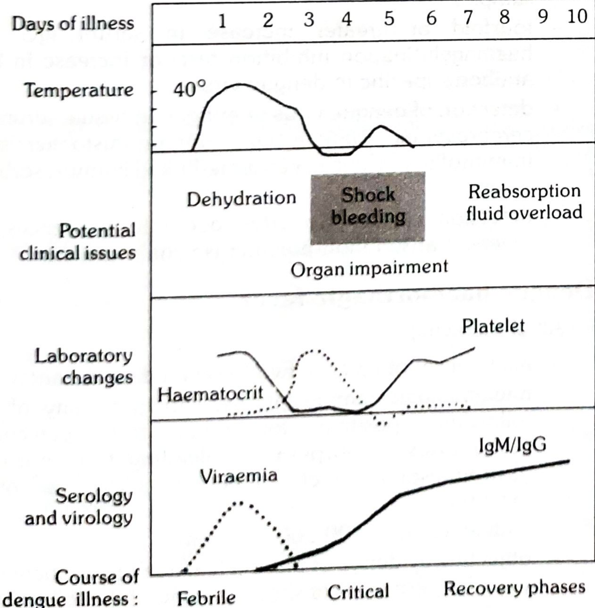
Phase 1: Febrile Phase (Days 1-3)
- Sudden high fever (39-40°C), facial flushing, severe headache
- Retroorbital pain, myalgia, arthralgia ("breakbone fever")
- Anorexia, nausea, vomiting, abdominal pain
- Maculopapular/rubelliform rash (less common than classical dengue)
- Infants: may have febrile convulsions, appear as mild upper respiratory illness
- Positive tourniquet test (≥10 petechiae per 1 inch²; in DHF usually ≥20)
- Labs: leukopenia, early thrombocytopenia
Phase 2: Critical Phase (Days 3-7, around defervescence)
- Temperature drops to ≤37.5-38°C - this is the danger window
- Capillary permeability increases → plasma leakage begins (lasts 24-48 hrs)
- Rising hematocrit (reflects severity of leakage)
- Thrombocytopenia nadir
- Pleural effusion (predominantly right-sided), ascites, gallbladder edema
- Shock when critical plasma volume lost: pulse pressure ≤20 mmHg in children, cold extremities, delayed capillary refill, tachycardia
- Note: systolic BP may remain normal in early compensated shock - the narrowed pulse pressure is the key early sign
Phase 3: Recovery Phase (Days 6-10)
- Reabsorption of extravascular fluid over 48-72 hours
- Diuresis, improving appetite
- Hematocrit falls (dilutional effect of reabsorbed fluid)
- Platelets begin to recover (after WBC recovery)
- Risk: fluid overload if excessive IV fluids were given → pulmonary edema, congestive heart failure
- "Isles of white in a sea of red" rash may appear; bradycardia common
Warning Signs (Require Hospitalization)
- Abdominal pain or tenderness
- Persistent vomiting
- Clinical fluid accumulation (ascites, pleural effusion, periorbital edema)
- Mucosal bleeding
- Lethargy or restlessness
- Liver enlargement >2 cm
- Rapidly rising hematocrit with rapid decline in platelet count
- Oliguria
Criteria for Severe Dengue
- Severe plasma leakage leading to shock (dengue shock syndrome) and/or fluid accumulation with respiratory distress
- Severe bleeding (massive GI bleed, intracranial hemorrhage)
- Severe organ impairment: liver (AST/ALT ≥1000 IU/L), CNS (impaired consciousness/encephalitis), heart (myocarditis), kidneys (AKI)
Diagnosis
Clinical Diagnosis - Dengue Fever (Probable):
Laboratory Confirmation (Red Book 2021):
| Timing | Test |
|---|---|
| Days 1-7 (febrile phase) | RT-PCR (dengue RNA) + NS1 antigen EIA |
| From Day 3-5 | Anti-dengue IgM (by EIA) - 99% positive by Day 10 |
| Combined NS1 + IgM | Identifies ≥90% of primary and secondary cases |
| Convalescent serology | 4-fold rise in IgG (acute vs. 15+ days) |
Note: A 2025 systematic review (PMID 40209729) confirmed RT-PCR has the highest sensitivity for acute dengue diagnosis, followed by NS1 ELISA.
Monitoring Labs:
- CBC with differential every 12-24 hours (hematocrit trend is the most useful guide to fluid management)
- Blood glucose, LFTs, renal function, coagulation studies
- Chest X-ray and abdominal ultrasound to detect plasma leakage
Management by Phase and Category
A. Dengue Without Warning Signs (Outpatient)
- Hydration: Encourage oral fluids (oral rehydration solution, fruit juices, coconut water). Maintain good urine output.
- Antipyretics: Paracetamol (acetaminophen) - drug of choice
- Dose: 10-15 mg/kg every 4-6 hours (max 5 doses/24h)
- AVOID: Aspirin, ibuprofen, other NSAIDs, and corticosteroids - these increase bleeding risk
- AVOID: Intramuscular injections (bleeding risk)
- Monitoring: Return immediately if warning signs appear (especially around defervescence, Days 3-7)
- Reassess daily; CBC every 24-48 hours
B. Dengue With Warning Signs (Hospitalize)
- Encourage oral/NG rehydration
- IV fluids at maintenance rate: 0.9% NaCl or Ringer's lactate (isotonic)
- Monitor urine output (target 0.5-1 mL/kg/hr)
- Check hematocrit every 4-6 hours
- IV crystalloid (0.9% NaCl or Ringer's lactate): 5-7 mL/kg/hr for 1-2 hours
- Reassess: if improving, reduce to 3-5 mL/kg/hr for 2-4 hours, then 2-3 mL/kg/hr
- Continue reducing based on clinical response and hematocrit trend
C. Severe Dengue / Dengue Shock Syndrome (ICU-level care)
- Isotonic crystalloid (0.9% NaCl or Ringer's lactate): 10 mL/kg IV over 15-30 minutes
- Reassess after each bolus (vital signs, capillary refill, urine output, hematocrit)
- Repeat bolus if still in shock (up to 10-20 mL/kg)
- If hematocrit falls (instead of rising) after bolus: suspect significant bleeding → transfuse packed red blood cells
- Switch to colloid (5% albumin or starch solutions): 10-20 mL/kg over 15-30 minutes
- Colloids are preferred over further crystalloids in refractory shock
- Packed RBCs: For significant/occult bleeding, falling hematocrit
- Platelet transfusion: NOT routinely given for thrombocytopenia alone; indicated only for active significant bleeding with very low platelet count
- Prophylactic platelet transfusion is generally not recommended even with counts <20,000/µL in the absence of bleeding
- Fresh frozen plasma / cryoprecipitate: Only for documented coagulation failure with bleeding
- Based on vital signs, urine output, hematocrit, bleeding, and signs of volume overload
- The narrowed pulse pressure (≤20 mmHg in children) is an early, key sign of shock
- Avoid excessive fluids - fluid overload is a leading cause of death in dengue shock
- Reduce IV fluids promptly once the patient stabilizes (especially entering recovery phase)
- Watch for signs of fluid overload: increasing respiratory distress, falling hematocrit (dilutional), new infiltrates on CXR
D. Recovery Phase Management
- Reduce IV fluid rate aggressively as patient stabilizes and diuresis begins
- Monitor for fluid overload: pulmonary edema, worsening respiratory distress, rising jugular venous pressure
- Hematocrit falling (not rising) = successful reabsorption of plasma
- If platelets are recovering and patient is stable, can be discharged with follow-up
Medications - What to Use and Avoid
| Drug | Use in Dengue | Notes |
|---|---|---|
| Paracetamol | YES - drug of choice | 10-15 mg/kg/dose q4-6h; sponge bathing for fever |
| Aspirin | CONTRAINDICATED | Worsens bleeding, Reye syndrome risk |
| Ibuprofen/NSAIDs | CONTRAINDICATED | Increases bleeding risk significantly |
| Corticosteroids | NOT recommended | No proven benefit; increases bleeding risk |
| Antibiotics | Only if secondary bacterial infection | Dengue is viral |
| DDAVP | No role | |
| Vitamin K | Consider if coagulopathy present |
Indications for Hospitalization
- Any warning sign present
- Severe dengue (shock, bleeding, organ failure)
- Social circumstances preventing close monitoring
- Infants (higher risk of severe disease)
- Pregnant girls
- Patients with comorbidities
- Rising hematocrit with rapidly falling platelets
Dengue Vaccine (Prevention)
- Approved for ages 9-16 years with laboratory-confirmed prior dengue infection
- 3-dose schedule: 0, 6, and 12 months
- Important: Contraindicated in dengue-naive children - increases risk of severe dengue in seronegative individuals
- Pre-vaccination screening for prior dengue infection is mandatory
A 2024 meta-analysis on risk factors for severe dengue (PMID 38263345) identified secondary infection (different serotype) as the strongest predictor of severe disease - confirming that seroprevalence screening before vaccination is clinically essential.
Summary Algorithm
Suspected Dengue in Child
         |
Febrile phase (Days 1-3)
→ Paracetamol, oral fluids, avoid NSAIDs/aspirin
→ CBC every 24-48h, watch for warning signs
         |
Approaching defervescence (Days 3-7) - DANGER WINDOW
         |
       Warning signs?
NO → Maintain oral hydration, daily review
YES → HOSPITALIZE
         |
    Hemodynamic status?
Stable → IV isotonic fluids 5-7 mL/kg/hr, adjust per hematocrit/UO
Shock → Bolus crystalloid 10 mL/kg x 15-30 min, reassess
    No response → Colloid, check for bleeding, consider blood products
         |
Recovery (Days 6-10)
→ Reduce IV fluids promptly
→ Watch for fluid overload
→ Monitor platelet recovery
Colloid solutions in pediatrics
colloid solutions pediatric fluid resuscitation
albumin versus crystalloid children sepsis outcomes
PMID: 38916738
Colloid Solutions in Pediatrics
What Are Colloids?
Classification of Colloids
1. Natural / Blood-Derived Colloids
A. Albumin
| Preparation | Concentration | Oncotic Pressure | Osmolality | Volume Expansion |
|---|---|---|---|---|
| 4% / 4.5% albumin | 4-4.5 g/dL | ~20 mmHg | ~274 mOsm/L | ~1:1 |
| 5% albumin | 5 g/dL | ~20 mmHg | ~309 mOsm/L | ~1:1 |
| 20% albumin | 20 g/dL | ~100 mmHg | ~330 mOsm/L | 4:1 (draws fluid) |
| 25% albumin | 25 g/dL | ~100 mmHg | ~330 mOsm/L | 4:1 (draws fluid) |
- 5% albumin: Isoncotic; used for volume expansion - expands plasma volume by approximately the volume infused
- 20-25% albumin: Hyperoncotic; used to mobilize edema and treat hypoalbuminemia; draws 3-4 mL of interstitial fluid per 1 mL infused - requires caution in hypovolemic patients
- Neonates and infants: remains the most frequently used plasma expander
- Premature infants with hypoalbuminemia
- Burns (after 24h for extensive injuries)
- Cardiac surgery pump priming
- Hepatic failure with ascites
- Severe septic shock refractory to crystalloids
B. Plasma Protein Fraction (PPF / Plasmanate)
- 5% solution containing albumin + α- and β-globulins
- Similar volume expansion to 5% albumin
- Important adverse effect: Hypotensive allergic reactions - especially with rapid infusion >10 mL/min (due to prekallikrein activator and kinins in the preparation)
- Less commonly used than pure albumin
C. Fresh Frozen Plasma (FFP)
- Contains all clotting factors and plasma proteins
- Not used primarily as a volume expander in pediatrics
- Indicated for documented coagulation factor deficiency with active bleeding, or prior to invasive procedures
- Risks: allergic reactions, TRALI (transfusion-related acute lung injury), infection transmission
2. Synthetic Colloids
A. Hydroxyethyl Starch (HES / Hetastarch)
| Type | MW | Molar Substitution | Example |
|---|---|---|---|
| High MW HES | 450-670 kD | 0.6-0.7 | Hetastarch (6% HES 450/0.7) |
| Medium MW HES | 200 kD | 0.5 | Pentastarch |
| Low MW HES | 130 kD | 0.4 | Voluven (3rd generation) |
- 3rd-generation HES (e.g., HES 130/0.4) revealed no serious adverse reactions (anaphylaxis, renal failure, clotting disorder) in children with normal renal and clotting function in a prospective observational safety trial
- Efficacy and safety of HES comparable to albumin in pediatric cardiac surgery
- However, HES is potentially nephrotoxic - contraindicated in patients with renal failure or at risk of AKI
- May prolong prothrombin time and decrease von Willebrand factor antigen
- Can cause hemorrhagic complications - do not exceed manufacturer's recommended dose
- Contraindicated in critically ill patients (based on adult trials showing increased mortality and AKI in the ICU)
- Not approved for use in critically ill adults in many countries; pediatric data remain insufficient
- Coagulopathy (proportional to MW and degree of molar substitution)
- AKI / nephrotoxicity
- Anaphylactoid reactions (rare)
- Tissue accumulation and pruritus (with high MW preparations)
B. Gelatins (Gelofusine, Haemaccel)
- Polypeptides produced from degradation of bovine collagen
- MW ~35,000 Da; shorter half-life (~2-3 hours) than HES or albumin
- Initial formulations had high incidence of hypersensitivity reactions (histamine-mediated)
- Newer formulations (succinylated gelatin) show improved safety in recent trials with no short- or long-term adverse effects
- Not available in the United States
- Used in some European and Asian centers for pediatric resuscitation
C. Dextrans
| Type | MW | Use |
|---|---|---|
| Dextran 70 (Macrodex) | 70,000 Da | Volume expansion |
| Dextran 40 (Rheomacrodex) | 40,000 Da | Microcirculatory flow, anti-thrombotic |
- Reduces blood viscosity
- Decreases von Willebrand factor antigen, platelet adhesion, and RBC aggregation
- Used to improve microcirculatory flow in microvascular surgery
- Interferes with blood typing and cross-matching
- Prolongs bleeding time
- Associated with AKI - do not use in renal disease
- High anaphylactic potential - among the highest of all colloids
- Negative coagulation effects
- Generally NOT recommended for pediatric use given coagulation and anaphylaxis risks
Colloids vs. Crystalloids in Pediatric Resuscitation
Current Evidence
| Comparison | Mortality | AKI |
|---|---|---|
| Colloids vs. Normal Saline | No significant difference | Very low certainty evidence |
| Balanced Crystalloids vs. Normal Saline | No significant difference | BC significantly lower AKI risk |
Miller's Anesthesia on the topic:
"There is currently no evidence supporting the role of crystalloids versus colloids in perioperative fluid loss replacement and volume expansion in neonatal and pediatric populations." - Miller's Anesthesia, 10e
Clinical Indications for Colloids in Pediatrics
| Indication | Preferred Colloid |
|---|---|
| Neonatal hypoalbuminemia with edema | 20-25% albumin (mobilizes fluid) |
| Severe septic shock refractory to crystalloids | 4-5% albumin |
| Cardiac surgery pump priming | 5% albumin or 4% albumin |
| Burns >24 hours (extensive) | Albumin (after initial crystalloid phase) |
| Plasma volume expansion in prematurity | 4.5% albumin |
| Dengue shock syndrome refractory to crystalloids | 5% albumin or HES (as rescue) |
Contraindications and Cautions
| Colloid | Contraindications / Cautions |
|---|---|
| Albumin (4%) | Traumatic brain injury (hypo-osmolar formulation worsens edema) |
| HES (any) | AKI/renal failure, coagulopathy, critically ill ICU patients |
| Dextran | Renal disease, coagulopathy, bleeding risk, blood typing interference |
| Gelatin | Anaphylaxis history, bovine product allergy |
| Hyperoncotic albumin (25%) | Severely hypovolemic patients without concurrent fluid replacement |
Practical Dosing in Pediatrics
| Agent | Dose | Notes |
|---|---|---|
| 4-5% albumin | 10-20 mL/kg IV over 15-30 min (resuscitation bolus) | Reassess after each bolus |
| 20% albumin | 1 g/kg (= 5 mL/kg) over 4 hours | For hypoalbuminemia/fluid mobilization |
| HES 130/0.4 (6%) | 10-20 mL/kg, max 33-50 mL/kg/day | Children with normal renal function only |
| Dextran 70 | 10-20 mL/kg; max 20 mL/kg/day | Risk of bleeding if exceeded |
Summary: Which Colloid in Which Pediatric Situation?
Pediatric Colloid Decision
         |
First - is crystalloid insufficient?
  → If NO: use isotonic crystalloid (first-line always)
  → If YES (refractory shock, severe hypoalbuminemia, capillary leak):
         |
    Neonates/Infants → 4.5% or 5% albumin (first choice)
    Cardiac surgery → 5% albumin or HES 130/0.4
    Mobilize edema → 20-25% albumin
    Burns >24h (extensive) → albumin
    Avoid → Dextran (coagulopathy + anaphylaxis risk)
    Avoid HES → if AKI risk, coagulopathy, or ICU-critically ill
    Avoid 4% albumin → TBI (hypo-osmolar, may worsen brain edema)
Current evidence reminder: No colloid has been shown to reduce mortality compared to crystalloids in pediatric septic shock. Balanced crystalloids are preferred over 0.9% saline as the first-line crystalloid due to lower AKI risk. Colloids are reserved for specific situations where crystalloids alone are insufficient.
Thiochchicoside tablet ingestion in pediatrics
thiocolchicoside overdose poisoning children
thiocolchicoside accidental ingestion pediatric management seizure toxicity
thiocolchicoside seizure toxicity case report
https://www.ema.europa.eu/en/documents/referral/thiocolchico…
PMID: 35083767
thiocolchicoside convulsions mechanism GABA
thiocolchicoside toxicity mechanism convulsions management activated charcoal benzodiazepine
thiocolchicoside pediatric accidental ingestion case report seizure treatment India
https://www.scielo.br/j/bjmbr/a/FfSkwhdNNKgFpzYggB4gzNf?lang…
thiocolchicoside seizure epilepsy treatment
Thiocolchicoside Tablet Accidental Ingestion in Pediatrics
What Is Thiocolchicoside?
Why This Is a Pediatric Emergency
Mechanism of Toxicity
| Receptor | Action | Clinical Effect |
|---|---|---|
| GABA-A receptors | Competitive antagonist (particularly cortical subtypes) | Pro-convulsant, CNS excitation |
| Glycine receptors | Antagonism at brainstem/spinal cord | Loss of inhibitory tone, hyperreflexia, muscle hyperexcitability |
| Nicotinic acetylcholine receptors (nAChRs) | Antagonism | Contributes to muscle effects |
Clinical Features of Toxicity in Children
Mild to Moderate Ingestion:
- Nausea, vomiting, abdominal pain
- Excessive drowsiness / somnolence
- Confusion, agitation
- Dizziness
- Vasovagal reactions
Severe / Overdose:
- Seizures (focal or generalized tonic-clonic; may progress to status epilepticus)
- Hyperreflexia, muscle rigidity or hypertonicity
- Non-convulsive status epilepticus (NCSE) - documented in adults, risk applies to children
- Acute toxic encephalopathy
- Liver injury (hepatotoxicity, cholestasis)
- Pancreatitis (rare)
- Rhabdomyolysis
- Blood cell disorders (thrombocytopenia)
- Severe skin reactions (Stevens-Johnson syndrome - rare)
- Immature blood-brain barrier = greater CNS drug penetration
- Higher body surface area-to-weight ratio = proportionally greater drug effect per tablet ingested
- No established safe dose exists for children <16 years
Emergency Management
Immediate Assessment - ABCDE Approach
Step 1: Gastrointestinal Decontamination
| Intervention | Recommendation | Condition |
|---|---|---|
| Activated charcoal (AC) | Give if within 1 hour of ingestion AND child is alert with intact airway | 1 g/kg orally (max 50 g); TCC adsorbs well to charcoal |
| Gastric lavage | Consider only if very recent large ingestion and AC cannot be given | Only with airway protection |
| Syrup of ipecac | Do NOT use - risk of aspiration if seizure occurs; no longer recommended | |
| Cathartics | Not routinely recommended |
Critical caution: If the child has already had a seizure or has a depressed level of consciousness, do NOT give activated charcoal orally without first securing the airway (intubation) - aspiration risk.
Step 2: Seizure Management
| Drug | Dose | Route |
|---|---|---|
| Diazepam | 0.2-0.5 mg/kg (max 10 mg) | IV slow; or 0.5 mg/kg per rectum |
| Midazolam | 0.1-0.15 mg/kg IV; or 0.2 mg/kg buccal/intranasal | IV/buccal/IN |
| Lorazepam | 0.05-0.1 mg/kg IV (max 4 mg) | IV |
- Phenobarbitone: 15-20 mg/kg IV loading dose (over 20-30 min) - preferred in infants; works independently of GABA-A receptor benzodiazepine binding site
- Phenytoin / Fosphenytoin: 15-20 mg/kg IV (fosphenytoin as phenytoin equivalents)
- Levetiracetam: 20-60 mg/kg IV - increasingly used as second-line; different mechanism (SV2A)
- Third-line: Propofol infusion, thiopentone/pentobarbital coma, or valproate IV
- EEG monitoring is essential to detect NCSE (non-convulsive status epilepticus), which can persist without overt motor activity
- ICU admission with continuous EEG monitoring
Step 3: Supportive Care
- Hydration: IV fluids to maintain perfusion and promote renal drug elimination
- Temperature monitoring: Hyperthermia from seizure activity - manage with cooling
- Monitor liver function serially (TCC can cause hepatotoxicity)
- Rhabdomyolysis monitoring: CK levels, urine output, urinalysis (myoglobinuria)
- Avoid: Flumazenil - not useful (TCC is not a BDZ); may actually lower seizure threshold further
Step 4: Observation and Monitoring Period
| Scenario | Disposition |
|---|---|
| Asymptomatic, small ingestion, >2 hours post-ingestion | Observe minimum 6-8 hours before discharge |
| Any neurological symptoms (drowsiness, confusion) | Hospital admission, 24-hour monitoring |
| Seizure occurred | Pediatric ICU admission; EEG monitoring |
| Status epilepticus | PICU, prolonged admission |
Dose Context: How Dangerous Is One Tablet?
| Tablet Strength | Adult Dose | Risk in a Toddler (10 kg) |
|---|---|---|
| 4 mg TCC | 1 tablet = therapeutic adult single dose | Equivalent to ~0.4 mg/kg - above any safe pediatric threshold |
| 8 mg TCC | Common adult dose | Equivalent to ~0.8 mg/kg - significant toxicity risk |
Key Points Summary
- TCC is contraindicated under age 16 (EMA-mandated) - no safe pediatric dose exists
- Primary danger is seizures (including status epilepticus) from GABA-A and glycine receptor blockade
- Even breastfeeding exposure from a mother taking TCC has caused neonatal seizures
- Activated charcoal within 1 hour if airway is safe - is the key decontamination step
- Benzodiazepines are first-line for seizures but have partial efficacy; may need phenobarbitone or levetiracetam
- No antidote exists - management is entirely supportive
- Monitor liver function, CBC, CK for secondary organ toxicity
- NCSE must be excluded by EEG in any obtunded child who had TCC exposure
Poison Control: Always contact your national poison control center for real-time guidance:
- India: 1800-11-6117 (AIIMS Poison Control, New Delhi)
- Global: www.poison.org
Lupus nephritis
lupus nephritis management treatment guidelines
PMID: 40127995
PMID: 41107121
Lupus Nephritis
Overview
Pathogenesis
- Loss of tolerance to nuclear antigens (dsDNA, histones, nucleosomes)
- Autoantibody production (anti-dsDNA, anti-Sm, antinuclear)
- Immune complex deposition in glomeruli → complement activation → inflammatory cascade
- Roles of complement (C1q, C3, C4) - "full house" pattern on immunofluorescence (IgG, IgA, IgM, C3, C1q) is pathognomonic
- T helper cell dysregulation (Th17), B cell hyperactivity
- Genetic susceptibility: >50 polymorphisms linked to LN, including CCR6 (Th17 marker variants in Asian populations)
Classification: ISN/RPS (2003, revised 2018)
| Class | Name | Light Microscopy | Glomeruli Affected | Clinical Significance |
|---|---|---|---|---|
| I | Minimal mesangial LN | Normal LM; deposits on IF/EM only | None visible | Benign; no specific treatment |
| II | Mesangial proliferative LN | Mesangial hypercellularity | Mesangial only | Usually mild; treat SLE; monitor |
| III | Focal LN | Endocapillary proliferation | <50% glomeruli | Active, moderate disease |
| IV | Diffuse LN | Global/segmental proliferation | ≥50% glomeruli | Most severe; requires aggressive treatment |
| V | Membranous LN | Subepithelial deposits; GBM thickening | Diffuse | Nephrotic syndrome; indolent but persistent |
| VI | Advanced sclerosing LN | >90% sclerosed | Global | Irreversible; no benefit from immunosuppression |
- IV-S (segmental): <50% of each glomerulus affected
- IV-G (global): >50% of each glomerulus affected
- Activity indices: Endocapillary proliferation, karyorrhexis/fibrinoid necrosis, cellular crescents, subendothelial hyaline deposits, interstitial inflammation
- Chronicity indices: Glomerular sclerosis, fibrous crescents, tubular atrophy, interstitial fibrosis
Renal Biopsy
- Proteinuria >0.5 g/24h (or urine protein:creatinine ratio >0.5)
- Active urine sediment (RBC casts, dysmorphic RBCs)
- Unexplained rise in serum creatinine
- Biopsy-proven LN is also a standalone SLICC classification criterion for SLE
Clinical Features
| Finding | Description |
|---|---|
| Hematuria | Usually microscopic; RBC casts highly specific |
| Proteinuria | Ranges from sub-nephrotic to nephrotic range (>3.5 g/day) |
| Nephrotic syndrome | Common in class V (membranous) |
| Hypertension | Frequent, especially in class III/IV |
| Renal insufficiency | Rising creatinine in active proliferative disease |
| Active urinary sediment | RBC casts = nephritis; WBC casts = interstitial nephritis |
| Systemic SLE features | Malar rash, arthritis, serositis, cytopenias |
Serological Markers
| Test | Role |
|---|---|
| ANA | Screening (sensitive, not specific) |
| Anti-dsDNA | Disease activity; titers correlate with nephritis flares |
| Anti-Sm | Associated with worse renal prognosis (48.8% of biopsy-proven LN in one Korean study) |
| C3, C4 | Fall during active nephritis (consumed by immune complexes) |
| Antiphospholipid antibodies | Poor prognosis; thrombosis risk |
| Urine PCR | Urine protein:creatinine ratio (spot) for monitoring |
Treatment
Foundational Principles (2024 ACR / 2025 EULAR)
- Kidney biopsy first - class-guided management is essential
- Hydroxychloroquine (HCQ) in ALL patients with LN - reduces flares, ESKD, and improves survival (dose: 200-400 mg/day; max 5 mg/kg/day based on ideal body weight)
- RAAS blockade (ACE inhibitor or ARB) for all patients with proteinuria - independent of BP
- SGLT2 inhibitors - now recommended as kidney-protective add-on therapy
- The 2024 ACR guideline shifts away from discrete "induction/maintenance" phases toward continuous therapy with the same agents at adjusting intensity, for 3-5 years after achieving complete renal response
- Treat-to-target: aim for complete renal response (proteinuria <0.5 g/day, stable/normal creatinine)
Treatment Algorithm (by Biopsy Class)
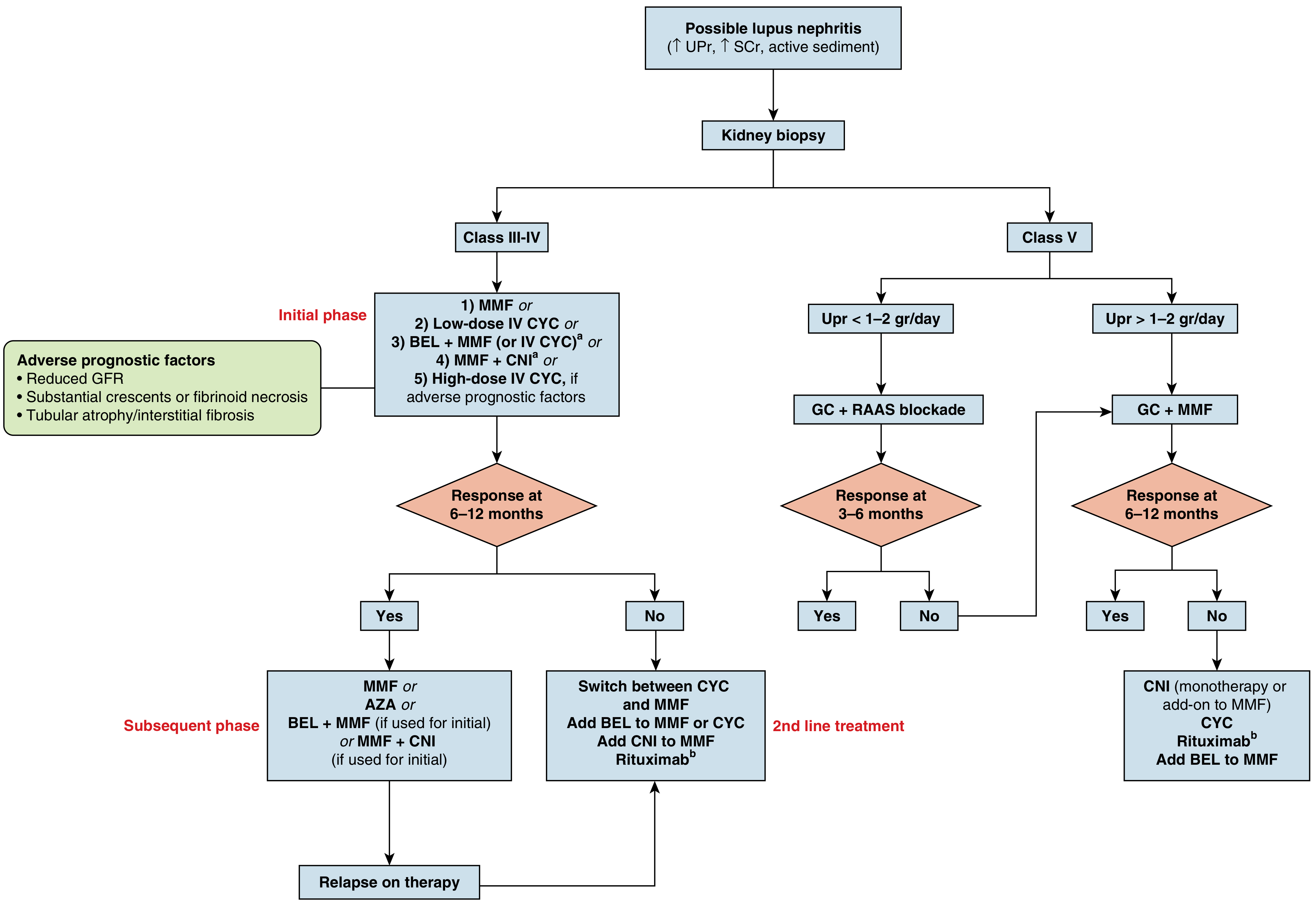
Class I and II: Minimal/Mesangial LN
- No specific immunosuppression for renal disease
- Treat extrarenal SLE manifestations
- HCQ + RAAS blockade
- Monitor: urine protein, sediment, serum creatinine, anti-dsDNA, complement every 3-6 months
Class III/IV: Focal and Diffuse Proliferative LN (Active Disease)
- Pulse IV methylprednisolone (MP): 500 mg - 1 g/day × 1-3 pulses
- Oral prednisone: 0.5-0.6 mg/kg/day × first 4 weeks, then taper (target ≤7.5 mg/day by 3-6 months; withdraw if possible)
| Regimen | Dose | Notes |
|---|---|---|
| MMF (Mycophenolate mofetil) | 2-3 g/day (target); MPA 1440 mg/day | First-line in most patients; may be more efficacious in African Americans |
| Low-dose IV CYC (Euro-Lupus) | 500 mg IV q2wk × 6 doses | Equivalent 10-year outcomes to high-dose; minimal gonadotoxicity |
| High-dose IV CYC (NIH) | 0.5-1 g/m² monthly × 6 doses, then quarterly × 2 years | Reserved for severe disease (crescents, fibrinoid necrosis, reduced GFR) |
| BEL + MMF (or BEL + low-dose CYC) | Belimumab 10 mg/kg IV q4wk (or 200 mg SC weekly) + MMF | FDA-approved 2020; preferred in high histologic activity or extrarenal disease |
| MMF + CNI (voclosporin or tacrolimus) | MMF 1-2 g/day + voclosporin 23.7 mg BID | Preferred for severe nephrotic-range proteinuria; FDA-approved 2021 |
- Reduced GFR at presentation
- Crescents or fibrinoid necrosis on biopsy
- Tubular atrophy / interstitial fibrosis
- Antiphospholipid syndrome
| Drug | Dose | Notes |
|---|---|---|
| MMF | 1.5-2 g/day | Preferred; superior to AZA in ALMS maintenance trial in non-white patients |
| Azathioprine (AZA) | 1.5-2 mg/kg/day | Alternative; comparable in MAINTAIN trial (European patients) |
| BEL + MMF | Continue if used initially | |
| MMF + CNI | Continue if used initially |
Class V: Membranous LN
- Glucocorticoids + RAAS blockade alone
- Reassess at 3-6 months
- Glucocorticoids + MMF (initial phase, 2-3 g/day)
- CNI monotherapy or add-on (tacrolimus or voclosporin) - particularly effective in membranous LN with nephrotic syndrome
- Belimumab may be added to MMF
Second-Line / Refractory Treatment
| Situation | Option |
|---|---|
| No response to MMF after 3-6 months | Switch to CYC (or vice versa) |
| Refractory despite CYC or MMF | Rituximab (anti-CD20) 1000 mg IV × 2 doses 2 weeks apart (despite failed primary RCT, nonrandomized evidence supports use; 2025 EULAR endorses) |
| Add-on for inadequate response | Belimumab added to MMF or CYC |
| Add-on for proteinuria | CNI (voclosporin or tacrolimus) added to MMF |
| Class III/IV refractory - very aggressive | Obinutuzumab (anti-CD20, next-generation) - 2025 EULAR recommendation |
Newer Approved Agents (Key Developments)
| Drug | Mechanism | Approval | Trial |
|---|---|---|---|
| Belimumab (Benlysta) | Anti-BLyS/BAFF monoclonal antibody | FDA 2020 for LN | BLISS-LN trial: significantly improved primary efficacy renal response vs placebo |
| Voclosporin (Lupkynis) | Novel CNI (less nephrotoxic than tacrolimus) | FDA 2021 for LN | AURORA 1 trial: superior complete renal response at 52 weeks vs placebo (both on MMF + GC) |
| Obinutuzumab | Type II anti-CD20 (more potent B-cell depletion than rituximab) | Investigational | NOBILITY trial: improved renal outcomes at 2 years vs rituximab-like comparator |
Response Definitions
| Category | Definition |
|---|---|
| Complete Renal Response (CRR) | Proteinuria <0.5 g/day + serum creatinine normal or ≤10% above baseline + inactive urine sediment |
| Partial Renal Response (PRR) | ≥50% reduction in proteinuria + creatinine stable (not more than 25% above baseline) |
| No response | <50% reduction in proteinuria or worsening creatinine |
| Renal relapse | Re-appearance of proteinuria ≥1 g/day or doubling of creatinine from nadir |
Supportive and Adjunct Measures
| Measure | Rationale |
|---|---|
| RAAS blockade (ACEi/ARB) | Reduces proteinuria and progression independent of BP |
| SGLT2 inhibitors | Kidney protection (cardiorenal benefit) - now included in 2025 EULAR recommendations |
| Hydroxychloroquine | ALL patients - reduces flares, ESKD, and cardiovascular risk |
| BP target <130/80 mmHg | Protects against progressive kidney disease |
| Statins | Accelerated atherosclerosis in SLE |
| Calcium/Vitamin D supplementation | Corticosteroid-associated bone loss |
| Vaccinations | Pneumococcal, influenza, COVID-19 before immunosuppression where possible |
| Avoid NSAIDs | Risk of aseptic meningitis, renal dysfunction, hypertension |
| Sun protection | UV light triggers SLE flares |
| Contraception | CYC and MMF are teratogenic; combined OCP may worsen thrombosis risk with APS |
Pregnancy in Lupus Nephritis
- CYC and MMF are contraindicated in pregnancy (MMF causes microtia, cleft palate, auditory canal atresia; stop ≥6 weeks before conception)
- Safe in pregnancy: HCQ, prednisone, azathioprine, tacrolimus, IV immunoglobulin
- Rituximab: not teratogenic but causes fetal B-cell depletion; avoid if possible
- Belimumab: insufficient safety data
- Target remission ≥6 months before attempting conception
- RAAS blockers must be stopped before conception (fetotoxic)
- Distinguish LN flare from preeclampsia (biopsy if needed in first/early second trimester)
- Low-dose aspirin for all (preeclampsia prevention)
Monitoring Schedule
| Parameter | Frequency |
|---|---|
| Urine protein:creatinine ratio, urinalysis | Every 1-3 months (active disease); every 3-6 months (remission) |
| Serum creatinine, electrolytes | Every 1-3 months |
| Anti-dsDNA, C3, C4 | Every 3 months (rising anti-dsDNA + falling complement = impending flare) |
| CBC (AZA/MMF/CYC toxicity) | Every 1-3 months |
| LFTs | Every 3 months |
| Blood pressure | Every visit |
| HCQ retinal screening | Annually after 5 years of use |
| Repeat kidney biopsy | For significant unexplained deterioration or reclassification |
Prognosis
- Class IV LN carries the worst prognosis; 10-year ESKD rate ~10-25% in treated patients
- Predictors of poor outcome: Black/Hispanic ethnicity, reduced GFR at presentation, high chronicity index (tubular atrophy, interstitial fibrosis), crescents, antiphospholipid antibodies, anti-Sm positivity, delayed treatment
- Asian patients have higher LN prevalence, more severe disease, but early treatment improves outcomes
- Overall 10-year renal survival with modern treatment: >80%
- Leading cause of death: infections (due to immunosuppression), not renal failure
2024 ACR & 2025 EULAR - Key paradigm shift: Lupus nephritis therapy is now conceptualized as continuous and ongoing (not discrete induction + maintenance), with the same drugs used throughout at varying intensity, and treatment duration of at minimum 3-5 years after achieving complete renal response - Sammaritano et al. 2025, PMID 40127995; Fanouriakis et al. 2026, PMID 41107121
Digeorge syndrome features
DiGeorge syndrome 22q11 deletion clinical features management
PMID: 36897497
DiGeorge Syndrome (22q11.2 Deletion Syndrome)
Nomenclature
| Synonym | Emphasis |
|---|---|
| DiGeorge syndrome | Thymic/parathyroid/cardiac defects |
| 22q11.2 deletion syndrome | Cytogenetic |
| Velocardiofacial syndrome (VCFS) | Velopalatal + cardiac + facial features |
| Conotruncal anomaly face syndrome | Cardiac outflow tract anomalies |
| CATCH-22 | Cardiac defects, Abnormal facies, Thymic hypoplasia, Cleft palate, Hypocalcemia, 22q11 deletion |
Genetics and Molecular Basis
- Prevalence: ~1 in 3,000 live births - one of the most common chromosomal microdeletion syndromes
- Genetics: ~90% have a hemizygous microdeletion of chromosome 22q11.2 (3 Mb region); rare cases involve 10p13 deletion
- Inheritance: Mostly de novo (sporadic); ~10% autosomal dominant (inherited from mildly affected parent)
- Key genes deleted:
- TBX1 (T-box transcription factor) - primary driver of cardiac, thymic, parathyroid, and facial anomalies
- HIRA and UFDIL - contribute to the phenotype
- CXCR4 signaling disruption - affects neural crest cell migration
- Variable expressivity: Patients with identical 22q11.2 deletions may have markedly different phenotypes
- Detection: Chromosomal microarray (gold standard); FISH is no longer recommended as first-line
Embryological Basis
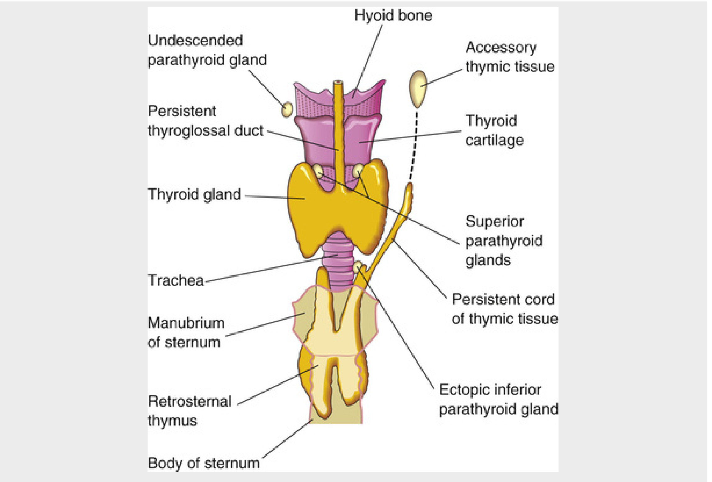
- Thymus (3rd pouch) → absent/hypoplastic → T-cell deficiency
- Parathyroid glands (3rd and 4th pouches) → absent/hypoplastic → hypocalcemia
- Portions of the face and aortic arch → via neural crest cell migration → conotruncal heart defects and facial anomalies
Clinical Features
1. Cardiac Defects (~75-90% of patients)
| Cardiac Lesion | Frequency (%) |
|---|---|
| Pulmonary atresia with VSD | 27.3 |
| Tetralogy of Fallot | 26.1 |
| Ventricular septal defect (VSD) | 17.0 |
| Truncus arteriosus | 11.4 |
| Interrupted aortic arch (type B) | 9.1 |
| Transposition of great arteries | 3.4 |
| Atrial septal defect (ASD) | 2.3 |
| Double outlet right ventricle | 1.1 |
| Double aortic arch | 1.1 |
Any infant with an interrupted aortic arch, truncus arteriosus, or tetralogy of Fallot should be screened for 22q11.2 deletion.
2. Hypocalcemia (17-60%)
- Caused by hypoparathyroidism from absent/hypoplastic parathyroid glands
- Presents as neonatal hypocalcemic tetany - often the first presentation in the newborn period
- May be transient (improves with age as ectopic parathyroid tissue compensates) or permanent
- Can cause hypocalcemic seizures - distinguishable from epilepsy
- Severity correlates with degree of parathyroid hypoplasia
3. Immunodeficiency (77%)
Spectrum:
| Form | Prevalence | T-cell Status | Clinical Significance |
|---|---|---|---|
| Partial DiGeorge | ~99.5% | Moderately reduced T cells; relatively normal T-cell function | Recurrent sinopulmonary infections, otitis media; most improve with age |
| Complete DiGeorge | <0.5% | Profound T-cell deficiency (SCID-like: CD3 <50/µL) | Life-threatening opportunistic infections; requires thymic transplantation |
Immune features:
- T cells: Reduced CD3+, CD4+, CD8+ counts; impaired T-cell function; naive T cell deficiency
- B cells and immunoglobulins: Generally normal; T-cell-dependent humoral function may be impaired
- Autoimmunity: Increased prevalence due to defective deletion of autoreactive T cells and impaired Tregs:
- Juvenile idiopathic arthritis
- Autoimmune hemolytic anemia
- Immune thrombocytopenia
- Autoimmune thyroid disease
- Atopy: Increased rates of eczema, food allergy, asthma
Infectious susceptibility:
- Partial DGS: Recurrent sinopulmonary infections, recurrent otitis media, oral/esophageal thrush
- Complete DGS: Opportunistic infections - Pneumocystis jirovecii pneumonia, CMV, disseminated candidiasis, viral infections
- Impaired defense against: viruses, fungi, protozoans, intracellular bacteria (T-cell mediated immunity)
4. Facial Features (~60-70%)
- Hypertelorism (wide-spaced eyes)
- Low-set, notched ears with abnormal helices
- Shortened philtrum of the upper lip
- Bulbous nasal tip with hypoplastic alae nasi
- Saddle nose deformity
- Mandibular hypoplasia (small jaw/micrognathia)
- Hooded eyelids / palpebral fissure anomalies
- Microcephaly (18-50%)
5. Palatal Abnormalities
| Anomaly | Frequency (%) |
|---|---|
| Velopharyngeal incompetence (VPI) | 27 |
| Submucosal cleft palate | 16 |
| Overt cleft palate | 11 |
| Bifid uvula | 5 |
| Cleft lip ± cleft palate | 2 |
| Infantile VPI | 8 |
- VPI and submucosal cleft palate → hypernasal speech (classic feature)
- Palatal anomalies contribute to feeding difficulties, aspiration risk, recurrent sinus disease
6. Neurodevelopmental and Psychiatric Features
| Feature | Frequency (%) |
|---|---|
| Developmental delay | Common |
| Learning disabilities | Common |
| Psychiatric disturbance (any) | 60 |
| Schizophrenia/psychosis | 25 |
| ADHD | ~35 |
| Anxiety disorders | 30-40 |
| Autism spectrum disorder | ~15 |
| Intellectual disability | Variable |
| Nonverbal learning disability | Common |
| Seizures (with or without hypocalcemia) | 7 |
22q11.2 deletion is the highest known single-gene risk factor for schizophrenia. Approximately 25% of 22q11DS patients develop schizophrenia or schizoaffective disorder, typically in late adolescence/early adulthood. Conversely, ~1-2% of all schizophrenia patients have 22q11.2DS.
7. Other Associated Features
| System | Features |
|---|---|
| Renal | Renal agenesis, horseshoe kidney, vesicoureteral reflux |
| Gastrointestinal | Feeding difficulties (36%), constipation (>50%), esophageal atresia, intestinal malrotation |
| Endocrine | Hypothyroidism, growth hormone deficiency |
| Hearing | Sensorineural and conductive hearing loss |
| Ophthalmologic | Posterior embryotoxon, tortuous retinal vessels |
| Musculoskeletal | Scoliosis, joint hypermobility |
| Laryngotracheoesophageal | Laryngeal web, tracheomalacia (contributes to aspiration) |
Summary: The CATCH-22 Mnemonic
| Letter | Feature |
|---|---|
| C | Cardiac defects (conotruncal) |
| A | Abnormal facies (hypertelorism, low-set ears, short philtrum) |
| T | Thymic hypoplasia → T-cell deficiency |
| C | Cleft palate / velopharyngeal anomalies |
| H | Hypocalcemia (hypoparathyroidism) |
| 22 | Chromosome 22q11.2 deletion |
Diagnosis
| Investigation | Purpose |
|---|---|
| Chromosomal microarray | Gold standard; detects 22q11.2 deletion (FISH no longer recommended) |
| Serum calcium, PTH | Hypocalcemia/hypoparathyroidism |
| Full blood count with differential | Lymphopenia |
| CD3, CD4, CD8 counts | T-cell subsets |
| Immunoglobulin levels | Humoral assessment |
| Diphtheria and tetanus antibodies | Functional humoral response |
| Renal ultrasound | Renal anomalies |
| Chest X-ray | Thymic shadow (absent in complete DGS), cardiac silhouette |
| Echocardiogram | Cardiac structural defects |
| Newborn screening (TRECs) | T-cell receptor excision circles - low/absent in DGS; increasingly detecting DGS on newborn screening |
Management
1. Cardiac
- Surgical repair as appropriate for specific defect
- Tetralogy of Fallot, truncus arteriosus, and interrupted aortic arch all require staged surgical correction
2. Hypocalcemia
- Oral calcium supplementation (calcium carbonate/citrate)
- Calcitriol (active vitamin D) to enhance calcium absorption
- IV calcium gluconate for acute hypocalcemic tetany
- Monitor calcium levels regularly; may resolve or persist with age
3. Immunodeficiency Management
- Hold all live vaccines until immune function is fully assessed (risk of vaccine-strain infection in complete DGS)
- Irradiated blood products for complete DGS (prevent transfusion-associated GVHD)
- PCP prophylaxis with cotrimoxazole for complete DGS
- IVIG replacement for significant humoral dysfunction
- Complete DGS: Thymic tissue transplantation (allogeneic thymus from an infant undergoing cardiac surgery) - successfully reconstitutes T-cell immunity; currently available at specialized centers
- Partial DGS: Most patients show immune improvement spontaneously with age - thymic transplant not usually required; monitor and treat infections
- Spontaneous immune reconstitution can occur even with initially severe T-cell lymphopenia in partial DGS
4. Developmental/Neurological
- Early speech therapy (velopharyngeal dysfunction, speech delay)
- Occupational therapy, physiotherapy for developmental delays
- Psychiatric screening from adolescence (schizophrenia risk)
- Educational support for learning disabilities
- Calcium monitoring for hypocalcemic seizures
5. Multidisciplinary Follow-up
- Genetics, cardiology, immunology, endocrinology, nephrology, ENT, speech pathology, developmental pediatrics, psychiatry (from adolescence)
- Annual thyroid function
- Audiological assessment
- Ophthalmology