
Tag pi and tag capsulotomy
Reading File
Now let me search the library for both topics in parallel.
Finding Sources
Finding Sources
Good - the context is ophthalmology. Let me read the relevant sections from Kanski's for both topics in parallel.
Reading File
Reading File
Let me read more of the LPI section and fetch the relevant images in parallel.
Reading File
Reading File
I now have comprehensive content for both topics. Let me also read the complications/closure part of the LPI section and the YAG capsulotomy complications more completely.
Reading File
I now have all the information needed. Let me fetch the key images for both topics in parallel.
Loading Image
Loading Image
I now have comprehensive information for both procedures. Here is the complete structured answer:
TAG PI (Laser Peripheral Iridotomy) and TAG Capsulotomy (Nd:YAG Posterior Capsulotomy)
TAG PI - Laser Peripheral Iridotomy (LPI)
What it is
A laser procedure that creates a small full-thickness hole in the peripheral iris to relieve pupillary block, allowing aqueous to bypass the pupil and flow freely from the posterior to the anterior chamber.
Indications
- Primary angle closure (PAC/PACG) - the main indication
- Primary angle closure suspect (PACS) - selectively; the ZAP trial showed LPI has only a small prophylactic effect over 6 years, so it should be offered only to those at highest risk
- Secondary angle closure with pupillary block
- Pigment dispersion syndrome (effectiveness under ongoing investigation)
- Prophylactic indications that may influence the decision:
- Symptoms of previous intermittent angle closure
- Systemic medication with atropine-like structure (risk of pharmacologic pupillary block)
- Need for frequent dilated fundal examination (e.g., diabetes, AMD)
- Occupational/geographic difficulty accessing urgent ophthalmic care
Technique
- Pre-procedure: Topical anaesthetic + apraclonidine (or brimonidine) prophylactically. Pupil constricted with topical pilocarpine 2% (one drop).
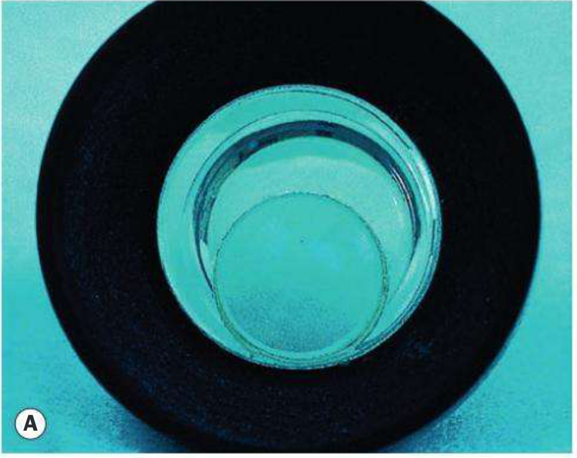
- Lens: Special iridotomy contact lens - Abraham lens or Volk MagPlus lens.
Fig. 11.67A - Abraham contact lens used for LPI:
-
Site: Between 11-1 o'clock (under upper eyelid) is standard. Some prefer 3 or 9 o'clock. The iridotomy should be in the outer third radially (to reduce lens damage risk). Targeting an iris crypt makes penetration easier.
- Highest risk of monocular dysphotopsia occurs when the iridotomy is half-covered by the lid margin.
-
Energy: Nd:YAG laser at 4-5 mJ (2-4 mJ for thin blue iris). Risk of lens damage increases above 5 mJ. Single or up to 3-pulse shots.
- Thick dark irides: pre-treat with argon/diode laser (600-900 mW, 50 µm spot, 0.03-0.05 s) before YAG application.
-
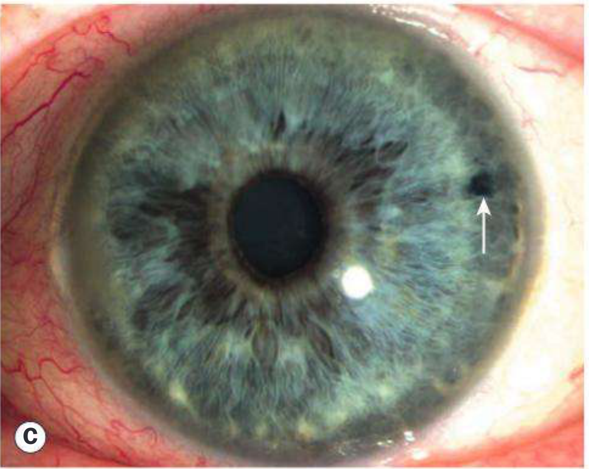
Endpoint: Gush of pigment debris confirms full-thickness penetration. Optimal size = 150-200 µm.
Fig. 11.67C - Temporal iridotomy (arrow showing patent PI):
- Post-procedure: Second drop of apraclonidine. IOP check at 1-2 hours. Topical steroid (e.g., dexamethasone 0.1%) every 10 min x 30 min, then hourly x 6-8 hours, then QID x 1 week. Review at 1-2 weeks.
Complications
| Complication | Notes |
|---|---|
| Bleeding | ~50% - usually mild, self-limiting; increase contact lens pressure if persistent |
| IOP spike | Usually early and transient; occasionally persistent |
| Iritis | More common in dark irides or with inadequate post-laser steroid therapy |
| Corneal burns | Usually heal without sequelae |
| Cataract | Localized opacity at treatment site; may accelerate age-related cataract |
| Linear dysphotopsia | Can occur regardless of location or size |
| Posterior synechiae | Laser-induced iritis complication |
TAG Capsulotomy - Nd:YAG Posterior Capsulotomy
What it is
A laser procedure to create an opening in the opacified posterior capsule following cataract surgery - treating posterior capsule opacification (PCO), also called "after-cataract" or "secondary cataract."
Background
PCO occurs due to:
- Pearl-type: Proliferation of equatorial lens epithelial cells (Elschnig pearls)
- Fibrosis-type: Fibroblastic metaplasia of epithelial cells with contractile qualities
- Soemmering ring: Whitish annular peripheral proliferation
Indications
- Reduced visual acuity or glare from PCO (primary indication)
- Inadequate fundus view impeding assessment/treatment of posterior segment pathology (e.g., diabetic retinopathy, AMD)
Technique
- Energy: Start at 1 mJ/pulse; increase only if necessary.
- Pattern: Cruciate or circumferential series of punctures using single-pulse shots.
- Size: Opening should approximate the physiologically dilated pupil under scotopic conditions - average 4-5 mm in the pseudophakic eye.
- Larger opening may be needed for persistent glare or retinal examination/treatment.
- Do not extend beyond the optic edge - vitreous may prolapse around the edge.
- Caution in high-risk eyes: Adopt higher threshold and minimize extent in:
- High myopia (risk of retinal detachment)
- History of uveitis (risk of CME)
- Pseudoexfoliation (risk of lens displacement)
- Energy limit: Total energy should be < 80 mJ to reduce risk of IOP spike.
- Timing: Delay at least 6 months after cataract surgery to reduce risk of CMO.
Complications
| Complication | Notes |
|---|---|
| IOL pitting | Usually visually inconsequential |
| IOP rise | Mild and transient; more significant in pre-existing glaucoma |
| Retinal tear / detachment | Myopic patients must be warned about PVD symptoms |
| CMO (cystoid macular oedema) | Less common when capsulotomy delayed ≥6 months post-cataract surgery |
| IOL subluxation/dislocation | Rare |
| Vitreous prolapse | If capsulotomy extends beyond optic edge |
Anterior capsulotomy (bonus point)
Capsulophimosis - contraction of the anterior capsular opening after continuous curvilinear capsulorhexis - may require Nd:YAG anterior capsulotomy. Risk factors: small capsulorhexis, pseudoexfoliation, retinitis pigmentosa, plate-haptic silicone IOL.
Source: Kanski's Clinical Ophthalmology: A Systematic Approach, 10th Ed., pp. 348, 428.
This is a shared conversation. Sign in to Orris to start your own chat.