
Olecranon bursitis surgical steps
Reading File
Finding Sources
Finding Sources
Searching PubMed
"olecranon bursitis" AND surgery
Reading File
Reading File
Reading File
Olecranon Bursitis: Surgical Management
Surgery is reserved for chronic, recalcitrant, or septic olecranon bursitis that fails conservative measures (activity modification, NSAIDs, aspiration ± antibiotics, compressive dressing). The two accepted surgical approaches are open bursectomy (excision) and arthroscopic bursectomy.
Indications for surgery
- Chronic/recurrent aseptic bursitis unresponsive to repeated aspiration and conservative care
- Septic bursitis with abscess formation, failed antibiotics, or purulent collection needing formal drainage
- Symptomatic calcific nodules within the bursal lining
- Cosmetically or functionally troublesome chronic swelling
(Bailey and Love's Short Practice of Surgery, 28th Ed, p. 2640; Campbell's Operative Orthopaedics, 15th Ed)
1. Open Bursectomy (standard approach)
This is the most commonly used technique for chronic recalcitrant disease:
- Position the patient with the elbow flexed and the extensor surface exposed; tourniquet control as needed.
- Make a longitudinal or curvilinear skin incision lateral (not directly over) the tip of the olecranon to avoid a poorly healing scar directly over the bony prominence.
- Carefully raise skin flaps, preserving as much subcutaneous tissue as possible to reduce wound-healing complications.
- Excise the bursal sac in its entirety, including any calcific nodules or thickened, chronically inflamed lining.
- If septic, send bursal tissue/fluid for culture; irrigate the wound thoroughly.
- Obtain meticulous hemostasis (the region is prone to hematoma).
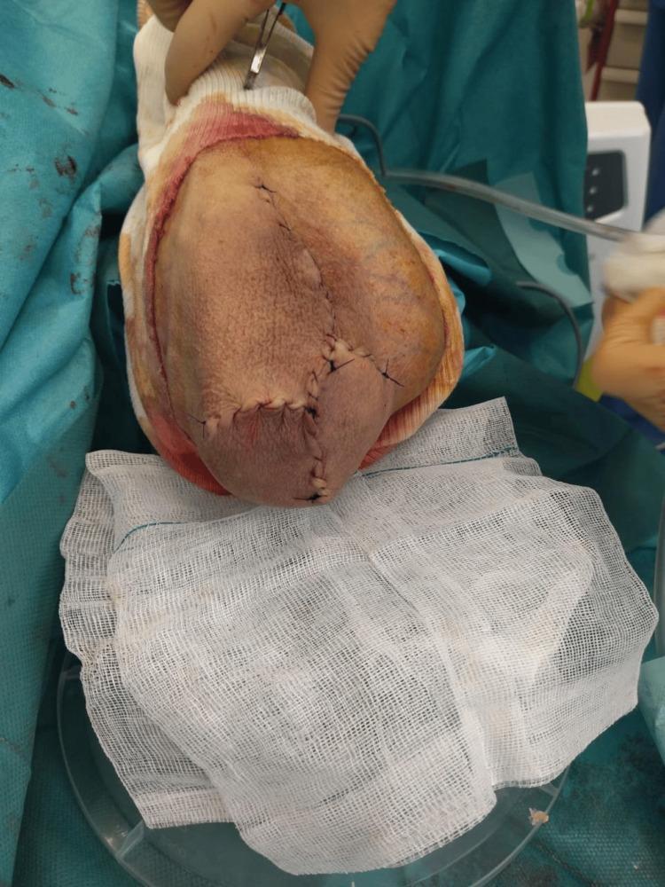
- Close skin in layers; a drain or compressive dressing is often used to obliterate dead space and prevent seroma/hematoma, a common complication.
- Postoperatively: compressive dressing, elbow immobilization briefly, then early gentle range-of-motion to avoid stiffness.
(Bailey and Love's Short Practice of Surgery, p. 2640; Campbell's Operative Orthopaedics 15th Ed, p. 5539)
Below is an intraoperative example showing wound closure after open excision of a chronic olecranon bursitis mass:
2. Arthroscopic Bursectomy (Baker and Cummings technique)
Campbell's Operative Orthopaedics describes this alternative, aimed at reducing wound-healing problems seen with open incisions directly over the olecranon:
- Positioning: After intubation, place the patient prone.
- Portals: Establish three portals - lateral, proximal central, and distal central - using a No. 11 blade and a hemostat to spread soft tissue bluntly. Avoid medial portals because of risk to the ulnar nerve.
- Bursectomy: Perform a total bursectomy, exchanging the arthroscope and instruments between the operative and viewing portals as needed. Resection is considered complete when increased light transillumination is visible through the skin and the triceps tendon/muscle can be seen through the resected bursal bed.
- Portal closure: Close the arthroscopic portals with 3-0 nylon sutures.
- Dressing: A compression dressing is applied; postoperative anesthetic injections are generally avoided so an immediate neurologic exam can be performed.
- Postoperative care: Compression dressing removed at 7-10 days; early mobilization of the extremity is encouraged immediately.
Evidence on arthroscopic bursectomy is limited, and complication rates are reported as similar to open technique, so its relative advantage remains unproven per Campbell's.
(Campbell's Operative Orthopaedics, 15th Ed, Technique 57.39, p. 5549-5565)
Key complications to counsel on
- Wound dehiscence / delayed healing (most common issue with direct posterior incisions)
- Recurrence
- Infection
- Hematoma/seroma
- Ulnar nerve injury (arthroscopic medial portals)
Recent literature note
A 2021 review in the Journal of Hand Surgery (Nchinda & Wolf, PMID 33840568) reinforces that surgery is reserved for refractory cases and that recurrence/wound complications remain the main surgical concerns. A 2024 case series (PMID 38299046) reported no wound healing complications or recurrences with endoscopic bursectomy for recalcitrant cases, suggesting outcomes may be improving with endoscopic technique, though this is a single small series and not yet practice-changing evidence. A 2021 Journal of Shoulder and Elbow Surgery study (PMID 33144225) identified factors associated with revision surgery after bursectomy - worth reviewing if you want failure-risk factors specifically.
If you want, I can pull the specific revision-risk factors from that 2021 study or expand on nonoperative/aspiration technique as a comparison.
Olecranon bursitis
Finding Sources
Searching PubMed
"olecranon bursitis" AND management
Searching Images
olecranon bursitis clinical photograph swollen elbow
Olecranon Bursitis
Definition and Anatomy
The olecranon bursa is a subcutaneous, synovial-lined sac situated between the skin and the olecranon process of the ulna, on the posterior/extensor aspect of the elbow. Normally thin-walled and containing minimal fluid, it becomes inflamed and distended with excess fluid in olecranon bursitis. It is the most frequent site of septic bursitis in the body.
(Roberts and Hedges' Clinical Procedures in Emergency Medicine, p. 3441; Firestein & Kelley's Textbook of Rheumatology)
Etiology
- Trauma - the leading cause, either a single acute impact or chronic repetitive friction/pressure (e.g., leaning on elbows at work - "student's elbow")
- Infection (septic bursitis) - most often Staphylococcus aureus introduced through minor skin breaks; trauma precedes roughly 70% of septic cases
- Inflammatory/crystal disease - gout (tophi, urate crystals) and rheumatoid arthritis are common causes of aseptic inflammatory bursitis
- Idiopathic
(Roberts and Hedges', p. 3462; ROSEN's Emergency Medicine, p. 458; Goldman-Cecil Medicine, p. 3622)
Clinical Features
- Painless or mildly painful swelling at the tip of the elbow, often described as a soft, fluctuant "goose egg"
- Elbow flexion-extension arc is typically preserved (30-130 degrees), which helps distinguish it from septic arthritis
- Warmth, erythema, marked tenderness, fever, or surrounding cellulitis suggest septic bursitis and warrant more urgent evaluation
- Tophi may be visible/palpable if gout is the cause
(Bailey and Love's Short Practice of Surgery, p. 2640)
Diagnosis
- Clinical exam is usually sufficient for uncomplicated cases
- Bursal aspiration is key when infection or crystal disease is suspected: send fluid for cell count, Gram stain, culture, and crystal analysis
- A low bursal WBC count does not reliably exclude septic bursitis
- Aspiration can yield urate paste in gout
- Steroid injection is absolutely contraindicated if septic bursitis has not been excluded
(Roberts and Hedges', pp. 3445-3513)
Treatment
Conservative (first-line for aseptic cases):
- Activity modification, protective padding, NSAIDs
- Aspiration with compressive dressing/elbow wrap
- Antibiotics if pyrexial or septic bursitis confirmed; a 2024 retrospective study examined optimal antibiotic duration after surgical management of septic bursitis (PMID 38779581)
- Intrabursal corticosteroid injection for refractory aseptic cases only (never if infection suspected)
- Newer non-surgical option: intrabursal doxycycline sclerotherapy has been studied for recurrent aseptic bursitis (PMID 39166200, 2024 case-control study)
Surgical (for chronic, recalcitrant, or complicated cases):
- Open bursectomy - complete excision of the bursal sac (including calcific nodules) through an incision placed lateral to the olecranon tip to protect wound healing; drains/compression to prevent hematoma
- Arthroscopic bursectomy (Baker and Cummings technique) - three portals (lateral, proximal central, distal central), avoiding medial portals to protect the ulnar nerve; total bursal resection confirmed by transillumination
I covered the detailed step-by-step surgical technique in my previous answer - happy to re-expand if useful here.
Complications
- Recurrence (occurs in a subset regardless of technique)
- Wound dehiscence/delayed healing, especially with incisions directly over the olecranon
- Chronic sinus/fistula formation
- Spread of septic bursitis to adjacent joint or soft tissue if untreated
Recent evidence to be aware of
- Nchinda & Wolf's 2021 review in the Journal of Hand Surgery (PMID 33840568) remains a solid overview of stepwise management.
- A 2024 Cureus case report highlights chronic bilateral olecranon bursitis as an atypical presentation worth considering in patients with systemic risk factors (PMID 39219896).
- A 2023 observational study (PMID 37724223) found open bursa excision effective for both septic and aseptic disease, supporting it as a reliable surgical option.
None of this recent literature contradicts the standard textbook approach; it mainly refines antibiotic duration and adds sclerotherapy as an emerging non-surgical alternative for recurrent aseptic cases.
Let me know if you'd like me to go deeper into any single aspect - for example, distinguishing septic vs. gouty bursitis, the aspiration/injection technique, or a closer look at the sclerotherapy evidence.
This is a shared conversation. Sign in to Orris to start your own chat.