
Give me how to examine hip for AVN.
Reading File
Now let me search the medical library for hip examination and AVN content in parallel.
Finding Sources
Finding Sources
Reading File
Reading File
Finding Sources
Now let me read more of the Bailey & Love hip examination section and search for specific clinical tests for AVN.
Reading File
Finding Sources
Finding Sources
Reading File
I now have comprehensive content. Let me fetch the key examination images for better visual reference.
Loading Image
Loading Image
I now have all the information needed to provide a thorough answer. Here is the complete clinical examination of the hip for AVN:
Hip Examination for Avascular Necrosis (AVN)
AVN of the femoral head causes cellular death of bone and cartilage due to disrupted blood supply. The most common risk factors are prolonged corticosteroid use (~30%), alcoholism (~40%), sickle cell disease, post-traumatic hip fracture, and caisson disease. Pain in the groin radiating to the anterior thigh is the hallmark symptom - present at rest but worse with weight bearing and hip movement.
1. History
Before examining, note:
- Risk factors: steroid use, alcohol, sickle cell/hemoglobinopathy, hyperuricemia, prior hip fracture or dislocation
- Pain character: groin pain ± radiation to anterior thigh/knee
- Aggravating factors: weight bearing, hip ROM
- Functional limitation: limp, need for walking aids
2. LOOK (Patient Standing)
Examine from the front, side, and back:
- Walking aids / heel raises in shoes - note before patient undresses
- Skin: scars, sinuses
- Muscle wasting: gluteal or thigh wasting from disuse due to pain
- Posture of the limb: adduction deformity (pelvis tilts, creating apparent leg shortening)
- Gait: watch the patient walk - look for:
- Antalgic gait - shortened stance phase on affected side (pain-avoidance)
- Trendelenburg gait (waddling) - if abductor muscles are weakened
3. FEEL (Patient Supine)
- Greater trochanter - tenderness here suggests trochanteric bursitis rather than AVN
- Inguinal region - palpate for lymphadenopathy or hernia
- Femoral artery - midpoint between ASIS and pubic tubercle (rule out vascular cause)
- Joint line - deep groin tenderness on palpation suggests intra-articular pathology (including AVN)
4. MOVE (Passive ROM - Patient Supine)
True hip movement ends when the pelvis begins to move. Place one hand on the contralateral ASIS while moving the hip to detect pelvic tilt.
| Movement | Normal Range | Finding in AVN |
|---|---|---|
| Flexion | 0-120° | Reduced, painful |
| Extension | 0-10° (prone) | Fixed flexion deformity |
| Abduction | 0-40° | Reduced |
| Adduction | 0-30° | Reduced |
| Internal rotation | 0-45° | Often the earliest and most restricted |
| External rotation | 0-45° | Reduced |
Key point: Loss of internal rotation (with the hip at 90° flexion) is typically the earliest sign of hip joint pathology including AVN. Pain at the extremes of rotation indicates intra-articular inflammation.
How to test rotation: With hip and knee both flexed to 90°, hold the knee and the foot. Move the foot outward = internal rotation; foot inward = external rotation. The angle the tibia makes with the vertical gives the ROM.
5. SPECIAL TESTS
Thomas's Test (Modified) - Fixed Flexion Deformity
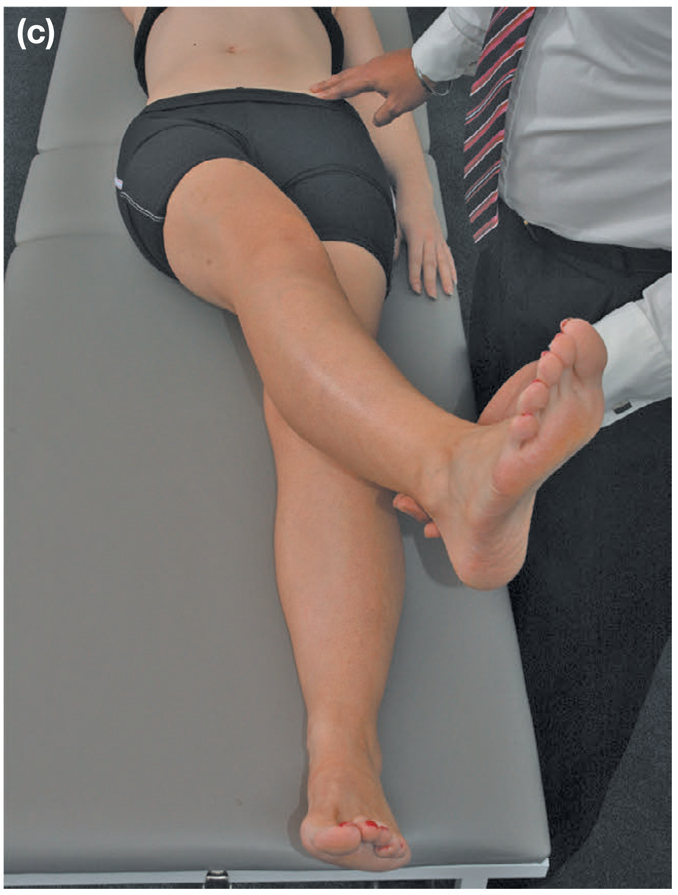
- Patient lies supine and draws both knees to the chest (flexing hips + spine fully)
- Patient holds the good leg at the knee while allowing the bad leg to drop onto the couch
- Positive: bad leg does not fully extend = fixed flexion deformity
- In advanced AVN, a fixed flexion deformity may be present
Trendelenburg Test - Abductor Weakness
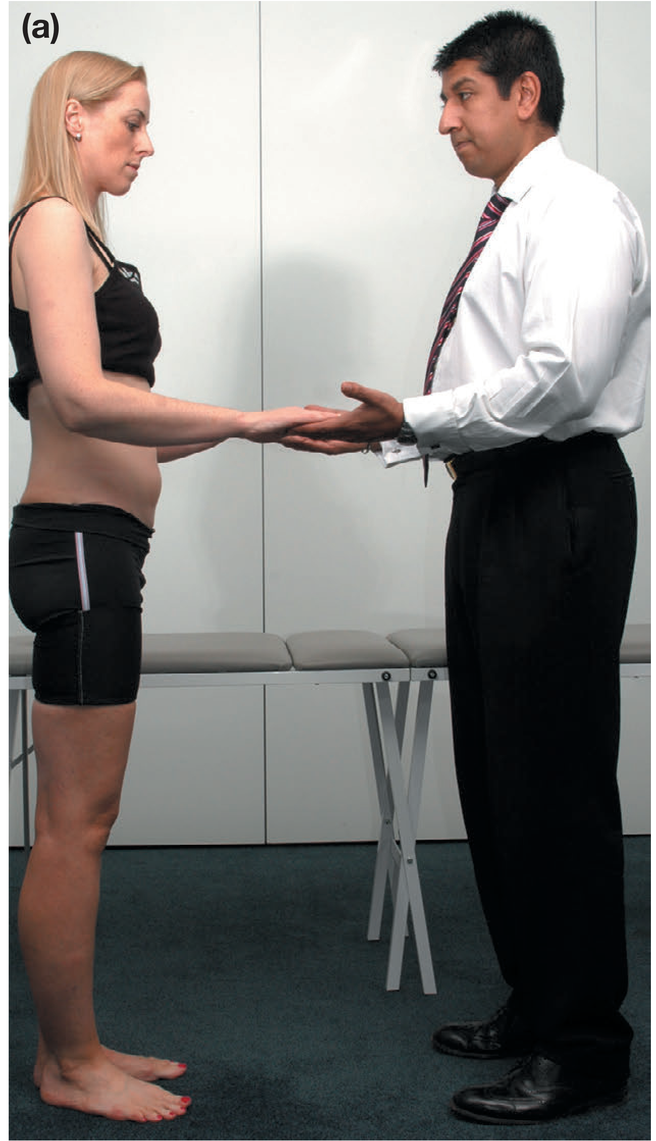
- Face the patient; they place their hands on your palms for support
- Ask patient to stand on one leg at a time
- Positive: increased pressure on the contralateral hand as they take weight through the weak/painful hip = abductor weakness (Trendelenburg positive)
- In AVN, this may be positive due to pain inhibition or secondary muscle wasting
Leg Length Discrepancy (LLD)
- Real LLD: measure ASIS to medial malleolus (true bone length difference)
- Apparent LLD: measure from xiphisternum/umbilicus to medial malleolus (due to pelvic tilt from deformity)
- In AVN with femoral head collapse, real shortening can develop due to structural loss of the femoral head
FADDIR Test (Anterior Impingement)
- Hip Flexion 90° + ADDuction + Internal Rotation
- Reproduces anterior groin pain - positive in femoroacetabular impingement (also seen in early AVN)
FABER Test (Patrick's Test)
- Hip Flexion + ABduction + External Rotation (foot resting on opposite knee)
- Reproduces groin pain from intra-articular pathology or sacroiliac joint
6. AVN Staging (Ficat / FCAT Classification)
| Stage | X-ray | MRI | Clinical Symptoms |
|---|---|---|---|
| 0 | Normal | Normal | Asymptomatic |
| I | Normal / minor osteopenia | Edema | Groin pain |
| II | Mixed osteopenia/sclerosis | Geographic defect | Pain + stiffness |
| III | Crescent sign, cortical collapse | Same as XR | Pain/stiffness + radiation to knee + limp |
| IV | End-stage, secondary OA changes | Same as XR | Pain + limp |
Crescent sign on X-ray = subchondral fracture = hallmark of Stage III AVN
7. Summary of AVN-Specific Examination Findings
| Finding | Significance |
|---|---|
| Groin pain on ROM (esp. internal rotation) | Most sensitive clinical sign |
| Antalgic gait | Pain during weight-bearing |
| Fixed flexion deformity (Thomas's test +ve) | Advanced disease |
| Real leg shortening | Femoral head collapse |
| Trendelenburg +ve | Abductor weakness from disuse/pain |
| Positive FADDIR | Anterior impingement from head irregularity |
| Restricted all planes of motion | Progressive joint destruction |
8. Investigations After Examination
- X-ray (AP + lateral): normal in early stages; late stages show sclerosis, crescent sign, flattening, joint space loss
- MRI (no contrast): investigation of choice - detects early edema (Stage I) before X-ray changes; assesses % femoral head involvement and presence of subchondral collapse
- Bone scan: increased uptake in Stages I-III
Sources: Bailey and Love's Short Practice of Surgery 28th Edition, p. 510-511; Textbook of Family Medicine 9th Edition, p. 827
This is a shared conversation. Sign in to Orris to start your own chat.