Rheumatoid arthritis
Reading File
Asking for Preferences
Finding Sources
Searching PubMed
"rheumatoid arthritis"[MeSH Terms] AND "drug therapy"[MeSH Terms]
Finding Sources
Searching PubMed
"rheumatoid arthritis" AND treatment
Finding Sources
Finding Sources
Finding Sources
Reading File
Reading File
Reading File
Reading File
Loading Image
Reading File
I now have comprehensive, high-quality content from Goldman-Cecil Medicine and Textbook of Family Medicine. I can compile a thorough answer.
Rheumatoid Arthritis: Management & Treatment
Treatment Goals
The primary goal is clinical remission (or at minimum low disease activity), assessed by ACR/EULAR criteria. When treated early, remission is achievable in >50% of patients and low disease activity in at least another 35%. Therapy must be escalated rapidly — if insufficient improvement (<50% reduction in disease activity) is seen after 3 months, the plan should be changed. Sustained remission is defined as ≥6 months.
All RA patients should receive a DMARD — this is non-negotiable. Three categories of drugs are used:
- NSAIDs (symptom relief only)
- Glucocorticoids (bridge/adjunct)
- DMARDs — conventional (csDMARDs), biologic (bDMARDs), and targeted synthetic (tsDMARDs)
Management Algorithm
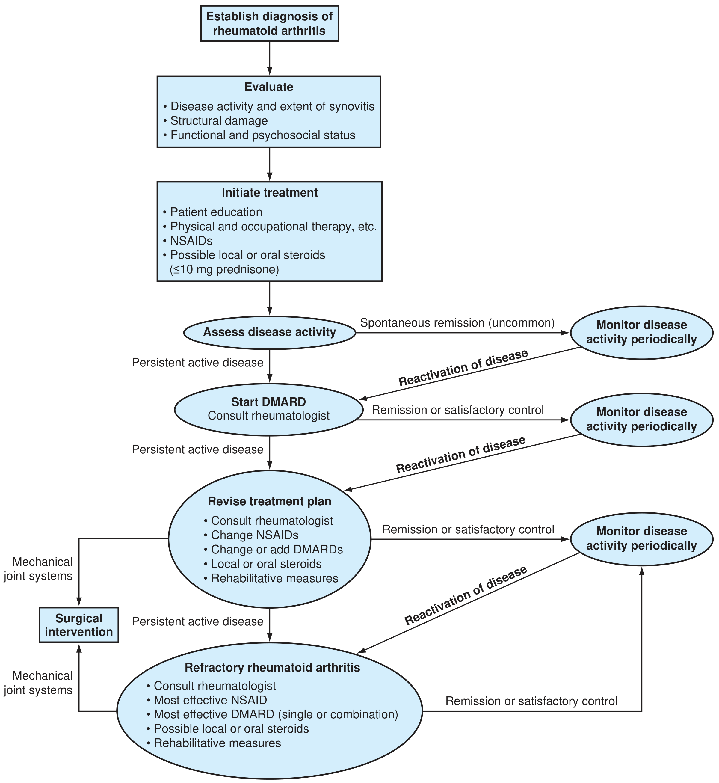
Figure: Algorithm for management of rheumatoid arthritis (ACR guidelines)
1. NSAIDs
NSAIDs provide symptomatic relief only — they have minimal or no effect on the underlying disease process, cannot halt joint erosion, and should never be used as sole therapy without a DMARD.
- COX-2 selective agents (e.g., celecoxib): less GI bleeding risk, but potential cardiovascular toxicity is a concern since RA patients already have elevated CV risk. Keep at low dose.
- Add a proton pump inhibitor in all RA patients taking NSAIDs.
- Monitor for reduced renal blood flow and elevated blood pressure.
2. Glucocorticoids
Glucocorticoids provide rapid, dramatic symptomatic and anti-inflammatory effect, and significantly reduce radiographic progression — but long-term toxicity is extensive:
- ≥25% increased risk of serious infection at doses as low as 5 mg/day
- Risk doubles at 5–10 mg/day
Key principles:
- Use as bridge therapy while slower-acting DMARDs take effect (2–6 months onset)
- Prednisone ≤10 mg/day — rarely exceed this for articular manifestations
- Starting prednisone at DMARD initiation reduces erosive damage, disease activity, disability, and the need for biologic escalation at 2 years
- Taper to the lowest effective dose as soon as possible
- Intramuscular depot injections and intra-articular injections (ultrasound-guided for difficult joints) are useful for flares
- Always prescribe osteoporosis prophylaxis (bisphosphonates, unless childbearing age)
3. Conventional DMARDs (csDMARDs)
| Drug | Key Points |
|---|---|
| Methotrexate | First-line anchor DMARD for most patients. Economical, rare serious toxicity, enhances all other DMARDs. Start 7.5–15 mg/week PO, can escalate to 25 mg/week SC/IM. Add folic acid 1 mg/day to reduce mouth sores. Monitor CBC + AST/ALT every 8–12 weeks. Contraindicated in pregnancy. Avoid alcohol. |
| Hydroxychloroquine | Keep dose <5.0 mg/kg/day. Annual ophthalmology exam after 5 years of therapy (retinal toxicity). |
| Sulfasalazine | 2–3 g/day in two divided doses. Check CBC + LFTs monthly for 1 month, then every 4–6 weeks. Sulfa allergy must be excluded. Widely used in Europe/Asia. |
| Leflunomide | CBC + AST/ALT every 4–8 weeks. Long half-life — cholestyramine washout needed if stopped. Contraindicated in pregnancy. |
| Azathioprine | Second- or third-line; causes bone marrow suppression. |
| Triple therapy | Methotrexate + sulfasalazine + hydroxychloroquine — most frequently used csDMARD combination per EULAR. |
The critical issue is not which DMARD to start first, but starting it early. — Goldman-Cecil Medicine
4. Biologic DMARDs (bDMARDs)
Used when csDMARDs provide inadequate response. Mandatory pre-treatment TB screening before starting any biologic.
| Class | Drugs |
|---|---|
| TNF inhibitors | Adalimumab, etanercept, infliximab, certolizumab, golimumab |
| IL-6 receptor inhibitors | Tocilizumab, sarilumab |
| T-cell co-stimulation blocker | Abatacept |
| B-cell depletion | Rituximab (anti-CD20; especially under certain conditions) |
Important caveats:
- Biologics must not be combined with each other — significantly increases infection risk
- If fever or infectious symptoms develop, stop biologic until resolved
- TNF inhibitors can precipitate CHF, demyelinating syndromes, or lupus-like syndromes
- Biosimilars (bsDMARDs) approved by EMA/FDA are interchangeable options
5. Targeted Synthetic DMARDs — JAK Inhibitors (tsDMARDs)
Oral small molecules targeting the JAK-STAT signaling pathway. Used after inadequate DMARD response.
| Drug | Dose | Notes |
|---|---|---|
| Tofacitinib | 5 mg twice daily | Effective with methotrexate; also after TNF inhibitor failure |
| Baricitinib | Varies | Superior to placebo and adalimumab in MTX-inadequate responders |
| Upadacitinib | 15 mg daily | Selective JAK1 inhibitor; effective as monotherapy or add-on |
| Filgotinib | 100–200 mg daily | Effective in MTX-inadequate or intolerant patients |
Shared toxicity concerns (all JAK inhibitors): infections (including TB), malignancies, cardiovascular events, herpes zoster reactivation. Monitor CBC and LFTs.
⚠️ Efficacy and safety of bDMARDs after JAK inhibitor failure, or JAK inhibitor after another JAK inhibitor, are not fully established.
6. Order of Therapy (EULAR 2019 Framework)
Step 1: Early RA → Start methotrexate ± short-term glucocorticoid bridge
         ↓  (reassess at 3 months; change if <50% improvement)
Step 2: csDMARD inadequate → Add/switch to another csDMARD (triple therapy)
         OR
         Add a bDMARD (TNF inhibitor, IL-6Ri, abatacept, rituximab)
         OR
         Add a tsDMARD (JAK inhibitor)
         ↓  (reassess; target remission by 6 months)
Step 3: Refractory RA → Most effective DMARD single or combination
         Consider surgical intervention for mechanical joint problems
7. Non-Pharmacological Measures
- Patient education — essential at diagnosis
- Physical therapy — preserve joint function, prevent deformity
- Occupational therapy — adaptive equipment, joint protection
- Rehabilitative measures — throughout the disease course
- Surgical intervention — for persistent active disease with mechanical joint failure (synovectomy, joint replacement)
Monitoring Summary
| Drug | Key Monitoring |
|---|---|
| Methotrexate | CBC + AST/ALT every 8–12 weeks (stable dose) |
| Hydroxychloroquine | Annual ophthalmology after 5 years |
| Sulfasalazine | CBC monthly × 1 month, then every 4–6 weeks |
| Leflunomide | CBC + AST/ALT every 4–8 weeks |
| TNF inhibitors | Screen for TB; stop if any infectious symptoms |
| JAK inhibitors | CBC + LFTs; screen for TB and zoster |
| Glucocorticoids | Osteoporosis prophylaxis; taper to lowest dose |
Sources: Goldman-Cecil Medicine (International Edition) | Textbook of Family Medicine, 9e | Katzung's Basic & Clinical Pharmacology, 16e
Postgraduate students are presenting a seminar on “approach to a patient of snake bite”. Formulate some interesting questions that can be asked
Reading File
Finding Sources
Finding Sources
Reading File
Finding Sources
Reading File
Finding Sources
Reading File
Reading File
Reading File
Finding Sources
Reading File
I now have rich, authoritative content from multiple textbooks (Sabiston Surgery, Tintinalli's, Goldman-Cecil) to formulate highly targeted, intellectually stimulating seminar questions. Let me craft them:
Seminar Questions: Approach to a Patient of Snake Bite
These questions are organized by topic depth — from fundamentals to clinical traps to controversial/advanced — ideal for grilling postgraduate presenters.
🔬 Venom Pharmacology & Classification
1. Snake venoms are often loosely labelled "neurotoxic" or "hemotoxic" — but Sabiston's Textbook of Surgery states "all snakes have varying compositions of venoms." Given this, how does classifying a snake purely as "neurotoxic" vs "hemotoxic" mislead your clinical approach? What is the correct framework?
2. The dry weight of pit viper venom is >90% proteinaceous. Name the key enzymes in crotalid venom — phospholipases, metalloproteases, hyaluronidase, collagenase — and explain the specific pathological consequence each produces clinically (e.g., why does hyaluronidase accelerate local spread of edema?).
3. Elapids have fixed, grooved fangs that wick venom, while viperids have long, hollow, retractable fangs that inject venom like a hypodermic needle. How does this mechanical difference influence the depth of envenomation, the latency of symptom onset, and your first-aid approach?
🚨 First Aid — Myths vs Evidence
4. A farmer is bitten in a field, 2 hours from the nearest hospital. He asks you what to do. List the evidence-based first-aid measures. Now tell him what he must not do — and for each harmful intervention (incision-suction, electric shock/TASER, tourniquet with ice immersion, crosscut), explain the physiological reason it causes harm rather than helping.
5. Pressure immobilization bandaging (PIB) is recommended for elapid bites but is contraindicated in crotaline (pit viper) bites. Explain the pathophysiological basis for this distinction. What does PIB achieve in neurotoxic envenomation that it cannot achieve (and in fact worsens) in cytotoxic envenomation?
6. A patient arrives with a tight tourniquet that has been on for 90 minutes. Do you remove it immediately? Walk us through your decision — considering the systemic bolus of venom that may be released, hemodynamic implications, and what you must have ready before removal.
🏥 Clinical Assessment & Grading
7. Define a "dry bite" and give its approximate incidence in viperid vs elapid bites. How does the concept of a dry bite change your observation period and discharge criteria? If 50% of elapid bites are dry, does that mean you can discharge a coral snake bite victim who is completely asymptomatic at 2 hours?
8. Walk us through the Dart grading system for envenomation severity (Grade 1, 2, 3). A patient presents with fang marks on the hand, swelling up to the wrist, nausea, and mild tachycardia — what grade is this, and what is your immediate management decision?
9. You have a patient with a pit viper bite. Swelling is at the ankle. You mark the margins with a pen every 30 minutes. After two markings, the swelling line has not advanced, but the platelet count has dropped from 180,000 to 60,000. Does this patient require antivenom? Justify using the criteria for "progression of envenomation."
10. What is the "look-alike" problem in snakebite? A non-venomous snake bite can mimic an early envenomation — and vice versa. What clinical and morphological features of the snake (if available) and what early lab parameters help you distinguish true envenomation from a dry or non-venomous bite within the first 4–6 hours?
💉 Antivenom — Indications, Administration & Complications
11. A pregnant woman, 28 weeks, is bitten by a rattlesnake with moderate envenomation. Her family refuses antivenom, citing fear of the foreign protein harming the fetus. How do you counsel them? Is pregnancy a contraindication to antivenom therapy?
12. Antivenom is the definitive treatment — but when exactly do you give it? The textbook states antivenom should be given for "progression" of local injury, systemic effects, or hematologic abnormalities. Critically, why is "worsening coagulopathy without clinical bleeding" still an indication? Why should you not wait for the patient to bleed before acting?
13. Your patient develops urticaria, bronchospasm, and hypotension 10 minutes into the antivenom infusion. Describe your stepwise management. After stabilization, do you rechallenge with antivenom — and if so, how? What is the pathophysiological difference between this acute reaction and serum sickness that appears 7–14 days later, and how does each get treated differently?
14. Serum sickness occurs in 13–16% of patients treated with ovine-derived antivenom (CroFab). Explain the immunological mechanism (Type III hypersensitivity) and the timeline of symptoms. A patient comes to clinic 10 days after discharge complaining of joint pain, urticaria, and fever — how do you confirm and treat this?
🫀 Systemic Complications
15. A crotalid envenomation patient has PT >30 sec, INR 4.2, fibrinogen 60 mg/dL, and platelet count 22,000 — but no active bleeding. A colleague wants to immediately give FFP and platelet transfusions. What is your stance? When are blood products actually indicated, and why does antivenom take priority even over blood products in this setting?
16. Acute kidney injury (AKI) is a recognized complication of snake envenomation. Name at least three distinct mechanisms by which snakebite causes AKI (e.g., direct nephrotoxin, myoglobinuria from rhabdomyolysis, DIC-mediated microthrombi, hypotension/shock, hemolysis). How does the mechanism influence your management strategy?
17. Coagulopathy from viper envenomation can recur up to 2 weeks after apparently successful antivenom treatment, even after laboratory values normalize. Explain the pharmacokinetic basis for this — the "venom-antivenom redistribution" phenomenon — and what surveillance protocol does this mandate after discharge?
🧠 Neurotoxic Envenomation (Elapids)
18. A patient bitten by a cobra presents with drooping eyelids and difficulty swallowing 4 hours after the bite, with no local swelling and no pain at the bite site. Why is the absence of local tissue reaction not reassuring in elapid envenomation? How do post-synaptic (α-bungarotoxin type) and pre-synaptic (β-bungarotoxin type) neurotoxins differ, and which is more reversible with antivenom?
19. Neuromuscular paralysis from an elapid bite leads to respiratory failure. Antivenom is given. How soon do you expect clinical improvement? If there is no improvement in respiratory function after adequate antivenom doses, what is the role of anticholinesterase drugs (neostigmine + atropine), and for which type of neurotoxin are they useful?
⚗️ Controversial & Higher-Order Questions
20. A systematic review showed that prophylactic antibiotics are not routinely recommended for snakebite wounds. Yet most field practitioners give them empirically. Present both sides of this argument — what is the microbiological basis for potential wound infection, and what does the evidence say about prophylactic antibiotic use?
21. Fasciotomy for compartment syndrome in snakebite — is it ever indicated? Tintinalli's and Sabiston's both note that fasciotomy worsens myonecrosis in animal models of crotaline envenomation. What is the correct algorithm — antivenom first, then compartment pressure measurement, and then fasciotomy only if pressures remain critically elevated after adequate antivenom?
22. "The vast majority of snakebite victims will recover uneventfully with supportive treatment alone." — Sabiston's Textbook of Surgery. Does this statement justify watchful waiting without antivenom? Construct an argument for why this statistic is being used to rationalize undertreatment, and define the patient subgroups where conservative management is genuinely appropriate.
🌍 Epidemiology & Global Perspective
23. The WHO estimates 2.5 million envenomations and >100,000 deaths annually from snakebite — yet snakebite was only re-listed as a Neglected Tropical Disease (NTD) by the WHO in 2017. What are the systemic barriers (supply chain of antivenom, cold chain requirements, lack of species-specific antivenoms in endemic regions, cost) that prevent reduction of this mortality, especially in South Asia and sub-Saharan Africa?
Sources: Sabiston Textbook of Surgery | Tintinalli's Emergency Medicine | Goldman-Cecil Medicine | Rosen's Emergency Medicine
How neostigmine and atropine administration help in cobra bite
Finding Sources
Finding Sources
Reading File
Reading File
Reading File
Finding Sources
Reading File
Reading File
Reading File
I have all the mechanistic and clinical detail needed. Here is the comprehensive answer:
Neostigmine + Atropine in Cobra Bite: Mechanism & Clinical Use
Step 1 — Understanding the Target: The Neuromuscular Junction (NMJ)
To understand why this combination works, you need to understand what cobra venom does to the NMJ.
Under normal conditions:
- A motor nerve impulse triggers release of acetylcholine (ACh) from the presynaptic terminal into the synaptic cleft
- ACh diffuses across and binds to nicotinic ACh receptors (nAChR) on the postsynaptic muscle end plate
- This opens ion channels → depolarization → muscle contraction
- ACh is rapidly degraded by acetylcholinesterase (AChE) in the cleft, terminating the signal
Step 2 — What Cobra Venom Does
Cobra (Naja species) venom contains two principal neurotoxins, both targeting the NMJ, but at different sites:
α-Neurotoxins (Postsynaptic) — the dominant toxin in cobras
- These are long-chain polypeptides (α-bungarotoxin family)
- They bind competitively to the nicotinic ACh receptor on the muscle end plate
- They physically occupy the receptor, blocking ACh from binding
- The NMJ is structurally intact — the receptor exists, the nerve terminal is undamaged — but ACh cannot activate it
- Result: flaccid paralysis — progressive, descending, beginning with cranial nerves (ptosis, ophthalmoplegia) → bulbar → respiratory muscles
β-Neurotoxins / Phospholipase A₂ (Presynaptic) — more prominent in kraits, some cobras
- These destroy the presynaptic nerve terminal — degrading the vesicle release machinery
- The axon must physically regrow to recover
- This damage is irreversible by any drug — recovery takes days to weeks
- Antivenom must be given before these toxins cause axon damage
Goldman-Cecil Medicine: "Once damaged by presynaptic neurotoxins, the axon must regrow, a process that can take days to weeks... In contrast, postsynaptic neurotoxins bind to the acetylcholine receptor on the muscle end plate external to the cell, so antivenom may reverse this type of paralysis."
Step 3 — The Pharmacological Rationale for Neostigmine
Neostigmine is an acetylcholinesterase inhibitor (anticholinesterase).
It works by blocking the enzyme AChE, which normally breaks down ACh in the synaptic cleft.
By inhibiting AChE:
- ACh accumulates in the cleft and remains there much longer
- The elevated concentration of ACh competitively displaces the α-neurotoxin from the nAChR
- With enough ACh molecules present, some bind to any unoccupied or partially blocked receptors
- Net result: partial restoration of neuromuscular transmission → improvement in muscle strength
This is precisely why it works only against postsynaptic (α-) neurotoxins — there are still functional receptors to rescue. Against presynaptic β-neurotoxins (which have already destroyed the nerve terminal), there is no ACh being released and no receptor to compete at — neostigmine has nothing to work with.
This is the same principle as using anticholinesterases in myasthenia gravis, where antibodies block nAChRs — cobra α-neurotoxin essentially produces a pharmacological myasthenia gravis.
Step 4 — Why Atropine Must Be Given First
Neostigmine is non-selective — it inhibits all cholinesterases, not just those at the NMJ. This means ACh accumulates at muscarinic receptors throughout the body as well, producing unwanted parasympathomimetic (SLUD) effects:
| Muscarinic Excess Effect | Manifestation |
|---|---|
| ↑ Salivation | Hypersalivation, drooling |
| ↑ Lacrimation | Excessive tearing |
| ↑ Urination | Incontinence |
| ↑ Defecation/GI motility | Nausea, vomiting, diarrhea, cramping |
| Bradycardia | Dangerous hypotension |
| Bronchospasm + ↑ secretions | Worsens respiratory compromise |
| Miosis |
Atropine is a competitive muscarinic receptor antagonist — it blocks all these peripheral muscarinic effects without blocking the nicotinic receptors at the NMJ that you want neostigmine to work on.
By pre-treating with atropine, you:
- Prevent life-threatening bradycardia
- Prevent bronchospasm (critical in a patient already at risk of respiratory failure)
- Reduce secretions that could compromise the airway
- Allow neostigmine to exert its therapeutic effect at the NMJ freely
Step 5 — Clinical Protocol (Harrison's Principles, Table 471-2)
Indication: Clear, objective evidence of neurotoxicity — ptosis, inability to maintain upward gaze, bulbar weakness.
Step-by-step administration:
1. Pretreat with Atropine:
- Adults: 0.6 mg IV
- Children: 0.02 mg/kg IV (minimum 0.1 mg)
2. Test dose — Neostigmine (or Edrophonium):
- Neostigmine: 0.02 mg/kg IV or IM (children: 0.04 mg/kg)
- Alternative: Edrophonium 10 mg IV (shorter acting, used as a diagnostic test dose)
3. Assess at 30 minutes:
- If objective improvement is seen (e.g., ptosis improves, power returns) → respond to maintenance dosing
- If no improvement → presynaptic toxin is likely dominant; drug will not help further; focus on ventilatory support
4. Maintenance (if test positive):
- Neostigmine: 0.5 mg IV/IM/SC every 1 hour as needed
- Atropine: 0.6 mg as continuous IV infusion over 8 hours (children: 0.02 mg/kg over 8 h)
5. Airway monitoring throughout — neostigmine is an adjunct, not a substitute for intubation when needed.
Summary: What Each Drug Does
| Drug | Mechanism | Purpose |
|---|---|---|
| Neostigmine | Inhibits acetylcholinesterase → ↑ACh in synaptic cleft → competes with α-neurotoxin for nAChR binding | Partially reverses postsynaptic neuromuscular blockade |
| Atropine | Blocks muscarinic receptors | Prevents bradycardia, bronchospasm, hypersecretion caused by excess ACh at muscarinic sites |
Critical Limitations
- Works only for postsynaptic neurotoxins (α-type) — cobra is a good candidate; krait (predominantly β-neurotoxin) responds poorly
- Not a substitute for antivenom — antivenom should always be given first if available; neostigmine buys time or supplements antivenom effect
- Not a substitute for intubation — if the airway is at risk, secure it; neostigmine cannot be relied upon to maintain ventilation
- Edrophonium test (short-acting) can be used diagnostically first — if there is clear improvement within minutes, it confirms postsynaptic mechanism and guides you to proceed with neostigmine maintenance
Sources: Harrison's Principles of Internal Medicine 22e | Goldman-Cecil Medicine | Sabiston Textbook of Surgery | Bradley & Daroff's Neurology in Clinical Practice
Which drugs can cause dystonia?
Finding Sources
Finding Sources
Finding Sources
Reading File
Finding Sources
Finding Sources
Reading File
Reading File
Excellent — rich content from Katzung, Stahl's, Goldman-Cecil, Kaplan & Sadock. I now have a comprehensive picture. Let me compile a well-organized answer.
Drugs That Can Cause Dystonia
Dystonia is involuntary, sustained or intermittent muscle contraction causing twisting movements or abnormal postures. Drug-induced dystonia can be acute (within hours to days) or tardive (after months to years of exposure), and the underlying mechanism shapes both the causative drug class and the treatment.
Pathophysiological Basis
The key mechanism is dopamine–acetylcholine imbalance in the striatum. Normally, dopaminergic and cholinergic activity are balanced to coordinate smooth movement. When dopamine D2 receptor blockade is uncompensated by muscarinic blockade, acetylcholine activity becomes dominant → abnormal involuntary contractions. This is why anticholinergic drugs (e.g., benztropine, diphenhydramine) are the antidote for acute drug-induced dystonia.
Drug Classes Causing Dystonia
1. Antipsychotics (Most Common Cause)
Typical (First-Generation) Antipsychotics — highest risk:
| Drug | Notes |
|---|---|
| Haloperidol | Highest risk; pure D2 blocker with no anticholinergic/serotonergic offset |
| Fluphenazine | High extrapyramidal burden |
| Chlorpromazine, Trifluoperazine | Phenothiazines — significant risk |
| Perphenazine, Thiothixene | Similar risk profile |
| Prochlorperazine | Often forgotten — used as an antiemetic but a potent D2 blocker |
Atypical (Second-Generation) Antipsychotics — lower but not absent risk:
| Drug | Notes |
|---|---|
| Risperidone, Lurasidone | Higher D2 occupancy among atypicals; notable risk per Goldman-Cecil |
| Olanzapine, Quetiapine, Clozapine | Lower extrapyramidal risk due to anticholinergic/serotonergic offset |
| Ziprasidone, Aripiprazole | Intermediate risk |
Stahl's: "Exposure to D2 blockers, especially those with neither serotonergic nor anticholinergic properties, can cause dystonia, often upon first exposure."
Timing:
- Acute dystonia — within 48 hours of starting or rapid dose increase; responds to anticholinergics
- Tardive dystonia — after >1 year of chronic use; anticholinergics are less effective and may worsen it
2. Antiemetics (D2 Antagonists — Frequently Overlooked)
| Drug | Context |
|---|---|
| Metoclopramide | Very common cause; used for gastroparesis, nausea, reflux |
| Promethazine | Phenothiazine antiemetic |
| Prochlorperazine | Used for nausea/vertigo |
| Domperidone | Peripheral D2 blocker; less CNS penetration but risk exists |
| Droperidol | Butyrophenone used peri-operatively |
These are clinically important because the drug is prescribed for a benign indication, dystonia catches clinicians off guard, and patients are rarely warned about this risk.
3. Dopaminergic Agents (Paradoxical / Dose-Related)
| Drug | Mechanism |
|---|---|
| Levodopa | Dose-related dyskinesia/dystonia in Parkinson disease; peak-dose or off-period dystonia |
| Dopamine agonists (pramipexole, ropinirole, cabergoline) | Similar dose-related phenomenon |
Here, the mechanism is the opposite — excess dopaminergic stimulation → dyskinesia/dystonia, not deficiency. Off-period dystonia occurs when levodopa levels drop.
4. Antiepileptics
| Drug | Notes |
|---|---|
| Carbamazepine | Can cause dystonia and chorea |
| Phenytoin | Chorea more commonly, dystonia reported |
| Valproic acid | Tremor more typical; dystonia less common |
5. Antidepressants
| Drug | Notes |
|---|---|
| SSRIs (fluoxetine, sertraline, paroxetine, etc.) | Rare; can precipitate, aggravate, or sometimes improve dystonia; also reported: torticollis, opisthotonos, oculogyric crisis |
| Tricyclic antidepressants (TCAs) | Rare dystonia |
6. Mood Stabilizers
| Drug | Notes |
|---|---|
| Lithium | Can cause dystonia; also postural tremor and chorea |
7. Stimulants / Drugs of Abuse
| Drug | Notes |
|---|---|
| Cocaine | Depletes dopamine → increased vulnerability to dopamine antagonist-induced dystonia; also direct dystonic effects |
| Amphetamines | Chorea more common; dystonia reported |
8. Other Drugs
| Drug | Notes |
|---|---|
| Cinnarizine / Flunarizine | Calcium channel blockers (antivertiginous); well-known cause of tardive dystonia and parkinsonism |
| Reserpine / Tetrabenazine | Dopamine-depleting agents; can cause dystonia |
| Antiretrovirals | Some reports with certain HIV drugs |
Quick Reference Table
| Category | Key Drugs | Onset |
|---|---|---|
| Typical antipsychotics | Haloperidol, fluphenazine, chlorpromazine | Acute or tardive |
| Atypical antipsychotics | Risperidone, lurasidone | Acute or tardive |
| Antiemetics (D2 blockers) | Metoclopramide, prochlorperazine, promethazine | Acute |
| Dopaminergics | Levodopa, dopamine agonists | Dose-related |
| Antiepileptics | Carbamazepine, phenytoin | Variable |
| SSRIs | Fluoxetine, sertraline | Rare, variable |
| Mood stabilizers | Lithium | Variable |
| Stimulants/drugs of abuse | Cocaine, amphetamines | Acute |
| Ca²⁺ channel blockers | Cinnarizine, flunarizine | Tardive |
Acute Drug-Induced Dystonia: Treatment
| Agent | Dose | Route |
|---|---|---|
| Diphenhydramine | 25–50 mg | IV (first-line in emergency) |
| Benztropine | 2 mg | IV or IM |
| Biperiden | 2–5 mg | IV or IM |
| Diazepam | 10 mg | IV (refractory cases) |
Response is typically seen within 20 minutes of anticholinergic administration.
Tardive dystonia: Anticholinergics are less effective. Use VMAT2 inhibitors (valbenazine, deutetrabenazine) or botulinum toxin for focal forms. Stop/reduce the offending drug whenever possible.
Risk factors for acute dystonia: young males, high-potency D2 blockers, rapid dose escalation, recent cocaine use (depletes endogenous dopamine), and prior dystonic reactions.
Sources: Katzung's Basic & Clinical Pharmacology 16e | Stahl's Essential Psychopharmacology | Goldman-Cecil Medicine | Kaplan & Sadock's Comprehensive Textbook of Psychiatry | Harrison's Principles of Internal Medicine 22e
Trihexyphenydyl renal clearance
Finding Sources
Searching the Web
trihexyphenidyl renal clearance pharmacokinetics elimination
Finding Sources
Searching PubMed
trihexyphenidyl pharmacokinetics renal clearance
Finding Sources
Searching PubMed
trihexyphenidyl pharmacokinetics metabolism
Reading Article
Reading Article
Reading File
I now have what I need from both textbook sources and primary pharmacokinetic literature.
Trihexyphenidyl: Renal Clearance & Pharmacokinetics
Short Answer
Trihexyphenidyl is not primarily renally cleared. It undergoes extensive hepatic metabolism, and the parent drug along with its metabolites is excreted through both urine and bile. Formal renal clearance data are sparse — this is a recognized gap in its pharmacokinetic literature.
What Is Known About Its Pharmacokinetics
Absorption
- Rapidly absorbed from the GI tract after oral administration
- Onset of action: within 1 hour of oral dosing
- Peak plasma levels: 2–3 hours post-dose
- Duration of action: 6–12 hours
Distribution
- Highly lipophilic → crosses the blood–brain barrier readily
- Significant CNS penetration within 30–60 minutes
- Large volume of distribution (Vd) — characteristic of the entire anticholinergic class used in Parkinson disease
- Plasma protein binding: poorly characterized
Metabolism
- Extensively metabolized — primarily via N-dealkylation and hydroxylation
- Relatively low clearance relative to hepatic blood flow (i.e., it is not a high-extraction hepatic drug — metabolism is capacity-limited rather than flow-limited)
Elimination
- Parent drug and metabolites excreted in both urine and bile (dual route)
- Elimination half-life: ~3.7 hours (Burke & Fahn, 1985 — the key primary PK study in dystonic patients)
- Elimination follows first-order kinetics
- No significant accumulation demonstrated; half-life independent of age, peak level, or duration of therapy in the Burke & Fahn study
The Renal Clearance Data Gap
The most authoritative pharmacokinetic review of anticholinergic drugs used in Parkinsonism (Brocks, J Pharm Pharm Sci, 1999 — PMID 10952768) explicitly states:
"There is little pharmacokinetic information available for these drugs... the available information suggests that excretion of parent drug and metabolite is via the urine and bile."
Kaplan & Sadock's Comprehensive Textbook of Psychiatry echoes this:
"There is limited information available regarding the pharmacokinetics of these drugs, especially among older adults."
What this means practically:
- Specific renal clearance values (mL/min) have not been formally established for trihexyphenidyl in the published literature
- Unlike drugs where renal clearance dominates (e.g., lithium, digoxin, aminoglycosides), trihexyphenidyl's primary elimination route is hepatic, not renal
- Urinary excretion represents a secondary route for metabolites, not selective glomerular filtration of intact drug
Clinical Implications
| Situation | Implication |
|---|---|
| Renal impairment | Dose adjustment guidelines are not formally established; however, since hepatic metabolism dominates, mild-to-moderate renal failure has less impact than with renally cleared drugs |
| Elderly patients | Reduced renal function + altered hepatic metabolism → caution; PK data in elderly specifically lacking |
| Hepatic impairment | More likely to be clinically relevant than renal impairment, given hepatic metabolic dominance |
| Monitoring | No plasma drug level monitoring is routinely used; anticholinergic activity does not correlate linearly with serum levels (100-fold inter-individual variability in drug levels) |
Summary
| Parameter | Value/Status |
|---|---|
| Route of elimination | Hepatic (primary) + renal & biliary (metabolites) |
| Elimination half-life | ~3.7 hours |
| Renal clearance (specific value) | Not established in literature |
| Volume of distribution | Large (high lipophilicity) |
| Plasma protein binding | Poorly characterized |
| Dose adjustment in renal failure | No formal guidelines; likely minor impact |
The absence of formal renal clearance data for trihexyphenidyl is a known and acknowledged gap in the pharmacokinetic literature for this drug class. If renal clearance is clinically critical (e.g., in severe renal failure), conservative dosing and careful monitoring of anticholinergic toxicity signs (confusion, urinary retention, tachycardia) are advisable.
Sources: Kaplan & Sadock's Comprehensive Textbook of Psychiatry | Brocks DR, J Pharm Pharm Sci 1999 [PMID: 10952768] | Burke RE & Fahn S, Ann Neurol 1985 [PMID: 4037749]
This is a shared conversation. Sign in to Orris to start your own chat.