

162e6a19-6404-4169-bd6f-a6d739823045.jpg
Analyse
analyse_media
Reading File
Now let me search the medical library for ILD/pulmonary fibrosis content and look for relevant images in parallel.
Finding Sources
Searching PubMed
"interstitial lung disease" AND "chest radiograph" AND diagnosis
Now let me read the key textbook sections on ILD CXR findings:
Reading File
Reading File
I have sufficient information to provide a thorough analysis. Let me fetch one of the textbook ILD images:
Loading Image
Loading Image
I now have a comprehensive analysis. Here is the full radiological report:
Chest X-Ray Analysis
Patient: SALBIAH | DOB: 18/12/1957 (approx. age 68)
View: AP Supine
Study Date: 2026
Technical Note
This is an AP supine projection photographed from a monitor (note the moiré/color banding artifact from the camera). This view inherently magnifies the cardiac silhouette and can underestimate lung field aeration. True PA erect comparison is advisable.
Radiological Findings
1. Lung Fields - PRIMARY ABNORMALITY
Diffuse bilateral coarse reticulonodular opacities are present throughout both lung fields, involving upper, mid, and lower zones. The pattern is:
- Bilateral and largely symmetric
- Characterized by a "dirty lung" or shaggy interstitial texture
- More confluent and dense in the mid-to-lower zones
- The lung markings are increased, irregular, and thickened - consistent with chronic interstitial thickening
- No focal lobar consolidation or discrete lung mass is identified
- No pneumothorax
As described in Fishman's Pulmonary Diseases and Disorders: "The reticular interstitial lung pattern, most marked at the bases, is characteristic of idiopathic pulmonary fibrosis or collagen vascular disease."
Comparison with reference ILD chest X-ray (UIP pattern):
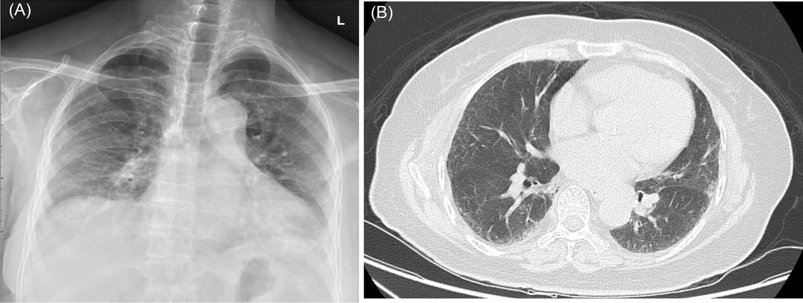
The pattern in this patient closely resembles UIP-type ILD as seen above, though more diffuse and pronounced.
Reference: UIP in rheumatoid arthritis (CXR + honeycomb HRCT):
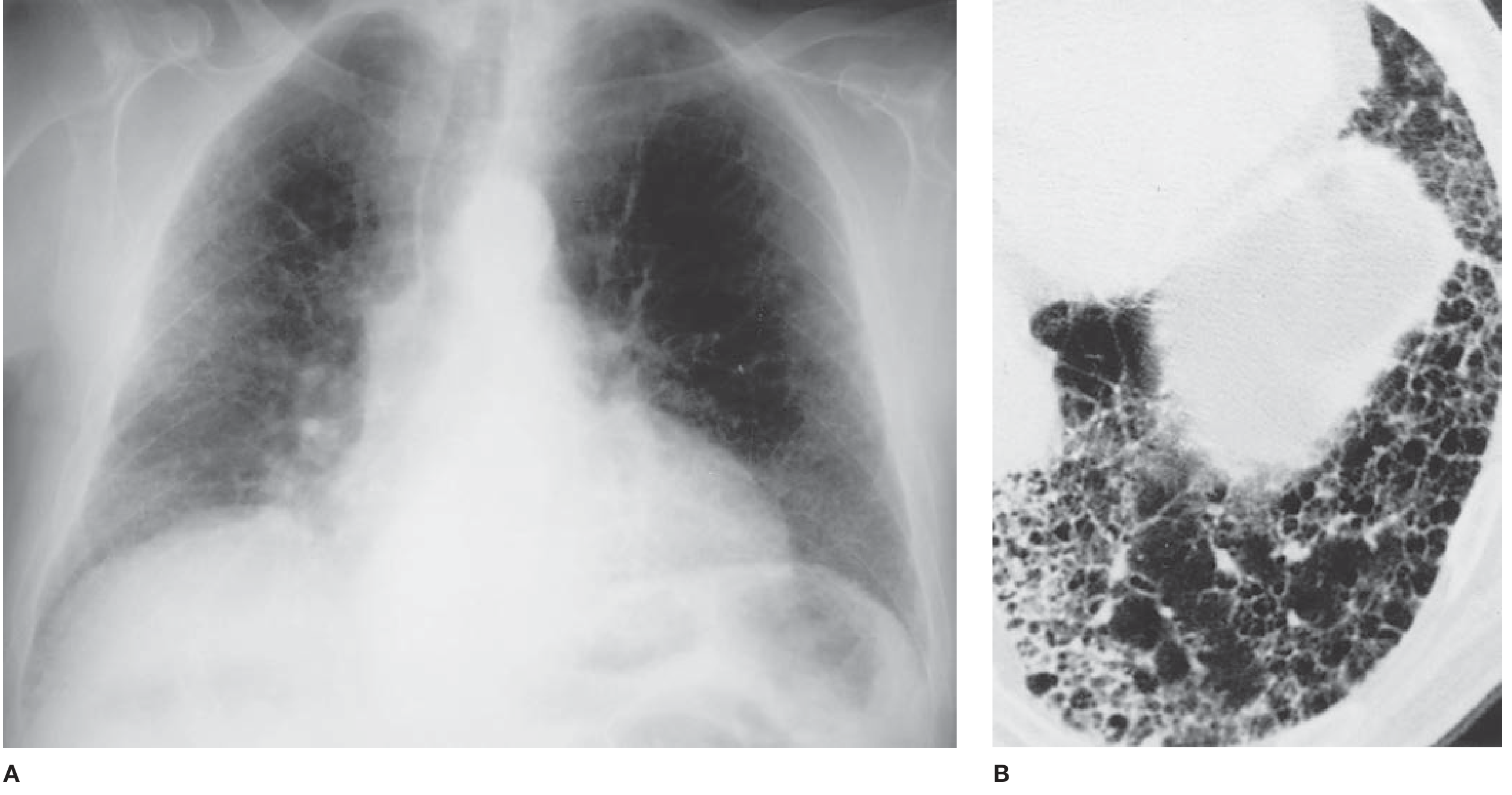
2. Heart
- Cardiac silhouette appears mildly to moderately enlarged
- Given the AP supine projection, some magnification is expected - true cardiomegaly cannot be confirmed without PA erect film
- The cardiac borders are ill-defined and shaggy, a consequence of adjacent dense parenchymal disease
3. Mediastinum
- Trachea is midline - no mediastinal shift
- Mediastinal width appears within acceptable limits for a supine AP film
- Bilateral hilar regions appear mildly prominent, possibly reflecting pulmonary vascular changes, reactive lymphadenopathy, or simply parenchymal overlay
4. Pleura
- No large pleural effusion identified
- No obvious pneumothorax
- Costophrenic angles are partially obscured by breast soft tissue and basilar parenchymal disease - small effusions cannot be fully excluded
5. Bones and Soft Tissues
- Visualized ribs, clavicles, and scapulae appear intact
- No acute fractures or aggressive bony lesions seen
- Mild degenerative changes in the thoracic spine (consistent with age)
- Bilateral breast shadows present (female patient)
- No subcutaneous emphysema
Impression
| Finding | Significance |
|---|---|
| Diffuse bilateral reticulonodular opacities | HIGH - Chronic interstitial lung disease |
| Mid-to-lower zone predominance | Favors UIP/IPF or CTD-ILD over sarcoidosis |
| Apparent cardiomegaly | AP supine effect + possible true enlargement |
| Prominent hila | Secondary pulmonary hypertension vs. parenchymal overlay |
| No focal consolidation or mass | Against acute pneumonia, lobar collapse |
Primary Impression: Diffuse bilateral interstitial lung disease (ILD), chronic pattern
Differential Diagnosis
Given the age (68F), pattern (bilateral reticulonodular, lower-zone predominant), and chronicity implied by the extent of disease:
- Idiopathic Pulmonary Fibrosis (IPF/UIP) - Most likely; typical demographics and radiographic pattern
- Connective tissue disease-associated ILD (RA-ILD, Systemic Sclerosis/Scleroderma-ILD, Sjogren's-ILD) - Scleroderma produces an identical reticulonodular pattern on CXR
- Chronic Hypersensitivity Pneumonitis - Can produce coarse bilateral reticular opacities with upper-zone predominance, but lower-zone disease is seen in fibrotic HP
- Nonspecific Interstitial Pneumonia (NSIP) - More ground-glass predominant, but reticular pattern can overlap
- Drug-induced ILD - Important to review medication history (nitrofurantoin, amiodarone, methotrexate, chemotherapy)
- Occupational/Environmental ILD (asbestosis, silicosis) - Requires occupational history; calcified pleural plaques would favor asbestosis
Less likely: lymphangitic carcinomatosis (typically more linear/Kerley B lines), miliary TB (more uniformly nodular, finer pattern), pulmonary edema (would be more acute with Kerley lines and vascular redistribution).
Recommended Next Steps
- HRCT Thorax (high-resolution CT) - Gold standard for ILD pattern characterization (UIP vs NSIP vs HP). Identifies honeycombing, traction bronchiectasis, ground-glass opacity, and exact distribution
- Pulmonary Function Tests (PFTs) - Expected restrictive pattern (reduced TLC, FVC, DLCO) in fibrotic ILD
- Erect PA CXR - For accurate cardiac size assessment and comparison
- Connective tissue disease serology: ANA, anti-CCP, RF, anti-Scl-70 (topoisomerase I), anti-centromere, anti-Jo1 (myositis-associated ILD)
- 6-minute walk test / SpO2 on exertion - Functional assessment
- Multidisciplinary discussion (radiologist + pulmonologist + rheumatologist) if CT confirms fibrotic ILD
- BAL or lung biopsy if HRCT pattern is indeterminate
Disclaimer: This is an AI-assisted educational analysis. All imaging findings should be interpreted in clinical context by a qualified radiologist and treating physician. This does not constitute a formal radiology report.
The 2025 review in Radiology (PMID 40590695) confirms that HRCT remains the cornerstone for characterizing fibrotic ILD subtypes and guiding treatment decisions - chest X-ray findings such as these are a strong indication for urgent CT correlation.
This is a shared conversation. Sign in to Orris to start your own chat.