


Teach me all these topics for Post graduation theory exams of MS Ophthalmology Basics Concepts Recent advances Everything as an ophthalmologist I should know
- Cataract and IOLs: FLACS, capsular tension ring (CTR), phakic IOLs, modern IOLs, accommodative IOLs
- Retina and vitreous: BRVO management, ARMD, OCT, ERG, ROP, retinal imaging, FFA, pars plana vitrectomy, Pascal laser, silicone oil
- Cornea and ocular surface: stem-cell therapy, ocular surface squamous neoplasia, amniotic membrane, collagen cross-linking/C3R, keratoprosthesis, lamellar keratoplasty, pterygium surgery, toxic contact lens reactions
- Glaucoma: neuroprotection, glaucoma implants, OCT in glaucoma
- Anterior segment / iris / diagnostic imaging: pupilloplasty, UBM
- General ophthalmic technology and materials: OVDs, lasers, dyes, tissue adhesives/glues
Which module should I teach first in full MS theory-answer format?
- Cataract and IOLs: FLACS, capsular tension ring (CTR), phakic IOLs, modern IOLs, accommodative IOLs
- Retina and vitreous: BRVO management, ARMD, OCT, ERG, ROP, retinal imaging, FFA, pars plana vitrectomy, Pascal laser, silicone oil
- Cornea and ocular surface: stem-cell therapy, ocular surface squamous neoplasia, amniotic membrane, collagen cross-linking/C3R, keratoprosthesis, lamellar keratoplasty, pterygium surgery, toxic contact lens reactions
- Glaucoma: neuroprotection, glaucoma implants, OCT in glaucoma
- Anterior segment / iris / diagnostic imaging: pupilloplasty, UBM
- General ophthalmic technology and materials: OVDs, lasers, dyes, tissue adhesives/glues
Okay
- Cataract and IOLs: FLACS, CTR, phakic IOLs, modern/accommodative IOLs
- Retina and vitreous: BRVO, ARMD, OCT, ERG, ROP, FFA, retinal imaging, vitrectomy, lasers, silicone oil
- Cornea and ocular surface: stem cells, amniotic membrane, OSSN, C3R, keratoplasty, KPro, pterygium, contact-lens toxicity
- Glaucoma: neuroprotection, drainage implants, OCT
- Imaging, lasers and materials: UBM, dyes, OVDs, tissue glues, lasers, pupilloplasty
Start with cataract and IOLs
femtosecond laser cataract surgery
phakic intraocular lens
2025 AAO cataract preferred practice pattern femtosecond laser cataract surgery premium intraocular lenses phakic IOL
PMID: 40731148
PMID: 40369812
https://www.aao.org/education/clinical-statement/laser-surge…
capsular tension ring cataract
PMID: 40504499
diagram femtosecond laser assisted cataract surgery capsulotomy lens fragmentation and corneal incisions
diagram capsular tension ring inserted in capsular bag zonular weakness cataract surgery
Cataract and IOLs for MS Ophthalmology Theory
- Femtosecond laser-assisted cataract surgery (FLACS)
- Capsular tension ring (CTR)
- Phakic intraocular lenses
- Modern pseudophakic IOLs, including toric, multifocal, EDOF and accommodating IOLs
- IOL selection, biometry and high-yield complications
1. Foundation: IOL basics
Definition
Parts
- Optic: central refracting portion
- Haptics: peripheral supporting elements
- Overall diameter: optic + haptic-to-haptic length
- Optic diameter: commonly 6 mm for standard adult posterior chamber IOLs
Ideal IOL properties
- Biocompatible and inert
- Stable fixation and centration
- Optically clear, scratch-resistant
- Minimal inflammation, posterior capsule opacification (PCO), dysphotopsia and glistenings
- Injectable through a small incision
- Predictable effective lens position (ELP)
Classification
| Basis | Categories |
|---|---|
| Site | Posterior chamber IOL (PCIOL), anterior chamber IOL (ACIOL), iris-fixated/iris-claw IOL, scleral-fixated IOL |
| Material | PMMA, silicone, hydrophobic acrylic, hydrophilic acrylic |
| Optic | Spherical, aspheric, monofocal, multifocal, extended-depth-of-focus (EDOF), toric, accommodating |
| Construction | One-piece or three-piece; rigid or foldable |
| Fixation | In-the-bag, sulcus, iris-claw, scleral fixation |
Materials: exam comparison
| Material | Advantages | Limitations |
|---|---|---|
| PMMA | Excellent optics, stable, inexpensive | Rigid, needs large incision |
| Silicone | Foldable, small incision | Silicone-oil adherence, therefore generally avoid if future retinal surgery with silicone oil is likely |
| Hydrophobic acrylic | Commonest modern material, foldable, low PCO with square edge, good capsular adhesion | Glistenings or surface light scatter may occur in some models |
| Hydrophilic acrylic | Flexible, good injector delivery | Greater calcification risk in some settings, including exposure to intraocular gas/air in susceptible lenses |
Design features that prevent PCO
- Sharp square posterior optic edge produces a capsular bend and contact inhibition of lens epithelial cell migration.
- In-the-bag placement and meticulous cortical clean-up further reduce PCO.
- PCO, if visually significant, is managed by Nd:YAG posterior capsulotomy.
2. FLACS: Femtosecond Laser-Assisted Cataract Surgery
Definition
- Corneal incisions
- Anterior capsulotomy
- Lens fragmentation/softening
- Arcuate or limbal-relaxing incisions for astigmatism
Principle
Essential components
- Docking interface: applanation or liquid-filled interface
- Image-guidance system: commonly OCT-based
- Laser delivery platform
- Patient interface and suction mechanism
- Integrated or adjacent phacoemulsification system
Steps of FLACS
- Pharmacological dilation and sterile preparation.
- Docking of the laser interface to the eye.
- Imaging and treatment planning.
- Laser creation of:
- Primary and side-port corneal incisions
- Precisely centered capsulotomy
- Lens fragmentation pattern
- Arcuate keratotomy if planned
- Transfer to operating microscope.
- Opening of laser capsulotomy and removal of free capsule disc.
- Hydrodissection, phacoaspiration and cortical clean-up.
- IOL implantation in the capsular bag.
Benefits
- Highly reproducible capsulotomy size, circularity and centration
- Precise corneal incisions
- Lens prefragmentation may reduce effective phaco time and cumulative dissipated energy
- Astigmatic arcuate incisions can be planned precisely
- Useful in selected challenging cases:
- Shallow anterior chamber
- Dense cataract
- Low endothelial reserve
- White/intumescent cataract, with careful case selection
- Premium IOL procedures where centration is especially important
Limitations
- High capital, consumable and maintenance costs
- Extra docking and workflow time
- Requires pupil dilation and adequate corneal clarity
- Not always feasible in deep-set eyes, marked kyphosis, severe tremor, poor cooperation, inability to lie flat, or significant conjunctival scarring
- Incomplete capsulotomy, capsular tags and incomplete fragmentation can occur
- Does not replace surgical judgement or conventional phaco skill
Complications
During docking
- Subconjunctival hemorrhage
- Corneal folds and imaging artefacts
- Transient rise in IOP from suction
- Loss of suction and incomplete treatment
During surgery
- Incomplete corneal incision
- Capsular tags, microadhesions or incomplete capsulotomy
- Anterior capsular tear if the capsule is pulled before identifying and releasing tags
- Incomplete nuclear fragmentation
- Miosis due to prostaglandin release
- Rare capsular block syndrome if hydrodissection is forceful in a gas-fragmented lens
FLACS versus conventional phaco: what to write as “recent evidence”
3. Capsular Tension Ring (CTR)
Definition
Design
- Usually made of PMMA
- Open ring with eyelets at both ends
- Inserted into the capsular bag after capsulorhexis and preferably after adequate hydrodissection
- Available in different diameters
Principle
Indications
Zonular weakness or dialysis
- Pseudoexfoliation syndrome
- Traumatic zonular dialysis
- High myopia
- Marfan syndrome and other ectopia lentis states
- Previous vitreoretinal surgery
- Mature/hypermature cataract with weak zonules
- Lens subluxation of limited extent
- Prior acute angle-closure attack with zonulopathy, in selected cases
Prevention of capsular contraction and IOL decentration
- Pseudoexfoliation
- Retinitis pigmentosa
- High myopia
- Uveitis, selectively
- Conditions predisposed to capsular phimosis
Types
| Device | Main use |
|---|---|
| Standard CTR | Mild-to-moderate diffuse zonular weakness or limited zonular dialysis |
| Modified CTR, Cionni ring | Significant or progressive zonular loss; has one or two fixation eyelets for scleral suturing |
| Capsular tension segment (CTS) | Localized zonular weakness; can be scleral fixated, often used with CTR in extensive dialysis |
| Ahmed capsular tension segment | Segmental capsular support and scleral fixation |
Surgical technique
- Create a well-centered continuous curvilinear capsulorhexis.
- Use dispersive OVD to maintain the bag and protect endothelium.
- Perform gentle hydrodissection, avoiding extension of zonular damage.
- Place capsular hooks or iris retractors, if severe focal dialysis.
- Insert the CTR slowly into the capsular bag using an injector or forceps.
- Ensure the leading eyelet does not engage or tear the capsulorhexis margin.
- Complete phacoemulsification with reduced stress on the weak zonular area.
- Implant IOL in the bag if support is adequate.
- In major dialysis, use a Cionni-modified device or CTS with scleral fixation.
Timing of insertion: common viva point
- Early insertion: improves early bag stability but may trap cortex behind the ring and make cortical aspiration difficult.
- Late insertion: allows easier cortical clean-up but may allow further zonular stress during phaco.
Advantages
- Stabilizes capsular bag during surgery
- Reduces capsular folds and equatorial bag collapse
- Improves IOL centration
- May improve rotational stability of toric IOLs
- Reduces risk of late decentration in selected cases, although it cannot eliminate progressive zonulopathy
Limitations and complications
- Cortex may become trapped between ring and capsule
- Capsular tear may extend if the ring is inserted forcefully
- Can worsen a pre-existing zonular dialysis if incorrectly inserted
- Standard CTR is inadequate when zonular loss is extensive or progressive
- Late in-the-bag IOL-CTR complex subluxation can still occur, especially in pseudoexfoliation
- A CTR should not be used as a substitute for scleral fixation when support is grossly insufficient
Recent evidence
4. Phakic Intraocular Lenses (pIOLs)
Definition
Place in refractive surgery
- Refractive error is too high for safe corneal laser ablation
- Cornea is thin or topographically unsuitable for laser refractive surgery
- Accommodation should be preserved
- The patient is a young adult with stable refraction
Classification
| Type | Position | Examples / features |
|---|---|---|
| Angle-supported anterior-chamber pIOL | Anterior chamber angle | Largely historical due to endothelial and angle complications |
| Iris-fixated pIOL | Clipped to mid-peripheral iris | Artisan/Verisyse-type designs, anterior or retropupillary fixation |
| Posterior-chamber pIOL | Between posterior iris and anterior crystalline lens, supported in ciliary sulcus | ICL and related implantable phakic contact lens designs |
Posterior chamber phakic IOL / ICL
Central-port ICL
Indications
- Stable refraction, generally for at least one year
- Moderate-to-high myopia, with or without astigmatism
- Hyperopia in selected cases
- Age typically 21 years or older, according to device approvals and local practice
- Adequate anterior chamber depth
- Adequate endothelial cell density
- Healthy cornea, crystalline lens, retina and optic nerve
- Strong desire to preserve accommodation
Contraindications
- Progressive refraction or unstable keratoconus
- Shallow anterior chamber
- Low endothelial cell count
- Narrow/occludable angle
- Cataract or significant lens opacity
- Active uveitis
- Uncontrolled glaucoma
- Corneal endothelial disease
- Significant retinal pathology requiring treatment first
- Unrealistic expectations or inability to comply with follow-up
Preoperative work-up
- Manifest and cycloplegic refraction
- Corneal tomography and pachymetry
- Anterior chamber depth, measured from endothelium to anterior lens surface
- White-to-white and/or sulcus-to-sulcus measurement
- Endothelial cell count
- Gonioscopy
- Dilated retinal examination, especially in high myopia
- IOP, optic-nerve assessment and macular OCT if indicated
- Lens-vault prediction and sizing assessment using ultrasound biomicroscopy, anterior-segment OCT or device-specific nomograms
Vault
- Low vault: risk of anterior subcapsular cataract
- Excessive vault: angle crowding, pigment dispersion and secondary angle closure
- Ideal target varies by device and imaging method, but an adequate central vault with open angles is the goal.
Complications
| Complication | Mechanism / prevention |
|---|---|
| Cataract, especially anterior subcapsular | Low vault, lens touch, older designs; careful sizing and follow-up |
| Pupillary block | More relevant to older non-central-port designs; prevented by PI or central-port design |
| Raised IOP | Retained OVD, steroid response, pigment dispersion, angle crowding, pupillary block |
| Endothelial cell loss | More important with anterior chamber pIOLs; monitor ECD |
| Uveitis | Surgical trauma, pigment dispersion, malposition |
| Pupillary ovalization | Mainly iris-claw lenses |
| Toric pIOL rotation | Causes residual astigmatism; may require repositioning |
| Retinal tear/detachment | Related partly to high-myopia phenotype; do meticulous peripheral retinal evaluation |
| Glare, halos, dysphotopsia | Optical effects, residual refractive error |
Recent advances
- Central-port posterior chamber ICLs
- Toric pIOLs
- Improved vault prediction using AS-OCT and UBM
- Larger optic zones and customized sizing
- Diffractive phakic lenses for carefully selected presbyopic patients, but evidence remains limited
5. Pseudophakic IOLs: Modern IOL Options
A. Monofocal IOL
Advantages
- Best contrast sensitivity
- Lowest rate of halos and glare
- Broadest suitability in eyes with retinal disease, glaucoma, corneal irregularity or uncertain visual potential
- Predictable and economical
Disadvantage
- Near spectacles are usually needed.
Aspheric monofocal IOL
B. Toric IOL
Principle
Indications
- Regular corneal astigmatism
- Cataract patient seeking reduced spectacle dependence
- Adequate capsular support
- Reliable keratometry and stable ocular surface
Contraindications / caution
- Irregular astigmatism, unstable keratoconus, corneal scar or severe dry eye
- Poor capsular support
- Unreliable biometry
- Marked zonulopathy unless support is secured
Key points
- Accurate biometry and posterior corneal astigmatism consideration are essential.
- Marking can be manual, image-guided or digitally guided.
- Rotation matters: each degree of toric IOL rotation causes roughly 3.3% loss of cylindrical correction. At 30 degrees, the intended astigmatic correction is effectively lost.
Complications
- Misalignment/rotation
- Residual refractive astigmatism
- IOL tilt or decentration
- Need for early surgical repositioning if substantial rotation occurs
C. Multifocal IOL
Principle
- Diffractive optics
- Refractive zones
- Hybrid designs
Advantages
- Greater spectacle independence at distance and near
Limitations
- Halos and glare
- Reduced contrast sensitivity, especially in dim illumination
- Neuroadaptation required
- Residual refractive error, dry eye, decentration or PCO can cause major dissatisfaction
Avoid or use cautiously in
- Significant ocular surface disease
- Corneal irregularity
- Macular disease, diabetic maculopathy, epiretinal membrane
- Advanced glaucoma or impaired contrast sensitivity
- Optic neuropathy
- Unrealistic patient expectations
- Occupations requiring excellent night contrast, depending on individual needs
D. Trifocal IOL
Advantages
- Stronger near and intermediate spectacle independence than many bifocal designs
Disadvantages
- Dysphotopsia and contrast trade-off remain
- Needs precise centration and refractive targeting
- Not ideal for eyes with retinal or optic-nerve disease
E. Extended Depth-of-Focus (EDOF) IOL
Principle
Advantages
- Often fewer halos than traditional multifocal lenses
- Good distance and intermediate vision
- Useful for computer users
Limitations
- Fine near tasks may still require spectacles
- Dysphotopsia and contrast effects can still occur
- Terminology and optical mechanisms vary between platforms
Important viva distinction
- Multifocal IOL: several discrete foci.
- EDOF IOL: elongated focus, usually better intermediate performance and often less severe dysphotopsia.
- Monofocal-plus/enhanced monofocal: improved intermediate range but not a true EDOF by all definitions.
F. Accommodating IOL
Definition
Mechanisms
- Single-optic movement: anterior movement of the optic during attempted accommodation.
- Dual-optic systems: relative movement of two optics changes overall power.
- Fluid-based or shape-changing designs: aim to alter curvature or refractive power.
Theoretical advantage
Limitations
- True, sustained objective accommodation has been difficult to demonstrate consistently.
- Capsular fibrosis and haptic restriction may reduce movement over time.
- Near outcomes may be partly due to depth of focus, pupil miosis or residual myopia rather than true accommodation.
- Current use is less widespread than monofocal, toric, multifocal, trifocal and EDOF designs.
Exam conclusion
6. IOL Power Calculation and Modern Biometry
Essential formula concept
- Axial length
- Corneal power
- Anterior chamber depth / lens position predictors
- Lens thickness
- White-to-white in some formulas
- Desired postoperative refraction
- Effective lens position (ELP)
Common formula evolution
| Generation | Examples | Main idea |
|---|---|---|
| First | SRK I | Regression-based |
| Second | SRK II | Axial-length correction |
| Third | SRK/T, Holladay 1, Hoffer Q | Uses predicted ELP |
| Fourth | Haigis, Holladay 2 | Multiple biometric variables |
| Modern theoretical/AI-assisted | Barrett Universal II, Kane, EVO, Hill-RBF, Olsen | More variables, ray tracing, large datasets or AI elements |
High-yield choices
- Short eyes: modern formulas such as Barrett Universal II, Kane, Hoffer QST and Holladay 2 may be useful.
- Long/high-myopic eyes: use modern formulas and consider axial-length adjustment where appropriate.
- Post-refractive surgery eyes: use no-history methods, tomography-derived corneal data and dedicated calculators where available.
- Toric IOLs: include posterior corneal astigmatism and surgically induced astigmatism.
Causes of refractive surprise
- Keratometry error, especially dry eye
- Incorrect axial length, especially posterior staphyloma
- Incorrect IOL constant
- Wrong IOL selection or implantation
- Unpredicted ELP
- Corneal edema or irregular astigmatism
- Prior refractive surgery
- IOL tilt, decentration or rotation
7. How to Select an IOL: Clinical Algorithm
Step 1: Determine visual potential
- Cornea and ocular surface
- Macula: OCT if indicated
- Optic nerve and glaucoma status
- Diabetic retinopathy
- Amblyopia and previous retinal surgery
Step 2: Determine refractive objective
- Distance target with reading glasses
- Monovision
- Distance + intermediate
- Maximum spectacle independence
Step 3: Assess corneal astigmatism
- Regular and significant: consider toric IOL
- Irregular: treat ocular surface/corneal condition first; avoid routine toric or multifocal decisions until measurements are reliable
Step 4: Assess suitability for presbyopia correction
Step 5: Assess capsular support
- Good support: in-the-bag PCIOL
- Mild zonulopathy: CTR + PCIOL
- Major zonulopathy: modified CTR/CTS with scleral fixation, or consider secondary fixation strategy
- No capsular support: scleral-fixated, iris-fixated or selected ACIOL depending on anatomy and surgeon expertise
8. Common Long-Answer Questions and Model Opening Lines
“Write a short note on FLACS.”
“Describe capsular tension ring.”
“Discuss phakic IOL.”
“Write a note on accommodating IOL.”
9. Rapid Revision Pearls
- FLACS: more precise capsulotomy and incisions, but no proven overall long-term superiority or cost-effectiveness over standard phaco.
- CTR: supports the bag, not the weak zonules themselves. Use a Cionni ring or CTS with scleral fixation if zonular loss is major.
- Standard CTR: can trap cortex. Do not insert forcefully.
- Phakic IOL: preserves accommodation; assess ACD, ECD, angle, retina and vault.
- Low pIOL vault: cataract risk.
- High pIOL vault: angle crowding/pigment/IOP risk.
- Toric IOL: 1 degree of rotation loses about 3.3% cylinder effect.
- Multifocal/trifocal IOL: better spectacle independence, but halos and lower contrast sensitivity.
- EDOF: distance-to-intermediate range with less near performance than trifocal in many designs.
- Accommodating IOL: variable evidence for sustained true accommodation.
- Hydrophobic acrylic square-edge IOL: standard modern choice for low PCO risk.
- Avoid silicone IOL when silicone-oil tamponade is likely in future retinal surgery.
Now retina nd vitreous
branch retinal vein occlusion treatment
age related macular degeneration treatment
retinopathy of prematurity treatment
pars plana vitrectomy retinal detachment
AAO preferred practice pattern retinal vein occlusion age related macular degeneration retinopathy prematurity 2025 2026
PMID: 38815844
PMID: 39842716
PMID: 42265442
optical coherence tomography angiography retinal imaging
fundus fluorescein angiography branch retinal vein occlusion sectoral hemorrhages macular edema image
optical coherence tomography neovascular age related macular degeneration subretinal fluid pigment epithelial detachment image
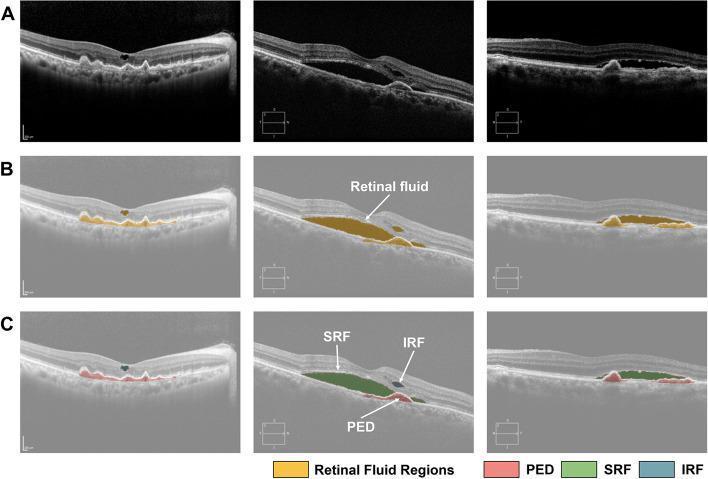
This composite diagnostic image showcases optical coherence tomography (OCT) retinal scans across three rows, demonstrating convolutional neural network (CNN)-based fluid segmentation in neovascular age-related macular degeneration (nAMD).
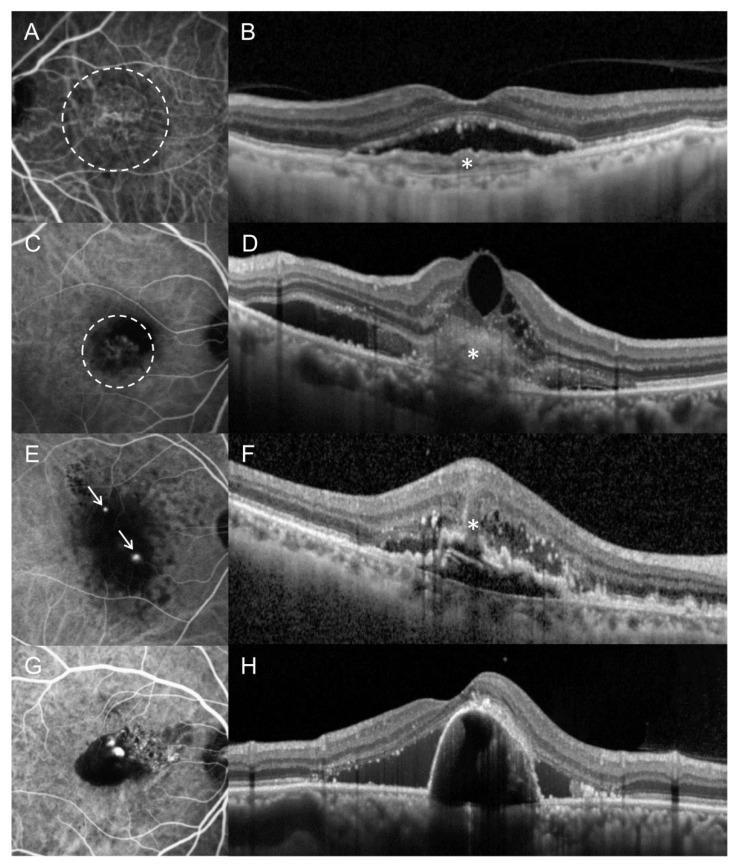
This composite diagnostic image displays paired Indocyanine Green Angiography (ICGA) and Optical Coherence Tomography (OCT) scans illustrating four subtypes of macular neovascularization (MNV) in age-related macular degeneration. (A-B) Type 1 MNV: ICGA shows a diffuse neovascular complex (dashed circle) while OCT demonstrates the complex confined beneath the retinal pigment epithelium (RPE). (C-D) Type 2 MNV: ICGA reveals a compact hyperfluorescent complex (dashed circle) with OCT showing subretinal hyperreflective material (SHRM) above the RPE. (E-F) Type 3 MNV (Retinal Angiomatous Proliferation): ICGA highlights focal punctate hyperfluorescent spots (arrows), and OCT shows an intraretinal hyperreflective lesion with associated intraretinal edema and RPE disruption. (G-H) Polypoidal Choroidal Vasculopathy (PCV): ICGA demonstrates distinct nodular hyperfluorescent polyps, while OCT shows characteristic peaked serous pigment epithelial detachment (PED) and subretinal fluid. The images are essential for ophthalmological training in distinguishing neovascular subtypes based on anatomical location and vascular morphology using multimodal imaging.
https://www.aao.org/Assets/7d362287-0a40-430a-9fe7-fd4380521…
Retina and Vitreous: MS Ophthalmology Theory Revision
- Branch retinal vein occlusion (BRVO)
- Age-related macular degeneration (AMD/ARMD)
- OCT and OCT angiography
- Electroretinography (ERG)
- Retinopathy of prematurity (ROP)
- Retinal lasers, including Pascal
- Pars plana vitrectomy (PPV)
- Silicone oil and other endotamponades
- Retinal imaging modalities
- Fundus fluorescein angiography (FFA)
1. Branch Retinal Vein Occlusion (BRVO)
Definition
Pathogenesis
- Venous narrowing
- Turbulent blood flow
- Endothelial injury
- Thrombus formation
- Venous obstruction
- Raised intraluminal venous pressure
- Capillary leakage causing macular edema
- Retinal hemorrhage
- Capillary nonperfusion and ischemia
- Increased VEGF production, causing macular edema and neovascularization
Risk factors
Ocular
- Hypertension-related arteriosclerosis
- Primary open-angle glaucoma
- Raised IOP
- Short axial length, reported in some populations
Systemic
- Hypertension
- Diabetes mellitus
- Dyslipidemia
- Smoking
- Obesity
- Renal disease
- Hyperhomocysteinemia
- Myeloproliferative disorders
- Thrombophilia, especially in young, bilateral, recurrent, or atypical RVO
Classification
By site
- Major BRVO: first-order branch vein occlusion, commonly superotemporal
- Macular BRVO: smaller macular venous branch involved
- Hemispheric retinal vein occlusion: one hemiretina involved, often considered intermediate between BRVO and CRVO
By perfusion
- Perfused BRVO
- Ischemic BRVO: extensive capillary nonperfusion, higher risk of retinal/disc neovascularization
Clinical features
Symptoms
- Painless unilateral decrease in vision
- Metamorphopsia
- Central or paracentral scotoma
- Often incidentally detected if macula is spared
Signs
- Dilated, tortuous vein
- Flame-shaped and dot-blot retinal hemorrhages
- Cotton-wool spots
- Retinal edema
- Macular edema if macular circulation is affected
- Collateral vessels later
- Neovascularization of retina or disc in ischemic disease
- Vitreous hemorrhage as a late complication
Investigations
| Investigation | Role |
|---|---|
| Visual acuity, IOP, slit-lamp and dilated fundus exam | Baseline assessment |
| OCT macula | Detects and follows macular edema, subretinal fluid, DRIL, outer retinal damage |
| OCT-A | Demonstrates superficial/deep plexus nonperfusion and collateral circulation, but does not show leakage |
| FFA | Defines ischemia, macular leakage, macular perfusion, neovascularization and capillary nonperfusion |
| Widefield FFA | Better for peripheral ischemia and targeted laser planning |
| Systemic work-up | Detects cardiovascular/metabolic risk factors |
Management
A. Treat systemic risk factors
- BP control
- Diabetes control
- Lipid management
- Smoking cessation
- Assessment for glaucoma
- Selective hematologic work-up where indicated
B. Macular edema
- Ranibizumab
- Aflibercept
- Bevacizumab, widely used off-label in many settings
- Faricimab, a bispecific antibody targeting VEGF-A and angiopoietin-2
- Initial loading followed by pro re nata regimen
- Treat-and-extend protocol
- Individualized OCT-guided treatment
C. Intravitreal corticosteroid
- In pseudophakia
- When anti-VEGF response is suboptimal
- If injection burden is difficult
- In anti-VEGF contraindication or selected inflammatory phenotypes
- IOP elevation
- Cataract, particularly in phakic eyes
- Need for repeat treatment
D. Laser photocoagulation
- Grid laser for macular edema: historical role, now usually secondary to anti-VEGF therapy.
- Sector scatter laser: indicated for retinal/disc neovascularization associated with nonperfusion, not prophylactically merely because ischemia exists.
Prognosis
- Baseline VA
- Duration/severity of macular edema
- Foveal ischemia
- Integrity of ellipsoid zone and external limiting membrane on OCT
- Disorganization of retinal inner layers, DRIL
- Development of neovascularization/vitreous hemorrhage
Recent advances
BRVO treatment is directed primarily at macular edema and neovascularization, while systemic evaluation reduces future ocular and cardiovascular risk.
2. Age-Related Macular Degeneration (AMD/ARMD)
Definition
Risk factors
Non-modifiable
- Increasing age
- Family history/genetic susceptibility
- White ethnicity
- Complement-pathway gene variants, including CFH and ARMS2/HTRA1 associations
Modifiable
- Smoking, the strongest modifiable risk factor
- Hypertension/cardiovascular risk factors
- Obesity
- Poor diet and low antioxidant intake
- Excess ultraviolet exposure is less clearly established
Classification
1. Early AMD
- Medium drusen
- Mild RPE pigmentary abnormalities
- Usually no visual symptoms
2. Intermediate AMD
- Large drusen, typically at least 125 micrometers
- Numerous medium drusen
- Noncentral geographic atrophy
3. Late AMD
A. Dry AMD
- Drusen and RPE dysfunction
- Geographic atrophy (GA): sharply demarcated RPE and photoreceptor loss
B. Neovascular or wet AMD
- Subretinal fluid
- Intraretinal fluid
- Pigment epithelial detachment
- Hemorrhage
- Fibrosis/disciform scar
Classification of macular neovascularization
| Type | Location | Typical imaging |
|---|---|---|
| Type 1 MNV | Sub-RPE | Irregular fibrovascular PED, sub-RPE flow on OCT-A |
| Type 2 MNV | Subretinal, above RPE | Subretinal hyperreflective material, classic leakage on FFA |
| Type 3 MNV | Intraretinal, formerly retinal angiomatous proliferation | Intraretinal cysts/hyperreflective foci, often with PED |
| Polypoidal choroidal vasculopathy | Aneurysmal type 1 neovascularization | Orange nodules, peaked PED, ICGA polypoidal lesions |
Clinical features
- Painless central visual loss
- Metamorphopsia
- Micropsia
- Difficulty recognizing faces and reading
- Central scotoma
- Reduced contrast and dark adaptation
Investigations
| Investigation | Key use |
|---|---|
| OCT | First-line diagnosis and monitoring of exudation |
| OCT-A | Noninvasive MNV visualization, no leakage information |
| FFA | Leakage pattern, classic/occult MNV, activity in difficult cases |
| ICGA | Particularly useful in PCV and type 1 MNV |
| Fundus autofluorescence | RPE health and geographic atrophy mapping |
| Color/widefield fundus photography | Baseline documentation and serial comparison |
Management
Dry AMD
- Smoking cessation
- Control cardiovascular risk factors
- Amsler monitoring and urgent review for new distortion/scotoma
- Low-vision support where necessary
- AREDS2 supplementation for selected intermediate AMD or advanced AMD in one eye
- Vitamin C
- Vitamin E
- Zinc
- Copper
- Lutein
- Zeaxanthin
Geographic atrophy: major advance
- Pegcetacoplan, a C3 inhibitor
- Avacincaptad pegol, a C5 inhibitor
Neovascular AMD
- Ranibizumab
- Aflibercept
- Bevacizumab, off-label in many regions
- Brolucizumab, used cautiously because of intraocular inflammation/retinal vasculitis concerns
- Faricimab: VEGF-A and Ang-2 inhibition
- Higher-dose aflibercept formulations in some regions for interval extension
- Fixed dosing
- Pro re nata
- Treat-and-extend, widely used in routine practice
Polypoidal choroidal vasculopathy
- Anti-VEGF is foundational.
- Photodynamic therapy may be added in selected cases, especially persistent polypoidal lesions or recurrent hemorrhage.
Recent advances
- OCT-guided treat-and-extend regimens
- Faricimab and high-dose aflibercept for reducing injection burden in selected patients
- Complement inhibition for geographic atrophy
- Home monitoring and AI-assisted fluid detection
- Long-acting delivery systems and gene therapy remain areas of active development rather than routine universal care
Dry AMD is not simply “benign.” It can progress to geographic atrophy or neovascular AMD. New metamorphopsia in a patient with dry AMD is an urgent symptom of possible conversion to MNV.
3. Optical Coherence Tomography (OCT)
Definition
Principle
- A reference beam
- A sample beam reflected from ocular tissues
Types
| Type | Main feature |
|---|---|
| Time-domain OCT | Older, slower, lower resolution |
| Spectral-domain OCT | Faster and higher resolution; common clinical platform |
| Swept-source OCT | Longer wavelength, deeper penetration through pigment, hemorrhage and media opacity; better choroid imaging |
| Enhanced-depth imaging OCT | Better choroidal visualization |
| OCT angiography | Flow-based visualization of retinal and choroidal vasculature without dye |
Retinal OCT applications
- Diabetic macular edema
- BRVO/CRVO macular edema
- Neovascular AMD
- Central serous chorioretinopathy
- Epiretinal membrane
- Vitreomacular traction
- Macular hole
- Myopic maculopathy
- Retinal dystrophies
- Hydroxychloroquine monitoring
- Optic nerve/RNFL and ganglion-cell analysis in glaucoma
Important OCT signs
| OCT finding | Clinical implication |
|---|---|
| Intraretinal cysts | Macular edema |
| Subretinal fluid | MNV, CSC, inflammatory choroidopathy, other causes |
| PED | AMD, PCV, CSC and other RPE disorders |
| Hyperreflective foci | RPE migration/inflammation, risk biomarker in some disorders |
| Subretinal hyperreflective material | Fibrovascular tissue, blood, MNV-associated material |
| Ellipsoid-zone disruption | Photoreceptor injury and poorer visual prognosis |
| DRIL | Associated with poorer VA in macular edema |
| VMT | Partial vitreous separation exerting foveal traction |
| Full-thickness macular hole | Defect from ILM to RPE with elevated margins |
OCT-Angiography
Principle
Advantages
- Noninvasive
- Rapid
- Layer-by-layer vascular segmentation
- Identifies nonexudative MNV
- Useful in diabetic retinopathy, AMD, retinal vein occlusion, macular telangiectasia and inherited retinal disease
Limitations
- Does not show leakage
- Motion artifact
- Projection artifact
- Segmentation errors, especially in edema/PED
- Poor images with media opacity or poor fixation
- Slow-flow lesions may be missed
FFA shows dynamic leakage and perfusion. OCT-A shows flow architecture but no leakage.
4. Fundus Fluorescein Angiography (FFA)
Definition
Principle
- Is a water-soluble orange dye
- Absorbs blue light near 490 nm
- Emits yellow-green fluorescence near 530 nm
- Is largely protein-bound intravascularly
- Is excreted by the kidneys
Phases of FFA
- Choroidal flush: patchy background choroidal fluorescence
- Arterial phase
- Arteriovenous/capillary phase
- Early venous phase: laminar flow
- Late venous phase
- Recirculation phase
Hyperfluorescence: causes
| Pattern | Meaning |
|---|---|
| Window defect | RPE atrophy allows increased choroidal fluorescence; early and stable intensity/size |
| Leakage | Increasing intensity and area with fuzzy margins |
| Pooling | Dye accumulation in an anatomical space, for example subretinal fluid/PED |
| Staining | Late fluorescence of tissue such as scar, drusen, disc or vessel wall |
Hypofluorescence: causes
| Pattern | Cause |
|---|---|
| Blocked fluorescence | Hemorrhage, pigment, exudate |
| Filling defect | Nonperfusion, arterial occlusion, choriocapillaris defect |
Indications
- Diabetic retinopathy and macular edema
- BRVO/CRVO evaluation
- Macular ischemia
- Neovascular AMD
- Central serous chorioretinopathy
- Retinal vasculitis
- Choroiditis
- Cystoid macular edema
- Retinal neovascularization
- Unexplained visual loss with suspected vascular/retinal pathology
Contraindications and adverse effects
Relative contraindications
- Previous severe fluorescein reaction
- Pregnancy, depending on risk-benefit analysis
- Severe asthma or major allergy history requires caution
Complications
- Nausea and vomiting, common minor effects
- Yellow discoloration of skin
- Bright yellow urine
- Extravasation pain/tissue irritation
- Urticaria
- Bronchospasm
- Anaphylaxis, rare but potentially fatal
FFA versus ICGA
| Feature | FFA | ICGA |
|---|---|---|
| Dye | Sodium fluorescein | Indocyanine green |
| Best visualized circulation | Retinal circulation | Choroidal circulation |
| Blocked by blood/pigment | More affected | Less affected because infrared light penetrates pigment/blood better |
| Major uses | DR, RVO, CME, leakage | PCV, occult/type 1 MNV, choroidal inflammatory disorders |
5. Electroretinography (ERG)
Definition
Principle
Components
| Wave | Main origin |
|---|---|
| a-wave | Photoreceptors, mainly photoreceptor hyperpolarization |
| b-wave | Bipolar cells and Müller-cell contribution |
| Oscillatory potentials | Inner retina, especially amacrine-cell activity |
| c-wave | RPE-photoreceptor complex, less often used clinically |
Types
Full-field ERG
- Retinitis pigmentosa
- Cone-rod dystrophy
- Congenital stationary night blindness
- Drug toxicity
- Widespread retinal dysfunction
- Unexplained reduced vision when a diffuse retinal dystrophy is suspected
Pattern ERG
Multifocal ERG
- Occult macular dystrophy
- Hydroxychloroquine toxicity
- Macular dysfunction with normal fundus
- Early regional retinal dysfunction
Electro-oculography
- Best vitelliform macular dystrophy
Dark-adapted versus light-adapted responses
| Condition | Dominant tested system |
|---|---|
| Dark-adapted/scotopic ERG | Rod pathway |
| Light-adapted/photopic ERG | Cone pathway |
Classic ERG patterns
| Disease | ERG finding |
|---|---|
| Retinitis pigmentosa | Reduced/extinguished rod responses early, later cone involvement |
| Cone dystrophy | Markedly reduced photopic response |
| Congenital stationary night blindness | Electronegative ERG, reduced b-wave relative to a-wave |
| X-linked juvenile retinoschisis | Electronegative ERG |
| Central retinal artery occlusion | Markedly reduced b-wave with relatively preserved a-wave, negative ERG |
| Birdshot chorioretinopathy | May show diffuse retinal dysfunction |
| Hydroxychloroquine toxicity | mfERG may detect localized parafoveal dysfunction |
A negative/electronegative ERG means the b-wave is smaller than the a-wave, suggesting post-photoreceptor inner retinal dysfunction.
6. Retinopathy of Prematurity (ROP)
Definition
Pathogenesis: two-phase model
Phase 1: Hyperoxia and vaso-obliteration
- Relative hyperoxia suppresses VEGF and IGF-1
- Normal retinal vascular growth is interrupted
- Peripheral retina remains avascular
Phase 2: Hypoxia-driven neovascularization
- Avascular retina becomes hypoxic
- VEGF rises
- Pathological neovascularization and fibrovascular proliferation develop
- Traction can cause retinal detachment
Risk factors
- Lower gestational age
- Lower birth weight
- Prolonged supplemental oxygen exposure
- Sepsis
- Apnea/respiratory distress
- Poor postnatal weight gain
- Anemia/transfusions
- Intraventricular hemorrhage
- Poor neonatal care and oxygen monitoring
International Classification of ROP
Zones
- Zone I: circle centered on optic disc, radius twice disc-fovea distance
- Zone II: from outer edge of zone I to nasal ora serrata and toward temporal equator
- Zone III: residual temporal crescent of peripheral retina
Stages
| Stage | Finding |
|---|---|
| 1 | Demarcation line |
| 2 | Ridge |
| 3 | Extraretinal fibrovascular proliferation |
| 4A | Partial tractional RD, macula spared |
| 4B | Partial tractional RD, macula involved |
| 5 | Total retinal detachment |
Plus disease
Aggressive ROP
Treatment indication: Type 1 ROP
- Zone I, any stage with plus disease
- Zone I, stage 3 without plus disease
- Zone II, stage 2 or 3 with plus disease
- Zone I, stage 1 or 2 without plus
- Zone II, stage 3 without plus
Treatment
A. Laser photocoagulation
- Well-established treatment
- Definitive peripheral ablation
- Less concern about prolonged systemic VEGF suppression compared with anti-VEGF
- Technically demanding in small infants
- More myopia
- May be difficult in posterior zone I disease
- Peripheral field is ablated
- Does not allow normal peripheral vascularization
B. Intravitreal anti-VEGF
- Bevacizumab
- Ranibizumab
- Aflibercept in some settings
- Very effective in zone I/posterior aggressive disease
- Rapid regression
- Preserves more peripheral retina
- May induce less myopia than laser
- Useful where media opacity or poor pupil dilation makes laser difficult
- Late recurrence/reactivation can occur, sometimes months later
- Requires long-term follow-up until peripheral vascularization is complete
- Systemic absorption and long-term neurodevelopmental/systemic safety remain important concerns
- Optimal agent and lowest effective dose remain unsettled
C. Surgery
- Lens-sparing vitrectomy for selected stage 4 disease
- Vitrectomy with or without lensectomy for advanced tractional detachment
- Anatomical success and visual outcome worsen markedly in stage 4B and stage 5 disease
Current evidence
Laser remains a standard definitive treatment, especially for zone II disease. Anti-VEGF is particularly valuable for zone I, posterior and aggressive ROP, but mandates prolonged follow-up for reactivation.
7. Retinal Lasers
Principle of retinal photocoagulation
- Wavelength
- Power
- Spot size
- Duration
- Retinal pigmentation
- Media clarity
- Lens used
Common laser wavelengths
| Laser | Wavelength | Major uses |
|---|---|---|
| Argon green | 514 nm | Historical/common retinal photocoagulation |
| Frequency-doubled Nd:YAG green | 532 nm | Common retinal laser |
| Yellow | 561-577 nm | Good hemoglobin absorption, macular applications |
| Diode infrared | 810 nm | Transscleral cyclophotocoagulation, some retinal uses |
| Krypton red | 647 nm | Better penetration through blood/pigment, largely less common now |
Types of retinal laser
| Procedure | Main purpose |
|---|---|
| Focal laser | Treat focal leakage/microaneurysms |
| Grid laser | Diffuse macular edema, now less frequently primary therapy |
| Panretinal photocoagulation, PRP | Regress neovascular drive in proliferative retinopathies |
| Barrier/barrage laser | Surround retinal breaks, lattice with holes or localized detachment |
| Sector scatter laser | Neovascularization due to sectoral ischemia, such as BRVO |
| Macular laser | Limited modern use due to anti-VEGF dominance |
| Micropulse/subthreshold laser | Tissue-sparing treatment in selected macular diseases |
PRP: indications
- Proliferative diabetic retinopathy
- Ischemic CRVO with neovascularization or high risk
- Ocular ischemic syndrome
- Proliferative sickle retinopathy
- Selected retinal vasculitis and Eales disease
Complications of retinal laser
- Pain
- Iatrogenic retinal break, rare
- Macular edema
- Reduced peripheral field after PRP
- Reduced night vision
- Reduced color/contrast sensitivity
- Choroidal effusion, exudative RD, rare
- Accidental foveal burn
- Bruch membrane rupture/secondary CNV, rare
8. Pascal Laser
Full form
Definition
Principle
- 2 × 2
- 3 × 3
- 4 × 4
- Arc
- Grid
- PRP arrays
Advantages
- Faster delivery of PRP
- More uniform spot pattern
- Less total procedure time
- Often better tolerated
- Potentially less collateral thermal diffusion with short pulses
- Useful in PRP, sector laser and pattern macular treatment
Limitations
- Shorter pulse duration requires higher power to achieve the intended burn
- Lesion intensity must be titrated carefully
- Less flexibility in irregular peripheral anatomy in some situations
- Cost and availability limitations
- A dense or excessively intense pattern can still produce significant field effects
Pascal versus conventional laser
9. Pars Plana Vitrectomy (PPV)
Definition
Anatomical basis
- Ora serrata anteriorly
- Ciliary body posteriorly
- Phakic adult eye: approximately 3.5-4 mm posterior to limbus
- Pseudophakic/aphakic eye: approximately 3-3.5 mm posterior to limbus
- In children: distance is adjusted according to age and globe size
Instrument systems
- 20 gauge: older, larger, sutured
- 23 gauge
- 25 gauge
- 27 gauge: very small, less flow, useful in selected fine maneuvers
- Infusion cannula
- Vitreous cutter
- Illumination probe
Indications
Vitreous hemorrhage
- Non-clearing diabetic vitreous hemorrhage
- Dense hemorrhage preventing retinal evaluation/treatment
- Vitreous hemorrhage associated with retinal tear/detachment
- Selected trauma
Retinal detachment
- Pseudophakic RRD
- Giant retinal tear
- Posterior breaks
- RRD with PVR
- Nonvisualized breaks due to hemorrhage/media opacity
- Complex or recurrent RD
Diabetic retinopathy
- Tractional RD threatening or involving macula
- Combined tractional-rhegmatogenous RD
- Non-clearing vitreous hemorrhage
- Dense premacular/subhyaloid hemorrhage, selected cases
- Severe fibrovascular traction
Macular disease
- Epiretinal membrane
- Full-thickness macular hole
- Vitreomacular traction
- Selected myopic traction maculopathy
Other
- Endophthalmitis, depending on visual acuity/severity
- Retained lens fragments
- Intraocular foreign body
- Diagnostic vitreous biopsy
- Dislocated IOL or lens material
- Severe posterior segment trauma
Goals in retinal detachment surgery
- Remove vitreoretinal traction
- Identify and treat all retinal breaks
- Flatten retina and drain subretinal fluid if required
- Create chorioretinal adhesion using laser/cryo
- Maintain retinal apposition with internal tamponade
Basic surgical steps
- Preoperative retinal mapping and consent
- Conjunctival displacement in transconjunctival systems
- Create pars plana ports
- Confirm infusion cannula position before opening infusion
- Core vitrectomy
- Induce posterior vitreous detachment, if not already present
- Peripheral vitreous shaving with scleral depression
- Remove membranes where indicated
- Identify all breaks
- Drain subretinal fluid, often through a break or drainage retinotomy
- Fluid-air exchange
- Endolaser retinopexy
- Gas or silicone-oil tamponade, if needed
- Close/suture leaking ports
Complications
Intraoperative
- Iatrogenic retinal break
- Lens touch
- Suprachoroidal hemorrhage
- Choroidal detachment
- Retinal incarceration at port
- Incomplete membrane removal
- Infusion misdirection
Postoperative
- Cataract progression, especially nuclear sclerosis in phakic patients
- Elevated IOP or hypotony
- Endophthalmitis
- Corneal edema
- Cystoid macular edema
- Recurrent RD/PVR
- Epiretinal membrane
- Retinal toxicity, rare
- Visual-field defects
- Need for reoperation
10. Silicone Oil
Definition
Mechanism
- Supporting the retina against the RPE
- Preventing fluid from entering retinal breaks
- Maintaining chorioretinal adhesion while laser scars mature
Types
- Conventional silicone oil: commonly 1,000 or 5,000 centistokes
- Heavy silicone oil: denser than water, designed to tamponade inferior pathology, but has limited long-term use due to complications/emulsification concerns
Indications
- Complex RRD with PVR
- Giant retinal tears
- Recurrent retinal detachment
- Severe diabetic tractional/combined RD
- Retinal detachment with proliferative vitreoretinopathy
- Ocular trauma
- Cases requiring long-term tamponade
- Patients unable to posture for gas tamponade
- Need for early air travel or situations where gas is unsuitable
Advantages compared with gas
- Long-term support
- No expansion with nitrous oxide/altitude in the same manner as gas
- No strict prolonged face-down positioning in some cases, although positioning can still be clinically important
- Fundus can be examined through oil
- Appropriate for complex disease
Disadvantages
- Usually requires a second surgery for removal
- Less favorable visual outcomes than gas in uncomplicated RRD, partly because oil is used in more complex eyes
- Emulsification and anterior-segment complications
Complications
| Complication | Mechanism |
|---|---|
| Cataract | Common in phakic eyes |
| Raised IOP/glaucoma | Pupillary block, emulsified oil in trabecular meshwork, inflammation, steroid response |
| Hypotony | Ciliary body dysfunction, PVR/traction |
| Keratopathy/band keratopathy | Oil in anterior chamber, endothelial toxicity |
| Corneal decompensation | Endothelial damage |
| Emulsification | More likely with longer retention, inflammation and lower-viscosity oil |
| Recurrent RD after removal | Persistent PVR or unsealed breaks |
| Retinal toxicity/inner retinal thinning | Multifactorial, particularly with long-term oil |
Pupillary block prevention
Silicone oil removal
- Retina is stable
- Adequate chorioretinal adhesion is established
- Oil-related complications develop
- Risk of redetachment is acceptable
Use silicone oil primarily for complex retinal detachment or where long-term tamponade is needed. It is not the routine preferred tamponade for uncomplicated RRD.
11. Other Vitreous Substitutes
| Agent | Features | Main cautions |
|---|---|---|
| Air | Short duration, useful in selected simple breaks/macular holes | Rapid absorption |
| SF6 gas | Expansile, intermediate duration | No air travel or nitrous oxide anesthesia until fully absorbed |
| C3F8 gas | More expansile and longer acting | Longer visual recovery, IOP rise, strict no-fly/no-N2O advice |
| Perfluorocarbon liquid | Heavy liquid, intraoperative retinal flattening, giant tear management | Must be removed, retinal toxicity if retained |
| Silicone oil | Long-term tamponade | Emulsification, glaucoma, cataract, keratopathy |
A patient with intraocular expansile gas must not fly or receive nitrous oxide anesthesia until the gas is fully absorbed, due to risk of acute dangerous IOP elevation.
12. Retinal Imaging Modalities: Exam Table
| Modality | Main information | Main strengths | Limitations |
|---|---|---|---|
| Color fundus photo | Surface retinal appearance | Documentation, screening, serial comparison | No depth or leakage information |
| Red-free photo | Nerve fiber layer, hemorrhage, vessels | Enhances retinal detail | Limited depth information |
| Ultra-widefield imaging | Peripheral retina | RVO, DR, uveitis, peripheral lesions | Peripheral distortion/artifact possible |
| OCT | Retinal microstructure | Macular fluid, traction, photoreceptor integrity | No direct leakage/flow information |
| OCT-A | Flow architecture | Noninvasive vascular mapping | No leakage, artifact-prone |
| FFA | Retinal perfusion and leakage | RVO, DR, vasculitis, MNV activity | Invasive dye test |
| ICGA | Choroidal circulation | PCV, type 1 MNV, choroiditis | Invasive, less available |
| FAF | RPE metabolic status | Geographic atrophy, inherited retinal disease | Interpretation needs clinical context |
| B-scan ultrasonography | Posterior segment through opaque media | RD, VH, mass, PVD | Lower retinal detail than OCT |
13. High-Yield Recent Advances
- Anti-VEGF treatment remains first-line for vision-threatening macular edema due to BRVO and for neovascular AMD.
- Faricimab targets VEGF-A and Ang-2 and may allow longer treatment intervals in some RVO and AMD patients, but superiority in durability over existing agents is not yet established.
- OCT-A is valuable for noninvasive vascular mapping, but FFA remains important when leakage and dynamic perfusion assessment are needed.
- Swept-source OCT gives deeper choroidal and vitreoretinal imaging.
- Geographic atrophy complement inhibitors can slow lesion enlargement but require counseling about limited functional recovery and risk of neovascular conversion.
- ROP anti-VEGF preserves peripheral retina and may reduce myopia, but requires prolonged surveillance for late reactivation and raises systemic-safety questions.
- Small-gauge vitrectomy with high-speed cutters has improved surgical efficiency and reduced tissue traction.
- Heads-up 3D vitreoretinal surgery, intraoperative OCT, digital visualization, robotic assistance and AI-supported imaging are evolving adjuncts.
- Pascal and micropulse lasers aim for more efficient or tissue-sparing laser delivery, but appropriate case selection and endpoint titration remain central.
14. One-Minute Viva Answers
What is the difference between FFA and OCT-A?
What is the first-line treatment for BRVO macular edema?
What are the major complications of silicone oil?
What does a negative ERG indicate?
What is plus disease in ROP?
What is the role of PPV in diabetic retinopathy?
Teach me cornea and ocular surface next
limbal stem cell deficiency treatment
corneal collagen cross linking keratoconus
ocular surface squamous neoplasia treatment
amniotic membrane transplantation ocular surface
AAO cornea preferred practice pattern limbal stem cell deficiency ocular surface squamous neoplasia corneal cross linking 2025
PMID: 39539159
PMID: 39387749
PMID: 42047870
amniotic membrane transplantation ocular surface surgery corneal epithelial defect image
keratoconus corneal collagen cross linking demarcation line slit lamp image
Cornea and Ocular Surface: MS Ophthalmology Theory Revision
- Limbal stem-cell deficiency and stem-cell therapy
- Amniotic membrane transplantation
- Ocular surface squamous neoplasia
- Corneal collagen cross-linking / C3R
- Lamellar keratoplasty, especially DALK
- Keratoprosthesis
- Pterygium surgery
- Toxic contact-lens-related ocular surface disease
1. Limbal Stem Cells and Limbal Stem-Cell Deficiency (LSCD)
Normal limbal stem cells
Location
Functions
- Lifelong regeneration of corneal epithelium
- Maintenance of transparent, avascular corneal surface
- Healing after epithelial injury
- Prevention of conjunctivalization
Stem-cell niche
- Palisades of Vogt
- Limbal stromal fibroblasts
- Blood vessels and extracellular matrix
- Corneal nerves
- Melanocytes and immune cells
Limbal Stem-Cell Deficiency
Definition
Etiology
| Acquired, unilateral | Acquired, bilateral | Inherited/congenital |
|---|---|---|
| Chemical or thermal burns | Stevens-Johnson syndrome/toxic epidermal necrolysis | Aniridia |
| Contact-lens-related toxicity | Ocular cicatricial pemphigoid | Ectodermal dysplasia |
| Multiple limbal surgeries | Severe bilateral burns | PAX6-related disease |
| Cryotherapy, radiation | Severe atopy | Congenital erythropoietic porphyria |
| Mitomycin-C toxicity | Chronic topical drug toxicity | |
| Ocular surface tumors and their treatment | Graft-versus-host disease |
Clinical features
- Persistent or recurrent epithelial defects
- Reduced vision, photophobia, pain and redness
- Whorl-like or late fluorescein staining
- Loss of limbal palisades of Vogt
- Superficial corneal vascularization
- Conjunctivalization of cornea
- Fibrovascular pannus and scarring
- Recurrent erosions
- In advanced disease: keratinization and symblepharon
Diagnosis
- Fluorescein staining pattern
- Impression cytology showing conjunctival goblet cells on cornea
- In vivo confocal microscopy
- Anterior-segment OCT
- Corneal epithelial markers, where available
Staging concept
- Partial LSCD: a sector or portion of limbus affected
- Total LSCD: entire limbus affected
- Unilateral versus bilateral LSCD is critical because it determines the donor source.
Management of LSCD
Step 1: Restore the ocular surface
- Stop toxic topical medications and preservatives where possible
- Treat dry eye and lid disease
- Preservative-free lubricants
- Control inflammation: topical steroids, ciclosporin/tacrolimus in selected cases
- Manage exposure, lagophthalmos and trichiasis
- Treat infection and neurotrophic keratopathy if present
- Autologous serum tears or platelet-rich plasma in selected patients
- Scleral lens for surface protection and visual rehabilitation
Step 2: Stem-cell restoration
| Procedure | Best indication | Key issue |
|---|---|---|
| Conjunctival limbal autograft, CLAU | Unilateral total LSCD with healthy fellow eye | Larger limbal tissue harvest from fellow eye |
| Simple limbal epithelial transplantation, SLET | Unilateral LSCD | Small biopsy from fellow eye expanded in vivo on amniotic membrane |
| Cultivated limbal epithelial transplantation, CLET | Unilateral or selected bilateral disease | Ex vivo cell expansion, specialized facility |
| Living-related conjunctival limbal allograft, lr-CLAL | Bilateral LSCD | Systemic immunosuppression required |
| Keratolimbal allograft, KLAL | Severe bilateral LSCD | Cadaveric tissue and systemic immunosuppression |
| Cultivated oral mucosal epithelial transplantation, COMET | Bilateral LSCD when limbal autograft unavailable | Can provide epithelial stability, but optical quality may be limited |
SLET: high-yield answer
- Harvest a small limbal biopsy from healthy fellow eye.
- Secure cryopreserved or fresh amniotic membrane over recipient cornea.
- Divide donor biopsy into small explants.
- Place explants on membrane.
- Cover with bandage contact lens.
- Cells expand across the membrane and repopulate the corneal surface.
Important principle
2. Amniotic Membrane Transplantation (AMT)
Definition
Biological properties
- Promotes epithelial migration and adhesion
- Provides a basement-membrane scaffold
- Reduces inflammation and inflammatory-cell infiltration
- Reduces fibrosis and myofibroblast activation
- Inhibits angiogenesis
- Reduces pain by covering exposed corneal nerves
- Has antimicrobial and anti-scarring effects
Types
| Type | Method | Use |
|---|---|---|
| Cryopreserved AM | Preserves more biologically active components | Ocular-surface reconstruction, inflammatory conditions |
| Dehydrated AM | Shelf-stable | Office-based or surgical use |
| Fresh AM | Limited practical use because of infection/transmission concerns | Research or selected settings |
| Sutureless device, for example AM ring device | Self-retained | Persistent epithelial defect, dry eye, moderate surface inflammation |
| Sutured AM | Graft or patch | Burns, LSCD, tumor excision, perforation support |
Orientation: common
Quick answer
- AMT orientation, techniques, indications and complications
- OSSN
- C3R/corneal cross-linking
- DALK and keratoprosthesis
- Pterygium surgery
- Contact-lens toxicity
Cornea nd ocular surface
ocular surface squamous neoplasia treatment
limbal stem cell deficiency ocular surface
corneal collagen cross linking keratoconus
AAO ocular surface squamous neoplasia limbal stem cell deficiency corneal cross linking guidelines 2025
Cornea and Ocular Surface: MS Ophthalmology Theory Notes
Topics
- Limbal stem-cell deficiency and stem-cell therapy
- Amniotic membrane transplantation
- Ocular surface squamous neoplasia
- Corneal collagen cross-linking, CXL/C3R
- Lamellar keratoplasty, especially DALK
- Keratoprosthesis
- Pterygium surgery
- Toxic contact-lens-related ocular surface disease
1. Limbal Stem-Cell Deficiency and Stem-Cell Therapy
Normal limbus and stem-cell function
Functions
- Continuous renewal of corneal epithelium
- Healing after epithelial trauma
- Maintenance of a transparent, avascular corneal surface
- Barrier function preventing conjunctival epithelial migration onto cornea
Limbal stem-cell deficiency (LSCD)
Definition
Causes
| Category | Examples |
|---|---|
| Chemical/thermal injury | Alkali burns, acid burns, thermal burns |
| Inflammatory/cicatrizing disease | Stevens-Johnson syndrome, toxic epidermal necrolysis, ocular cicatricial pemphigoid, graft-versus-host disease |
| Iatrogenic/toxic | Repeated ocular surgery, cryotherapy, mitomycin-C, radiation, chronic preserved topical medication |
| Contact lens related | Chronic soft contact-lens overwear, solution toxicity |
| Genetic | Aniridia, PAX6 abnormalities, ectodermal dysplasia |
| Neoplastic | Extensive ocular-surface squamous neoplasia or its treatment |
| Others | Severe atopy, neurotrophic disease, chronic ocular surface inflammation |
Clinical features
- Persistent/recurrent epithelial defect
- Photophobia, irritation, pain, redness
- Reduced vision
- Late fluorescein staining in a whorl or vortex pattern
- Loss of palisades of Vogt
- Conjunctivalization of cornea
- Superficial corneal vascularization and fibrovascular pannus
- Recurrent erosions, scarring, calcification
- In severe disease: keratinization, symblepharon and dry eye
Diagnosis
- Fluorescein staining
- Slit-lamp evaluation of limbus and palisades
- Impression cytology: conjunctival goblet cells on the cornea strongly support LSCD
- In vivo confocal microscopy
- Anterior segment OCT
- Corneal epithelial phenotype markers, in specialist centers
Management of LSCD
Principle
Conservative management
- Stop toxic medication and minimize preservatives
- Preservative-free lubricants
- Treat blepharitis, meibomian-gland dysfunction and dry eye
- Control inflammation with carefully supervised topical steroid
- Topical ciclosporin or tacrolimus in selected inflammatory disease
- Autologous serum tears or platelet-rich plasma tears
- Punctal occlusion, tarsorrhaphy, epilation of trichiasis
- Scleral lenses for surface protection and optical rehabilitation
- Treat exposure, infection and neurotrophic keratopathy
Surgical restoration of limbal stem cells
| Procedure | Best use | Major limitation |
|---|---|---|
| Conjunctival limbal autograft, CLAU | Unilateral total LSCD | Requires relatively large limbal harvest from fellow eye |
| Simple limbal epithelial transplantation, SLET | Unilateral LSCD | Depends on healthy fellow-eye limbus |
| Cultivated limbal epithelial transplantation, CLET | Unilateral LSCD, selected bilateral cases | Laboratory infrastructure and cost |
| Living-related conjunctival limbal allograft, lr-CLAL | Bilateral LSCD | Systemic immunosuppression |
| Keratolimbal allograft, KLAL | Severe bilateral LSCD | Rejection and immunosuppression burden |
| Cultivated oral mucosal epithelial transplantation, COMET | Severe bilateral LSCD with no limbal donor | Surface may remain less optically clear than corneal epithelium |
A. Conjunctival limbal autograft
- Tissue is harvested from the healthy contralateral eye.
- Transplanted to affected eye after removing conjunctivalized corneal tissue.
- Suitable for unilateral complete LSCD.
- Donor-site damage is possible if excessive limbus is harvested.
B. SLET: Simple limbal epithelial transplantation
Steps
- Excise fibrovascular pannus and abnormal epithelium from recipient cornea.
- Place cryopreserved amniotic membrane over bare corneal surface.
- Harvest a small limbal biopsy from healthy contralateral eye.
- Divide biopsy into multiple small explants.
- Arrange explants over amniotic membrane.
- Secure with fibrin glue or sutures and apply a bandage contact lens.
Advantages
- Small donor biopsy
- Less risk to donor eye than CLAU
- Does not need a cell-culture laboratory
- Cost-effective
- Particularly practical for unilateral chemical-burn-related LSCD
C. CLET
D. Allograft procedures
- Living related donor limbal tissue, or
- Cadaveric keratolimbal allograft.
Emerging advances
- Cultivated epithelial sheets
- Oral-mucosal epithelial transplantation
- Induced pluripotent stem-cell approaches
- Mesenchymal stem-cell-derived exosomes
- Biomaterial scaffolds and 3D engineered limbal niches
2. Amniotic Membrane Transplantation (AMT)
Definition
Properties and mechanisms
- Promotes epithelial migration, adhesion and differentiation
- Provides basement-membrane substrate
- Suppresses inflammation
- Reduces fibroblast activation and scarring
- Reduces neovascularization
- Reduces pain by covering exposed corneal nerves
- Has anti-protease and anti-microbial properties
Types
- Cryopreserved amniotic membrane
- Dehydrated amniotic membrane
- Sutured graft
- Fibrin-glue-assisted graft
- Sutureless/self-retained membrane device
- Multilayer membrane for deep ulcers or perforation risk
Orientation: viva question
- For a graft/inlay, place the epithelial/basement-membrane side up, facing the regenerating corneal epithelium.
- The stromal side is placed against the host tissue.
- In an overlay/patch technique, the membrane acts mainly as a biological dressing, although standard orientation is still generally maintained.
Surgical techniques
1. Inlay or graft technique
- Persistent epithelial defect
- Corneal ulcer
- Stromal thinning
- Post-pterygium or post-tumor excision defect
- Partial LSCD
2. Overlay or patch technique
- Acute chemical injury
- Acute Stevens-Johnson syndrome
- Severe ocular-surface inflammation
- Extensive epithelial defect
3. Multilayer technique
- Corneal melt
- Descemetocele
- Small corneal perforation, often with tissue adhesive or bandage lens
Indications
| Category | Examples |
|---|---|
| Persistent epithelial defects | Neurotrophic keratopathy, post-infectious defect, exposure |
| Corneal ulcer/melt | Sterile melts, descemetocele, selected infectious ulcers after control |
| Acute burns | Moderate chemical/thermal burns |
| Acute SJS/TEN | Reduce inflammation, lid-margin and conjunctival cicatrization |
| LSCD | Partial LSCD, adjunct to epithelial debridement or SLET |
| Ocular-surface reconstruction | Post-OSSN excision, symblepharon release, fornix reconstruction |
| Corneal surgery | Adjunct in pterygium surgery, lamellar graft fixation, surface defects |
AMT in acute ocular burns
Complications
- Membrane displacement, folding or dissolution
- Infection, uncommon
- Pyogenic granuloma
- Incomplete epithelialization
- Recurrence of underlying inflammation
- Transmission risk is extremely low with screened, processed tissue but must be discussed
3. Ocular Surface Squamous Neoplasia (OSSN)
Definition
Histological spectrum
- Squamous epithelial dysplasia
- Conjunctival intraepithelial neoplasia, CIN
- Carcinoma in situ
- Invasive squamous cell carcinoma
Risk factors
- Ultraviolet-B exposure and outdoor work
- Older age
- Male sex in many populations
- HIV infection and immunosuppression
- HPV association, though causality and subtype contribution vary
- Xeroderma pigmentosum
- Chronic ocular-surface inflammation
- Smoking
- Prior irradiation
- Vitamin A deficiency in some settings
Clinical features
- Usually unilateral, interpalpebral, nasal limbal lesion
- Gelatinous, papilliform, leukoplakic or nodular mass
- Prominent feeder vessels
- Corneal epithelial extension may appear as a gray, translucent, elevated lesion
- May mimic pterygium, pinguecula, papilloma, actinic keratosis or amelanotic melanoma
Investigations
- Slit-lamp photography and lesion mapping
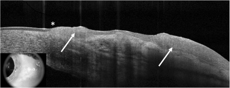
- High-resolution anterior-segment OCT:
- Thickened hyperreflective epithelium
- Abrupt transition between normal and abnormal epithelium
- Helpful to distinguish OSSN from pterygium
- Ultrasound biomicroscopy if intraocular extension suspected
- Impression cytology, selected cases
- Excision biopsy for histopathology where diagnosis is uncertain or lesion requires removal
- Orbital imaging if deep invasion is suspected
Management
A. Surgical excision: “no-touch technique”
Principles
- Avoid directly grasping tumor to prevent seeding.
- Wide conjunctival margins, commonly 3-4 mm of clinically normal tissue where feasible.
- Alcohol-assisted epitheliectomy for corneal component.
- Excise lesion with involved Tenon tissue if needed.
- Apply double freeze-thaw cryotherapy to conjunctival margins.
- Send specimen for histopathology.
- Reconstruct surface with conjunctival autograft or amniotic membrane if required.
B. Topical chemotherapy or immunotherapy
| Drug | Main strengths | Major limitations |
|---|---|---|
| Interferon alpha-2b | Generally well tolerated, useful topical/subconjunctivally | Longer treatment duration, cost/availability |
| 5-fluorouracil | Effective and relatively accessible | Epithelial toxicity, pain, hyperemia |
| Mitomycin-C | Effective for refractory/extensive disease | More surface toxicity, punctal stenosis, LSCD risk |
Choosing surgery versus medical treatment
- Histological diagnosis is needed
- Invasion is suspected
- Lesion is focal and easily excisable
- Patient may not comply with prolonged topical therapy
- Resources are limited
- Isolated corneal lesion requires diagnostic clarification
Follow-up
Major complication of treatment
- Large lesions
- More than 6 clock hours of limbal involvement
- Recurrent lesions
- Corneal involvement
- Repeated surgery or topical mitomycin-C
4. Corneal Collagen Cross-Linking: CXL / C3R
Definition
Main indications
- Documented progressive keratoconus
- Progressive post-LASIK or post-PRK ectasia
- Pellucid marginal degeneration, selected cases
- Keratoglobus or other ectasias, selected cases
- PACK-CXL: photoactivated chromophore for infectious keratitis, as adjunctive treatment in selected refractory infections
Evidence of progression
- Increase in Kmax
- Increase in manifest cylinder or myopia
- Progressive thinning
- Worsening corrected vision
- Change in posterior corneal curvature/elevation
- Serial topographic/tomographic worsening
Dresden protocol: conventional epithelium-off CXL
- Remove central 8-9 mm corneal epithelium.
- Instill 0.1% riboflavin in dextran solution for approximately 30 minutes.
- Confirm stromal saturation and adequate corneal thickness.
- Expose cornea to UVA at 370 nm, 3 mW/cm² for 30 minutes.
- Total radiant exposure is 5.4 J/cm².
- Apply antibiotic, bandage contact lens and postoperative anti-inflammatory regimen.
Safety criterion
Mechanism
Types
| Technique | Advantages | Limitations |
|---|---|---|
| Conventional epi-off CXL | Strongest evidence and deeper stromal effect | Pain, epithelial defect, infection risk, slower recovery |
| Accelerated CXL | Shorter procedure | Biological equivalence to conventional protocol is variable |
| Transepithelial/epi-on CXL | Less pain, faster healing | Riboflavin penetration and efficacy may be lower |
| Iontophoresis-assisted epi-on CXL | Improves riboflavin penetration | Long-term equivalence still uncertain |
| Contact-lens-assisted CXL | For thin cornea | Altered oxygen/UVA dynamics |
| Hypo-osmolar riboflavin protocol | Can swell thin corneas | Careful safety assessment required |
| Customized/topography-guided CXL | Targets cone region | Evolving evidence |
Complications
- Severe pain in early postoperative days
- Delayed epithelial healing
- Sterile infiltrates
- Infectious keratitis
- Corneal haze/scarring
- Endothelial damage in excessively thin cornea
- Herpes simplex keratitis reactivation
- Rare loss of corrected vision
Recent evidence
5. Lamellar Keratoplasty and DALK
Classification of corneal transplantation
| Procedure | Tissue replaced | Main indication |
|---|---|---|
| Penetrating keratoplasty, PK | Full-thickness cornea | Full-thickness scar, perforation, extensive disease involving endothelium |
| Superficial anterior lamellar keratoplasty, SALK | Anterior stroma | Superficial scar/dystrophy |
| DALK | Epithelium and stroma, preserves host Descemet membrane and endothelium | Keratoconus, stromal scar with healthy endothelium |
| DSAEK/DSEK | Posterior stroma, Descemet membrane and endothelium | Endothelial failure |
| DMEK | Descemet membrane and endothelium only | Endothelial disease, especially Fuchs dystrophy |
Deep anterior lamellar keratoplasty (DALK)
Definition
Indications
- Keratoconus
- Stromal corneal scars with healthy endothelium
- Stromal dystrophy
- Postinfectious stromal opacity after infection is controlled
- Some cases of corneal ectasia
Contraindications
- Endothelial dysfunction
- Significant Descemet membrane scarring
- Deep stromal scar adherent to Descemet membrane, relative contraindication
- Acute hydrops with severe Descemet membrane disruption, depending on case
Big-bubble technique
- Partial-depth trephination.
- Insert needle deeply into stroma.
- Inject air to create a cleavage plane between posterior stroma and Descemet membrane.
- Remove anterior stroma.
- Open and remove residual posterior stromal tissue.
- Place donor graft with donor Descemet membrane removed.
- Suture graft.
Advantages over PK
- Preserves host endothelium
- Very low risk of endothelial rejection
- Better long-term endothelial survival
- Reduced risk of catastrophic open-sky complications
- Stronger wound architecture
- Useful in young keratoconus patients
Disadvantages and complications
- Technically demanding
- Descemet membrane perforation
- Conversion to PK may be required
- Double anterior chamber if Descemet membrane detaches
- Interface haze
- Residual stromal bed can reduce optical quality
- Suture-related astigmatism and infection
- Recurrence of disease in graft, uncommon depending on disease
6. Keratoprosthesis
Definition
Main types
- Boston Type I keratoprosthesis
- Boston Type II keratoprosthesis
- Osteo-odonto-keratoprosthesis, OOKP
- Other specialist devices, including tibial osteokeratoprosthesis
Boston Type I KPro
Indications
- Multiple failed corneal grafts
- Severe bilateral corneal opacity with poor graft prognosis
- Chemical injury, selected cases
- Aniridia
- Severe herpetic disease, selected cases
- Some eyes with autoimmune ocular-surface disease, although outcomes are more guarded
Contraindications or poor-prognosis factors
- No light perception or poor optic-nerve/macular potential
- Uncontrolled glaucoma
- Active ocular inflammation
- Severe dry keratinized surface without adequate reconstruction
- Inability to comply with lifelong follow-up
Complications
- Glaucoma progression
- Retroprosthetic membrane
- Sterile keratolysis
- Infectious keratitis/endophthalmitis
- Retinal detachment
- Device extrusion
- Vitreous hemorrhage
- Need for lifelong antimicrobial prophylaxis and bandage contact lens in typical Boston Type I KPro care
7. Pterygium and Pterygium Surgery
Definition
Risk factors
- Chronic ultraviolet exposure
- Outdoor work
- Dust, wind and dry climate
- Chronic ocular-surface irritation
- Geographic “pterygium belt” exposure
Indications for surgery
- Progressive corneal encroachment threatening visual axis
- Induced irregular astigmatism or reduced vision
- Persistent inflammation/irritation despite conservative care
- Restricted motility or diplopia, uncommon
- Cosmetic concern after informed discussion
- Suspicion of dysplasia/OSSN, especially atypical, nodular, leukoplakic, rapidly growing or unusually vascular lesions
Surgical options
| Technique | Recurrence risk | Comments |
|---|---|---|
| Bare sclera excision | High | Avoid as routine modern technique |
| Primary conjunctival closure | Moderate | Limited role |
| Conjunctival autograft, CAG | Low | Preferred standard in many primary cases |
| Limbal conjunctival autograft | Low | Adds limbal barrier function |
| Amniotic membrane graft | Useful when conjunctiva must be preserved | Higher recurrence than CAG in many comparisons |
| Mitomycin-C adjunct | Reduces recurrence | Risk of scleral melt and delayed healing |
Conjunctival autograft technique
- Excise pterygium head from cornea.
- Remove fibrovascular body and Tenon tissue carefully.
- Polish residual corneal tissue as needed.
- Harvest superior bulbar conjunctival graft, often including limbal tissue.
- Place graft over bare sclera with limbal edge oriented toward limbus.
- Secure with sutures or fibrin glue.
Fibrin glue versus sutures
- Glue shortens operative time and improves comfort.
- Sutures are inexpensive and secure, but cause more postoperative inflammation and foreign-body sensation.
Mitomycin-C
- Delayed epithelial healing
- Scleral thinning or melt
- Necrotizing scleritis
- Secondary infection
- Corneal edema
- Cataract or glaucoma, rarely due to intraocular toxicity
8. Toxic Contact-Lens-Related Ocular Surface Disease
Definition
Major mechanisms
- Hypoxia: reduced oxygen transmission, especially with overnight wear
- Mechanical injury: tight lens, poor fit, edge trauma, lens deposits
- Solution toxicity: preservatives, hydrogen peroxide not neutralized, surfactants
- Inflammatory/hypersensitivity reaction
- Microbial infection: particularly Pseudomonas in contact-lens-associated keratitis
Clinical syndromes
| Condition | Features | Management principle |
|---|---|---|
| Contact-lens overwear syndrome | Diffuse SPK, edema, pain, photophobia | Stop lens wear, lubricate, review fit |
| Solution toxicity | Diffuse punctate keratitis, redness, burning | Stop product, preservative-free tears, change system |
| Hydrogen peroxide injury | Acute severe burning, epithelial defect if not neutralized | Immediate irrigation, stop lens use, treat epithelial injury |
| Superior epithelial arcuate lesion, SEAL | Arcuate superior epithelial lesion, tight/silicone hydrogel lens | Modify fit/lens, temporary cessation |
| Contact lens-induced acute red eye, CLARE | Acute unilateral red eye after overnight wear, infiltrates | Discontinue lens, exclude microbial keratitis |
| Contact-lens peripheral ulcer, CLPU | Peripheral infiltrate/ulcer, often with closed-eye wear | Stop lens, antibiotic where epithelial break |
| Infiltrative keratitis | Small peripheral infiltrates | Stop lens, assess infection risk |
| Giant papillary conjunctivitis, GPC | Itch, mucus, giant upper tarsal papillae | Stop/reduce lens wear, replace lens more often, mast-cell stabilizer |
| Contact-lens-induced LSCD | Superior conjunctivalization, whorl staining | Stop lens, manage surface, consider LSCD pathway |
| Microbial keratitis | Pain, infiltrate, epithelial defect, AC reaction | Emergency culture/treatment pathway |
Toxic keratitis: high-yield point
Management approach
- Stop contact-lens wear immediately.
- Remove lens and retain it/case for culture if microbial keratitis is suspected.
- Assess epithelial defect, infiltrate size/location, AC reaction and vision.
- Use preservative-free lubrication.
- Treat lid disease, dry eye and fit problems.
- Change to daily disposable lens or a preservative-free peroxide system only after complete recovery.
- Avoid topical steroid until infection is excluded or controlled.
- Educate: no overnight wear, no water exposure, no swimming/showering with lenses, strict hand hygiene and lens-case replacement.
Red flags for microbial keratitis
- Moderate/severe pain
- Reduced vision
- Central/paracentral infiltrate
- Epithelial defect over infiltrate
- Anterior-chamber reaction/hypopyon
- Rapid progression
- Contact-lens wearer with red painful eye
Rapid Exam Revision
One-line definitions
- LSCD: failure of limbal stem-cell function causing conjunctivalization and epithelial instability of cornea.
- AMT: biological ocular-surface graft or dressing that promotes epithelialization and reduces inflammation, fibrosis and angiogenesis.
- OSSN: spectrum from conjunctival epithelial dysplasia to invasive squamous cell carcinoma.
- CXL: riboflavin-UVA photochemical strengthening of corneal stromal collagen to arrest ectatic progression.
- DALK: stromal corneal transplant preserving host Descemet membrane and endothelium.
- KPro: artificial cornea for repeated graft failure or very poor prognosis for conventional grafting.
- Pterygium: fibrovascular conjunctival growth crossing limbus onto cornea.
Most important viva distinctions
| Question | Answer |
|---|---|
| What is the hallmark of LSCD? | Conjunctivalization of cornea, supported by goblet cells on corneal impression cytology |
| Why avoid early PK in LSCD? | The graft will fail unless stem-cell and tear-film environment are restored first |
| What is the standard CXL protocol? | Epi-off riboflavin plus UVA, 3 mW/cm² for 30 minutes, total 5.4 J/cm² |
| What is the key advantage of DALK over PK? | Preserved host endothelium and lower endothelial rejection risk |
| What is the preferred modern pterygium operation? | Excision with conjunctival, often limbal-conjunctival, autograft |
| What is the most dangerous contact-lens complication? | Microbial keratitis |
| What is the key treatment principle in OSSN surgery? | No-touch excision with adequate margins and cryotherapy, plus histopathology |
Recent-advance pearls
- SLET offers practical in vivo expansion of limbal epithelial cells using a small biopsy from the fellow eye.
- High-resolution anterior-segment OCT helps diagnose and monitor OSSN noninvasively.
- Topical interferon alpha-2b, 5-FU and mitomycin-C have shifted OSSN care from surgery-only to individualized medical-surgical management.
- Conventional epi-off CXL remains the reference treatment for progressive keratoconus.
- Accelerated, transepithelial and iontophoresis-assisted CXL are evolving alternatives, but may not have identical biomechanical effect.
- Cryopreserved amniotic membrane is particularly important in acute SJS/TEN and selected moderate ocular chemical burns.
Teach me glaucoma in the same exam-oriented format
glaucoma neuroprotection
glaucoma drainage device surgery
optical coherence tomography glaucoma
AAO primary open angle glaucoma preferred practice pattern 2025 glaucoma drainage devices OCT neuroprotection
glaucoma neuroprotection nicotinamide
minimally invasive glaucoma surgery
primary open angle glaucoma treatment
glaucoma OCT retinal nerve fiber layer optic disc analysis image
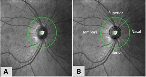
Diagnostic imaging showing fundus photographs with overlaid graphical markers for Retinal Nerve Fiber Layer (RNFL) measurement using Optical Coherence Tomography (OCT). Image A displays a green circular scan (peripapillary ring, typically 3.45 mm in diameter) centered on the optic disc, featuring a central crosshair and a directional arrow indicating the scan path. Image B adds an internal black circle delineating the optic disc margin and four diagonal lines that divide the peripapillary region into functional quadrants: superior, inferior, temporal, and nasal. The underlying anatomy shows the optic disc, emerging retinal vasculature, and subtle striations of the RNFL. This protocol is clinically used for quantifying nerve fiber loss in conditions such as glaucoma or diabetic retinopathy to assess neurodegeneration. The visualization demonstrates the methodology for regional thickness analysis relative to the optic nerve head.
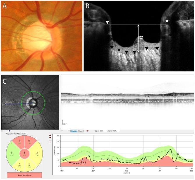
This composite image illustrates diagnostic findings of primary open-angle glaucoma through multimodal ophthalmic imaging. Panel A is a fundus photograph displaying a glaucomatous optic disc characterized by a large vertical cup-to-disc ratio, significant central excavation (cupping), and thinning of the neuroretinal rim, particularly in the superior and inferior poles. Panel B presents a vertical Optical Coherence Tomography (OCT) B-scan of the optic nerve head, measuring the distance (α = 406 µm) between the Bruch's membrane opening (BMO) reference line and the anterior surface of the lamina cribrosa. Panel C shows a circular OCT scan and accompanying quantitative analysis of the circumpapillary retinal nerve fiber layer (cpRNFL). The analysis indicates a significantly reduced mean cpRNFL thickness of 58 µm. The included TSNIT (Temporal, Superior, Nasal, Inferior, Temporal) thickness graph and color-coded sectoral map demonstrate thinning outside normal limits (red sectors), particularly in the superior and nasal regions. This clinical case highlights the correlation between structural optic disc changes and the quantitative loss of the retinal nerve fiber layer in glaucomatous neuropathy.
glaucoma drainage device tube shunt Ahmed Baerveldt diagram image
Glaucoma: MS Ophthalmology Theory Revision
Topics
- Basics of aqueous humor dynamics and IOP
- Classification of glaucoma
- Primary open-angle glaucoma (POAG)
- Primary angle-closure disease (PACD)
- Secondary glaucomas
- Glaucoma evaluation: gonioscopy, disc, fields and OCT
- Medical and laser treatment
- Trabeculectomy
- Glaucoma drainage devices / implants
- MIGS
- Neuroprotection in glaucoma
- Recent advances and rapid viva points
1. Fundamentals: Aqueous Humor and IOP
Aqueous humor production
Mechanisms
- Active secretion: major mechanism
- Ultrafiltration
- Diffusion
Functions
- Maintains IOP and globe shape
- Provides nutrition to avascular cornea and lens
- Removes metabolites
- Transports ascorbate and other substances
- Maintains optical clarity
Aqueous flow pathway
Conventional or trabecular pathway
Unconventional or uveoscleral pathway
- Ciliary muscle
- Supraciliary space
- Suprachoroidal space
- Sclera and venous circulation
Intraocular pressure
Goldmann equation
- F = aqueous formation rate
- C = outflow facility
- Pv = episcleral venous pressure
2. Definition of Glaucoma
- Retinal ganglion-cell death
- Retinal nerve fiber layer loss
- Characteristic optic-disc cupping
- Corresponding visual-field defects
3. Classification of Glaucoma
| Main group | Examples |
|---|---|
| Primary open-angle glaucoma | POAG, normal-tension glaucoma, ocular hypertension |
| Primary angle-closure disease | Primary angle-closure suspect, primary angle closure, primary angle-closure glaucoma |
| Congenital/developmental glaucoma | Primary congenital glaucoma, anterior-segment dysgenesis |
| Secondary open-angle glaucoma | Pseudoexfoliation, pigmentary, steroid-induced, traumatic angle recession, uveitic, lens-particle, ghost-cell, neovascular |
| Secondary angle-closure glaucoma with pupillary block | Phacomorphic, posterior synechiae, aphakic/pseudophakic block |
| Secondary angle-closure without pupillary block | Neovascular glaucoma, ICE syndrome, malignant glaucoma, plateau iris, ciliary-body tumor, choroidal effusion |
4. Primary Open-Angle Glaucoma (POAG)
Definition
Risk factors
| Risk factor | Importance |
|---|---|
| Raised IOP | Major modifiable risk factor |
| Increasing age | Strong association |
| Family history | Important genetic risk |
| African or Hispanic ancestry | Higher prevalence and often more severe disease |
| Thin central corneal thickness | Risk factor and may cause underestimation of IOP |
| Myopia | Especially moderate-to-high myopia |
| Diabetes, vascular factors | Association varies |
| Disc hemorrhage | Marker of progression risk |
| Low ocular perfusion pressure | Important in some patients |
| Steroid response | May reveal predisposition |
Pathogenesis
IOP-dependent mechanisms
- Increased resistance to aqueous outflow at trabecular meshwork
- Mechanical stress at lamina cribrosa
- Retinal ganglion-cell axonal compression
- Impaired axoplasmic flow
- Optic-nerve head ischemia
IOP-independent mechanisms
- Vascular dysregulation
- Low perfusion pressure
- Oxidative stress
- Mitochondrial dysfunction
- Glutamate excitotoxicity
- Neuroinflammation
- Genetic susceptibility
Clinical features
Symptoms
- Gradual peripheral-field loss
- Difficulty with dark adaptation
- Late tunnel vision
- Central vision affected only in advanced disease
Signs
- Raised IOP may be present
- Open angle on gonioscopy
- Optic-disc cupping
- Rim thinning/notching, especially inferotemporal and superotemporal
- Vertical cup enlargement
- RNFL wedge defects
- Disc hemorrhage
- Corresponding visual-field defects
Optic-disc changes
ISNT rule
Glaucomatous disc signs
- Progressive cup enlargement
- Vertical cup-to-disc asymmetry greater than about 0.2
- Focal rim notch
- Laminar-dot sign
- Bayonetting of vessels
- Nasal displacement of vessels
- Peripapillary atrophy
- Disc hemorrhage
5. Visual Field Defects in Glaucoma
Early defects
- Increased pattern standard deviation
- Paracentral scotoma
- Nasal step of Roenne
- Seidel scotoma
Established defects
- Arcuate scotoma of Bjerrum
- Double arcuate scotoma
- Temporal wedge defect
Advanced disease
- Central island
- Temporal island
- Tubular field or tunnel vision
Important rule
6. Diagnosis and Work-up of Glaucoma
- Visual acuity and refraction
- Slit-lamp examination
- Goldmann applanation tonometry
- Pachymetry
- Gonioscopy
- Dilated optic-disc assessment
- Disc photographs
- Visual-field testing
- OCT RNFL and macular ganglion-cell analysis
- Assessment of systemic risk, medications and family history
Gonioscopy
Why it is essential
- Open
- Narrow/occludable
- Closed
- Synechially closed
- Abnormally pigmented
- Neovascularized
- Recessed after trauma
Angle structures from anterior to posterior
Shaffer grading
| Grade | Angle width | Interpretation |
|---|---|---|
| 4 | 35-45 degrees | Wide open |
| 3 | 25-35 degrees | Open |
| 2 | About 20 degrees | Narrow, possible closure |
| 1 | About 10 degrees | Very narrow |
| 0 | Closed | No angle structures visible |
7. OCT in Glaucoma
Role
Main OCT parameters
| Parameter | Clinical use |
|---|---|
| Peripapillary RNFL thickness | Detects axonal loss around optic nerve |
| Ganglion-cell complex, GCC | Macular ganglion-cell and inner plexiform layer analysis |
| Ganglion-cell inner plexiform layer, GCIPL | Early central glaucomatous damage |
| Optic-nerve head parameters | Rim area, cup volume, BMO-MRW |
| Progression analysis | Event and trend analysis over serial scans |
| Anterior-segment OCT | Angle configuration, iris-lens relationship, post-LPI assessment |
RNFL pattern
- Superior peak
- Inferior peak
- Thinner nasal and temporal sectors
OCT interpretation: limitations
- Do not diagnose glaucoma from a color code alone.
- “Red disease” means false-positive abnormal classification.
- “Green disease” means falsely reassuring normal classification.
- Myopia, tilted disc, peripapillary atrophy, poor signal strength, segmentation error, retinal disease and media opacity can mislead.
- Always correlate OCT with disc appearance and visual field.
8. Medical Treatment of Glaucoma
Therapeutic goal
- Baseline IOP
- Severity of damage
- Rate of progression
- Age and life expectancy
- Fellow-eye status
- Corneal thickness
- Risk factors such as disc hemorrhage or low perfusion pressure
- Mild disease: 20%-30% reduction
- Moderate disease: 30%-40% reduction
- Severe/progressive disease: often 40%-50% or more
Topical anti-glaucoma drugs
| Drug group | Examples | Mechanism | Major adverse effects |
|---|---|---|---|
| Prostaglandin analogues | Latanoprost, travoprost, bimatoprost, tafluprost | Increase uveoscleral outflow | Hyperemia, iris darkening, periocular fat atrophy, eyelash growth, uveitis/CME risk |
| Beta blockers | Timolol, betaxolol | Reduce aqueous production | Bradycardia, bronchospasm, hypotension, fatigue |
| Alpha-2 agonists | Brimonidine | Reduces production and increases uveoscleral outflow | Allergy, fatigue, dry mouth; avoid in infants |
| Carbonic anhydrase inhibitors | Dorzolamide, brinzolamide; oral acetazolamide | Reduce aqueous formation | Topical burning; systemic paresthesia, acidosis, renal stones, sulfa-related cautions |
| Cholinergics | Pilocarpine | Increases trabecular outflow by ciliary-muscle contraction | Brow ache, miosis, induced myopia, retinal-detachment risk |
| Rho-kinase inhibitors | Netarsudil, ripasudil in some regions | Increases trabecular outflow, reduces episcleral venous pressure | Hyperemia, corneal verticillata, conjunctival hemorrhage |
First-line medical choice
- Strong efficacy
- Once-daily dosing
- Limited systemic effects
9. Laser Treatment
A. Selective Laser Trabeculoplasty (SLT)
Principle
Indications
- POAG
- Ocular hypertension
- Pseudoexfoliative glaucoma
- Pigmentary glaucoma, cautiously
- Alternative to first-line drops
- Poor adherence or intolerance to drops
- Add-on treatment
Advantages
- Outpatient procedure
- Repeatable in many cases
- Reduces dependence on drops
- Avoids preservative toxicity
Complications
- Transient IOP spike
- Mild anterior uveitis
- Peripheral anterior synechiae, rare
- Corneal edema, rare
- Limited response in heavily scarred or advanced angle disease
B. Argon Laser Trabeculoplasty
C. Laser Peripheral Iridotomy (LPI)
Indications
- Acute angle closure after initial IOP control
- Primary angle closure
- Occludable/narrow angle at risk of pupillary block
- Fellow eye of acute angle-closure attack
- Iris bombe from posterior synechiae
Mechanism
Complications
- IOP spike
- Inflammation
- Corneal burn
- Hyphema
- Dysphotopsia
- Closure of iridotomy
D. Laser Peripheral Iridoplasty
- Plateau iris
- Persistent appositional angle closure after LPI
- Some acute angle-closure settings when LPI cannot be done immediately
10. Primary Angle-Closure Disease
Classification
| Condition | Definition |
|---|---|
| Primary angle-closure suspect, PACS | Occludable angle, but no raised IOP, PAS or glaucomatous optic neuropathy |
| Primary angle closure, PAC | Occludable angle with raised IOP and/or PAS, but no glaucomatous damage |
| Primary angle-closure glaucoma, PACG | PAC plus glaucomatous optic neuropathy and visual-field loss |
Mechanisms
- Relative pupillary block
- Plateau iris configuration
- Thick/anterior lens
- Short axial length
- Hypermetropia
- Ciliary-body rotation
- Lens enlargement with age
Acute angle closure
Symptoms
- Severe ocular pain
- Headache
- Halos around lights
- Blurred vision
- Nausea and vomiting
Signs
- Markedly raised IOP
- Ciliary injection
- Corneal edema
- Shallow anterior chamber
- Mid-dilated fixed pupil
- Closed angle
Emergency management
- Analgesic and antiemetic
- Topical aqueous suppressants
- Systemic acetazolamide unless contraindicated
- Hyperosmotic agent such as mannitol if severe and medically suitable
- Topical steroid
- Pilocarpine once IOP has fallen enough for iris sphincter to respond
- Definitive LPI when cornea clears
- Prophylactic LPI in fellow eye, if indicated
11. Trabeculectomy
Definition
Indications
- Progressive glaucoma despite maximum tolerated medical therapy and/or laser
- Advanced glaucoma requiring very low target IOP
- Poor adherence to medical therapy
- Rapid progression
- Inadequate response to MIGS or prior treatment
- Selected pediatric or secondary glaucomas
Basic steps
- Conjunctival peritomy
- Hemostasis
- Mitomycin-C or 5-FU application where indicated
- Partial-thickness scleral flap
- Deep scleral block and internal ostium/sclerostomy
- Peripheral iridectomy
- Adjustable/releasable scleral flap sutures
- Conjunctival watertight closure
- Formation of diffuse posterior bleb
Antimetabolites
- Mitomycin-C: stronger anti-fibrotic effect
- 5-Fluorouracil: intraoperative or postoperative use
Complications
Early
- Hypotony
- Shallow/flat anterior chamber
- Choroidal detachment
- Hyphema
- Bleb leak
- Malignant glaucoma
- Suprachoroidal hemorrhage
- Bleb failure due to fibrosis
Late
- Bleb leak
- Bleb-related infection or blebitis
- Endophthalmitis
- Hypotony maculopathy
- Cataract
- Dysesthesia
- Encapsulated bleb
- Ptosis
12. Glaucoma Drainage Devices (GDDs)
Definition
Components
- Tube
- Plate/end plate
- Tube-covering graft: sclera, cornea, pericardium or synthetic material
- Conjunctival covering
Classification
| Type | Examples | Principle |
|---|---|---|
| Valved | Ahmed valve, Krupin valve | Valve provides early flow resistance and lowers hypotony risk |
| Non-valved | Baerveldt, Molteno, ClearPath | Tube is ligated initially until capsule forms; often lower long-term IOP |
Indications
- Previous failed trabeculectomy
- Extensive conjunctival scarring
- Neovascular glaucoma
- Uveitic glaucoma
- Post-keratoplasty glaucoma
- Aphakic/pseudophakic glaucoma
- Iridocorneal endothelial syndrome
- Epithelial ingrowth
- Complex pediatric glaucoma
- Traumatic glaucoma
- Refractory glaucoma after multiple surgery
Surgical principles
- Select quadrant, commonly superotemporal.
- Secure plate posteriorly beneath rectus muscles.
- Create a scleral tunnel.
- Insert tube into anterior chamber, ciliary sulcus, or pars plana.
- Cover tube with patch graft.
- Ensure watertight conjunctival closure.
Non-valved implant
Complications
| Early | Late |
|---|---|
| Hypotony and choroidal detachment | Tube erosion/exposure |
| Shallow AC | Endophthalmitis |
| Hyphema | Corneal endothelial loss/decompensation |
| Tube blockage by iris, vitreous or blood | Diplopia/strabismus |
| Hypertensive phase, especially Ahmed valve | Tube migration or retraction |
| Suprachoroidal hemorrhage | Encapsulated plate/capsular fibrosis |
| Malposition of tube | Persistent IOP elevation or hypotony |
Tube position
- Anterior chamber placement is standard in many eyes.
- Sulcus placement may reduce endothelial risk in pseudophakic eyes.
- Pars plana placement requires prior or concurrent adequate vitrectomy.
Ahmed versus Baerveldt: classic comparison
| Feature | Ahmed valve | Baerveldt implant |
|---|---|---|
| Valve | Yes | No |
| Early hypotony | Less common | Higher risk without ligature |
| Early IOP reduction | Faster | Delayed until ligature opens |
| Long-term IOP | May be slightly higher | Often lower in suitable eyes |
| Hypertensive phase | More common | Can occur but less typical |
| Use | Eyes where early hypotony avoidance matters | Need for lower long-term IOP in selected refractory eyes |
13. MIGS: Minimally Invasive Glaucoma Surgery
Definition
Main categories
| Target | Examples | Mechanism |
|---|---|---|
| Trabecular meshwork / Schlemm canal | iStent, Hydrus, Trabectome, Kahook Dual Blade, goniotomy | Bypass or remove trabecular resistance |
| Suprachoroidal space | Selected devices, evolving availability | Increase uveoscleral outflow |
| Subconjunctival pathway | XEN gel stent, PreserFlo MicroShunt | Create controlled bleb-forming outflow |
| Ciliary processes | Endocyclophotocoagulation | Reduces aqueous production |
Indications
- Mild-to-moderate open-angle glaucoma
- Cataract surgery combined with IOP-lowering intervention
- Medication intolerance or poor adherence
- Target IOP not extremely low
- Open angle with accessible trabecular meshwork
Limitations
- Conventional trabecular MIGS cannot lower IOP below episcleral venous pressure.
- Not generally suitable as sole therapy for severe rapidly progressive glaucoma needing very low IOP.
- Bleb-forming “MIGS” may achieve lower pressures but have bleb-related risks.
14. Neuroprotection in Glaucoma
Definition
Why it is needed
- Normal-tension glaucoma
- Advanced glaucoma
- Eyes with vascular dysregulation
- Eyes with disc hemorrhage or low ocular perfusion pressure
Proposed mechanisms of ganglion-cell damage
- Mechanical laminar stress
- Ischemia and reperfusion injury
- Oxidative stress
- Mitochondrial dysfunction
- Excitotoxicity from glutamate
- Calcium influx and apoptosis
- Neuroinflammation
- Reduced neurotrophic-factor support
Potential neuroprotective strategies
| Strategy | Rationale | Current status |
|---|---|---|
| IOP reduction | Reduces mechanical/ischemic injury | Only proven standard neuroprotective intervention |
| Brimonidine | Alpha-2 agonist, possible anti-apoptotic effects | Suggested benefit, not definitive independent proof |
| Calcium-channel blockers | Improve vascular dysregulation theoretically | Not standard glaucoma treatment |
| Memantine | NMDA antagonism, reduces excitotoxicity | Major trials did not establish routine clinical use |
| Citicoline | Mitochondrial/neurotransmitter support | Limited evidence, adjunct only |
| Nicotinamide, vitamin B3 | Supports NAD metabolism and mitochondrial resilience | Research stage, safety concerns at high dose |
| Coenzyme Q10 | Antioxidant/mitochondrial support | Insufficient evidence for routine use |
| Ginkgo biloba | Antioxidant/vascular effects | Inconsistent evidence and bleeding interactions |
| Gene/cell therapy | Retinal ganglion-cell survival/regeneration | Experimental |
Exam conclusion on neuroprotection
15. High-Yield Secondary Glaucomas
| Type | Key clue | Management principle |
|---|---|---|
| Pseudoexfoliative glaucoma | Pseudoexfoliative material, poor dilation, high fluctuating IOP | Often aggressive, laser may help temporarily, surgery often needed |
| Pigmentary glaucoma | Krukenberg spindle, mid-peripheral iris transillumination, heavy TM pigment | Treat IOP; LPI has limited established role |
| Steroid-induced glaucoma | Raised IOP after steroid use | Stop/reduce steroid if possible; treat IOP |
| Uveitic glaucoma | Inflammation plus steroid response | Control inflammation and IOP; avoid miotics; GDD often useful in refractory cases |
| Neovascular glaucoma | Rubeosis iridis, NVA, ischemic retina | Treat cause with PRP and anti-VEGF plus IOP control; often needs GDD/cyclodestruction |
| Traumatic angle recession | Broad ciliary-body band, history of trauma | Long-term monitoring; medical treatment then surgery as needed |
| Phacomorphic glaucoma | Intumescent lens, shallow AC | Control IOP then lens extraction |
| Phacolytic glaucoma | Hypermature cataract, macrophages in AC | Control inflammation/IOP then cataract extraction |
| Malignant glaucoma | Shallow AC despite patent PI, high or normal IOP after surgery | Cycloplegia, aqueous suppression, YAG hyaloidotomy or vitrectomy |
16. Recent Advances in Glaucoma
- Selective laser trabeculoplasty is increasingly used as first-line or early treatment for open-angle glaucoma.
- Preservative-free fixed combinations reduce drop burden and ocular-surface toxicity.
- Rho-kinase inhibitors offer a new pharmacological mechanism by improving trabecular outflow.
- Sustained-release delivery systems and drug-eluting implants are evolving, with availability varying by country.
- MIGS has expanded options for mild-to-moderate open-angle glaucoma, especially with cataract surgery.
- Bleb-forming microshunts occupy an intermediate space between trabeculectomy and traditional MIGS.
- Swept-source OCT, BMO-MRW, macular GCIPL and OCT angiography improve structural assessment, but do not replace optic-disc examination and perimetry.
- Artificial intelligence is being developed for disc-photo, OCT and visual-field interpretation, but needs clinician oversight and validation across populations.
- Neuroprotection through mitochondrial/metabolic pathways, including nicotinamide, remains investigational.
Rapid Viva Answers
What is the most important modifiable risk factor in glaucoma?
What is the definitive test to classify angle as open or closed?
What is the first-line treatment for most POAG patients?
What is the hallmark of glaucoma on OCT?
Why is a thin cornea important?
What is a trabeculectomy?
When do you prefer a tube shunt?
What is the only proven neuroprotective treatment?
What is the difference between PAC and PACG?
This is a 33-topic, syllabus-sized MS Ophthalmology revision plan. I will teach it as exam-ready modules: core concepts, indications, technique, complications, comparison tables, investigations, current evidence, and “recent advances” points. 1. **Cataract and IOLs**: FLACS, capsular tension ring (CTR), phakic IOLs, modern IOLs, accommodative IOLs 2. **Retina and vitreous**: BRVO management, ARMD, OCT, ERG, ROP, retinal imaging, FFA, pars plana vitrectomy, Pascal laser, silicone oil 3. **Cornea and ocular surface**: stem-cell therapy, ocular surface squamous neoplasia, amniotic membrane, collagen cross-linking/C3R, keratoprosthesis, lamellar keratoplasty, pterygium surgery, toxic contact lens reactions 4. **Glaucoma**: neuroprotection, glaucoma implants, OCT in glaucoma 5. **Anterior segment / iris / diagnostic imaging**: pupilloplasty, UBM 6. **General ophthalmic technology and materials**: OVDs, lasers, dyes, tissue adhesives/glues Each long-answer topic will follow a repeatable theory-answer structure: definition and relevance, classification, basic science, clinical indications, investigations, technique or management, complications, outcome/evidence, and advances. I will also flag where evidence is evolving or insufficient, particularly for neuroprotection and stem-cell therapies. Topic 5 and 6
Which topic should I teach in detail next?
- Cataract and IOLs: FLACS, capsular tension ring (CTR), phakic IOLs, modern IOLs, accommodative IOLs
- Retina and vitreous: BRVO management, ARMD, OCT, ERG, ROP, retinal imaging, FFA, pars plana vitrectomy, Pascal laser, silicone oil
- Cornea and ocular surface: stem-cell therapy, ocular surface squamous neoplasia, amniotic membrane, collagen cross-linking/C3R, keratoprosthesis, lamellar keratoplasty, pterygium surgery, toxic contact lens reactions
- Glaucoma: neuroprotection, glaucoma implants, OCT in glaucoma
- Anterior segment / iris / diagnostic imaging: pupilloplasty, UBM
- General ophthalmic technology and materials: OVDs, lasers, dyes, tissue adhesives/glues
- Cataract and IOLs: FLACS, CTR, phakic IOLs, modern/accommodative IOLs
- Retina and vitreous: BRVO, ARMD, OCT, ERG, ROP, FFA, retinal imaging, vitrectomy, lasers, silicone oil
- Cornea and ocular surface: stem cells, amniotic membrane, OSSN, C3R, keratoplasty, KPro, pterygium, contact-lens toxicity
- Glaucoma: neuroprotection, drainage implants, OCT
- Imaging, lasers and materials: UBM, dyes, OVDs, tissue glues, lasers, pupilloplasty
- Femtosecond laser-assisted cataract surgery (FLACS)
- Capsular tension ring (CTR)
- Phakic intraocular lenses
- Modern pseudophakic IOLs, including toric, multifocal, EDOF and accommodating IOLs
- IOL selection, biometry and high-yield complications
1. Foundation: IOL basics
Definition
Parts
- Optic: central refracting portion
- Haptics: peripheral supporting elements
- Overall diameter: optic + haptic-to-haptic length
- Optic diameter: commonly 6 mm for standard adult posterior chamber IOLs
Ideal IOL properties
- Biocompatible and inert
- Stable fixation and centration
- Optically clear, scratch-resistant
- Minimal inflammation, posterior capsule opacification (PCO), dysphotopsia and glistenings
- Injectable through a small incision
- Predictable effective lens position (ELP)
Classification
| Basis | Categories |
|---|---|
| Site | Posterior chamber IOL (PCIOL), anterior chamber IOL (ACIOL), iris-fixated/iris-claw IOL, scleral-fixated IOL |
| Material | PMMA, silicone, hydrophobic acrylic, hydrophilic acrylic |
| Optic | Spherical, aspheric, monofocal, multifocal, extended-depth-of-focus (EDOF), toric, accommodating |
| Construction | One-piece or three-piece; rigid or foldable |
| Fixation | In-the-bag, sulcus, iris-claw, scleral fixation |
Materials: exam comparison
| Material | Advantages | Limitations |
|---|---|---|
| PMMA | Excellent optics, stable, inexpensive | Rigid, needs large incision |
| Silicone | Foldable, small incision | Silicone-oil adherence, therefore generally avoid if future retinal surgery with silicone oil is likely |
| Hydrophobic acrylic | Commonest modern material, foldable, low PCO with square edge, good capsular adhesion | Glistenings or surface light scatter may occur in some models |
| Hydrophilic acrylic | Flexible, good injector delivery | Greater calcification risk in some settings, including exposure to intraocular gas/air in susceptible lenses |
Design features that prevent PCO
- Sharp square posterior optic edge produces a capsular bend and contact inhibition of lens epithelial cell migration.
- In-the-bag placement and meticulous cortical clean-up further reduce PCO.
- PCO, if visually significant, is managed by Nd:YAG posterior capsulotomy.
2. FLACS: Femtosecond Laser-Assisted Cataract Surgery
Definition
- Corneal incisions
- Anterior capsulotomy
- Lens fragmentation/softening
- Arcuate or limbal-relaxing incisions for astigmatism
Principle
Essential components
- Docking interface: applanation or liquid-filled interface
- Image-guidance system: commonly OCT-based
- Laser delivery platform
- Patient interface and suction mechanism
- Integrated or adjacent phacoemulsification system
Steps of FLACS
- Pharmacological dilation and sterile preparation.
- Docking of the laser interface to the eye.
- Imaging and treatment planning.
- Laser creation of:
- Primary and side-port corneal incisions
- Precisely centered capsulotomy
- Lens fragmentation pattern
- Arcuate keratotomy if planned
- Transfer to operating microscope.
- Opening of laser capsulotomy and removal of free capsule disc.
- Hydrodissection, phacoaspiration and cortical clean-up.
- IOL implantation in the capsular bag.
Benefits
- Highly reproducible capsulotomy size, circularity and centration
- Precise corneal incisions
- Lens prefragmentation may reduce effective phaco time and cumulative dissipated energy
- Astigmatic arcuate incisions can be planned precisely
- Useful in selected challenging cases:
- Shallow anterior chamber
- Dense cataract
- Low endothelial reserve
- White/intumescent cataract, with careful case selection
- Premium IOL procedures where centration is especially important
Limitations
- High capital, consumable and maintenance costs
- Extra docking and workflow time
- Requires pupil dilation and adequate corneal clarity
- Not always feasible in deep-set eyes, marked kyphosis, severe tremor, poor cooperation, inability to lie flat, or significant conjunctival scarring
- Incomplete capsulotomy, capsular tags and incomplete fragmentation can occur
- Does not replace surgical judgement or conventional phaco skill
Complications
During docking
- Subconjunctival hemorrhage
- Corneal folds and imaging artefacts
- Transient rise in IOP from suction
- Loss of suction and incomplete treatment
During surgery
- Incomplete corneal incision
- Capsular tags, microadhesions or incomplete capsulotomy
- Anterior capsular tear if the capsule is pulled before identifying and releasing tags
- Incomplete nuclear fragmentation
- Miosis due to prostaglandin release
- Rare capsular block syndrome if hydrodissection is forceful in a gas-fragmented lens
FLACS versus conventional phaco: what to write as “recent evidence”
3. Capsular Tension Ring (CTR)
Definition
Design
- Usually made of PMMA
- Open ring with eyelets at both ends
- Inserted into the capsular bag after capsulorhexis and preferably after adequate hydrodissection
- Available in different diameters
Principle
Indications
Zonular weakness or dialysis
- Pseudoexfoliation syndrome
- Traumatic zonular dialysis
- High myopia
- Marfan syndrome and other ectopia lentis states
- Previous vitreoretinal surgery
- Mature/hypermature cataract with weak zonules
- Lens subluxation of limited extent
- Prior acute angle-closure attack with zonulopathy, in selected cases
Prevention of capsular contraction and IOL decentration
- Pseudoexfoliation
- Retinitis pigmentosa
- High myopia
- Uveitis, selectively
- Conditions predisposed to capsular phimosis
Types
| Device | Main use |
|---|---|
| Standard CTR | Mild-to-moderate diffuse zonular weakness or limited zonular dialysis |
| Modified CTR, Cionni ring | Significant or progressive zonular loss; has one or two fixation eyelets for scleral suturing |
| Capsular tension segment (CTS) | Localized zonular weakness; can be scleral fixated, often used with CTR in extensive dialysis |
| Ahmed capsular tension segment | Segmental capsular support and scleral fixation |
Surgical technique
- Create a well-centered continuous curvilinear capsulorhexis.
- Use dispersive OVD to maintain the bag and protect endothelium.
- Perform gentle hydrodissection, avoiding extension of zonular damage.
- Place capsular hooks or iris retractors, if severe focal dialysis.
- Insert the CTR slowly into the capsular bag using an injector or forceps.
- Ensure the leading eyelet does not engage or tear the capsulorhexis margin.
- Complete phacoemulsification with reduced stress on the weak zonular area.
- Implant IOL in the bag if support is adequate.
- In major dialysis, use a Cionni-modified device or CTS with scleral fixation.
Timing of insertion: common viva point
- Early insertion: improves early bag stability but may trap cortex behind the ring and make cortical aspiration difficult.
- Late insertion: allows easier cortical clean-up but may allow further zonular stress during phaco.
Advantages
- Stabilizes capsular bag during surgery
- Reduces capsular folds and equatorial bag collapse
- Improves IOL centration
- May improve rotational stability of toric IOLs
- Reduces risk of late decentration in selected cases, although it cannot eliminate progressive zonulopathy
Limitations and complications
- Cortex may become trapped between ring and capsule
- Capsular tear may extend if the ring is inserted forcefully
- Can worsen a pre-existing zonular dialysis if incorrectly inserted
- Standard CTR is inadequate when zonular loss is extensive or progressive
- Late in-the-bag IOL-CTR complex subluxation can still occur, especially in pseudoexfoliation
- A CTR should not be used as a substitute for scleral fixation when support is grossly insufficient
Recent evidence
4. Phakic Intraocular Lenses (pIOLs)
Definition
Place in refractive surgery
- Refractive error is too high for safe corneal laser ablation
- Cornea is thin or topographically unsuitable for laser refractive surgery
- Accommodation should be preserved
- The patient is a young adult with stable refraction
Classification
| Type | Position | Examples / features |
|---|---|---|
| Angle-supported anterior-chamber pIOL | Anterior chamber angle | Largely historical due to endothelial and angle complications |
| Iris-fixated pIOL | Clipped to mid-peripheral iris | Artisan/Verisyse-type designs, anterior or retropupillary fixation |
| Posterior-chamber pIOL | Between posterior iris and anterior crystalline lens, supported in ciliary sulcus | ICL and related implantable phakic contact lens designs |
Posterior chamber phakic IOL / ICL
Central-port ICL
Indications
- Stable refraction, generally for at least one year
- Moderate-to-high myopia, with or without astigmatism
- Hyperopia in selected cases
- Age typically 21 years or older, according to device approvals and local practice
- Adequate anterior chamber depth
- Adequate endothelial cell density
- Healthy cornea, crystalline lens, retina and optic nerve
- Strong desire to preserve accommodation
Contraindications
- Progressive refraction or unstable keratoconus
- Shallow anterior chamber
- Low endothelial cell count
- Narrow/occludable angle
- Cataract or significant lens opacity
- Active uveitis
- Uncontrolled glaucoma
- Corneal endothelial disease
- Significant retinal pathology requiring treatment first
- Unrealistic expectations or inability to comply with follow-up
Preoperative work-up
- Manifest and cycloplegic refraction
- Corneal tomography and pachymetry
- Anterior chamber depth, measured from endothelium to anterior lens surface
- White-to-white and/or sulcus-to-sulcus measurement
- Endothelial cell count
- Gonioscopy
- Dilated retinal examination, especially in high myopia
- IOP, optic-nerve assessment and macular OCT if indicated
- Lens-vault prediction and sizing assessment using ultrasound biomicroscopy, anterior-segment OCT or device-specific nomograms
Vault
- Low vault: risk of anterior subcapsular cataract
- Excessive vault: angle crowding, pigment dispersion and secondary angle closure
- Ideal target varies by device and imaging method, but an adequate central vault with open angles is the goal.
Complications
| Complication | Mechanism / prevention |
|---|---|
| Cataract, especially anterior subcapsular | Low vault, lens touch, older designs; careful sizing and follow-up |
| Pupillary block | More relevant to older non-central-port designs; prevented by PI or central-port design |
| Raised IOP | Retained OVD, steroid response, pigment dispersion, angle crowding, pupillary block |
| Endothelial cell loss | More important with anterior chamber pIOLs; monitor ECD |
| Uveitis | Surgical trauma, pigment dispersion, malposition |
| Pupillary ovalization | Mainly iris-claw lenses |
| Toric pIOL rotation | Causes residual astigmatism; may require repositioning |
| Retinal tear/detachment | Related partly to high-myopia phenotype; do meticulous peripheral retinal evaluation |
| Glare, halos, dysphotopsia | Optical effects, residual refractive error |
Recent advances
- Central-port posterior chamber ICLs
- Toric pIOLs
- Improved vault prediction using AS-OCT and UBM
- Larger optic zones and customized sizing
- Diffractive phakic lenses for carefully selected presbyopic patients, but evidence remains limited
5. Pseudophakic IOLs: Modern IOL Options
A. Monofocal IOL
Advantages
- Best contrast sensitivity
- Lowest rate of halos and glare
- Broadest suitability in eyes with retinal disease, glaucoma, corneal irregularity or uncertain visual potential
- Predictable and economical
Disadvantage
- Near spectacles are usually needed.
Aspheric monofocal IOL
B. Toric IOL
Principle
Indications
- Regular corneal astigmatism
- Cataract patient seeking reduced spectacle dependence
- Adequate capsular support
- Reliable keratometry and stable ocular surface
Contraindications / caution
- Irregular astigmatism, unstable keratoconus, corneal scar or severe dry eye
- Poor capsular support
- Unreliable biometry
- Marked zonulopathy unless support is secured
Key points
- Accurate biometry and posterior corneal astigmatism consideration are essential.
- Marking can be manual, image-guided or digitally guided.
- Rotation matters: each degree of toric IOL rotation causes roughly 3.3% loss of cylindrical correction. At 30 degrees, the intended astigmatic correction is effectively lost.
Complications
- Misalignment/rotation
- Residual refractive astigmatism
- IOL tilt or decentration
- Need for early surgical repositioning if substantial rotation occurs
C. Multifocal IOL
Principle
- Diffractive optics
- Refractive zones
- Hybrid designs
Advantages
- Greater spectacle independence at distance and near
Limitations
- Halos and glare
- Reduced contrast sensitivity, especially in dim illumination
- Neuroadaptation required
- Residual refractive error, dry eye, decentration or PCO can cause major dissatisfaction
Avoid or use cautiously in
- Significant ocular surface disease
- Corneal irregularity
- Macular disease, diabetic maculopathy, epiretinal membrane
- Advanced glaucoma or impaired contrast sensitivity
- Optic neuropathy
- Unrealistic patient expectations
- Occupations requiring excellent night contrast, depending on individual needs
D. Trifocal IOL
Advantages
- Stronger near and intermediate spectacle independence than many bifocal designs
Disadvantages
- Dysphotopsia and contrast trade-off remain
- Needs precise centration and refractive targeting
- Not ideal for eyes with retinal or optic-nerve disease
E. Extended Depth-of-Focus (EDOF) IOL
Principle
Advantages
- Often fewer halos than traditional multifocal lenses
- Good distance and intermediate vision
- Useful for computer users
Limitations
- Fine near tasks may still require spectacles
- Dysphotopsia and contrast effects can still occur
- Terminology and optical mechanisms vary between platforms
Important viva distinction
- Multifocal IOL: several discrete foci.
- EDOF IOL: elongated focus, usually better intermediate performance and often less severe dysphotopsia.
- Monofocal-plus/enhanced monofocal: improved intermediate range but not a true EDOF by all definitions.
F. Accommodating IOL
Definition
Mechanisms
- Single-optic movement: anterior movement of the optic during attempted accommodation.
- Dual-optic systems: relative movement of two optics changes overall power.
- Fluid-based or shape-changing designs: aim to alter curvature or refractive power.
Theoretical advantage
Limitations
- True, sustained objective accommodation has been difficult to demonstrate consistently.
- Capsular fibrosis and haptic restriction may reduce movement over time.
- Near outcomes may be partly due to depth of focus, pupil miosis or residual myopia rather than true accommodation.
- Current use is less widespread than monofocal, toric, multifocal, trifocal and EDOF designs.
Exam conclusion
6. IOL Power Calculation and Modern Biometry
Essential formula concept
- Axial length
- Corneal power
- Anterior chamber depth / lens position predictors
- Lens thickness
- White-to-white in some formulas
- Desired postoperative refraction
- Effective lens position (ELP)
Common formula evolution
| Generation | Examples | Main idea |
|---|---|---|
| First | SRK I | Regression-based |
| Second | SRK II | Axial-length correction |
| Third | SRK/T, Holladay 1, Hoffer Q | Uses predicted ELP |
| Fourth | Haigis, Holladay 2 | Multiple biometric variables |
| Modern theoretical/AI-assisted | Barrett Universal II, Kane, EVO, Hill-RBF, Olsen | More variables, ray tracing, large datasets or AI elements |
High-yield choices
- Short eyes: modern formulas such as Barrett Universal II, Kane, Hoffer QST and Holladay 2 may be useful.
- Long/high-myopic eyes: use modern formulas and consider axial-length adjustment where appropriate.
- Post-refractive surgery eyes: use no-history methods, tomography-derived corneal data and dedicated calculators where available.
- Toric IOLs: include posterior corneal astigmatism and surgically induced astigmatism.
Causes of refractive surprise
- Keratometry error, especially dry eye
- Incorrect axial length, especially posterior staphyloma
- Incorrect IOL constant
- Wrong IOL selection or implantation
- Unpredicted ELP
- Corneal edema or irregular astigmatism
- Prior refractive surgery
- IOL tilt, decentration or rotation
7. How to Select an IOL: Clinical Algorithm
Step 1: Determine visual potential
- Cornea and ocular surface
- Macula: OCT if indicated
- Optic nerve and glaucoma status
- Diabetic retinopathy
- Amblyopia and previous retinal surgery
Step 2: Determine refractive objective
- Distance target with reading glasses
- Monovision
- Distance + intermediate
- Maximum spectacle independence
Step 3: Assess corneal astigmatism
- Regular and significant: consider toric IOL
- Irregular: treat ocular surface/corneal condition first; avoid routine toric or multifocal decisions until measurements are reliable
Step 4: Assess suitability for presbyopia correction
Step 5: Assess capsular support
- Good support: in-the-bag PCIOL
- Mild zonulopathy: CTR + PCIOL
- Major zonulopathy: modified CTR/CTS with scleral fixation, or consider secondary fixation strategy
- No capsular support: scleral-fixated, iris-fixated or selected ACIOL depending on anatomy and surgeon expertise
8. Common Long-Answer Questions and Model Opening Lines
“Write a short note on FLACS.”
“Describe capsular tension ring.”
“Discuss phakic IOL.”
“Write a note on accommodating IOL.”
9. Rapid Revision Pearls
- FLACS: more precise capsulotomy and incisions, but no proven overall long-term superiority or cost-effectiveness over standard phaco.
- CTR: supports the bag, not the weak zonules themselves. Use a Cionni ring or CTS with scleral fixation if zonular loss is major.
- Standard CTR: can trap cortex. Do not insert forcefully.
- Phakic IOL: preserves accommodation; assess ACD, ECD, angle, retina and vault.
- Low pIOL vault: cataract risk.
- High pIOL vault: angle crowding/pigment/IOP risk.
- Toric IOL: 1 degree of rotation loses about 3.3% cylinder effect.
- Multifocal/trifocal IOL: better spectacle independence, but halos and lower contrast sensitivity.
- EDOF: distance-to-intermediate range with less near performance than trifocal in many designs.
- Accommodating IOL: variable evidence for sustained true accommodation.
- Hydrophobic acrylic square-edge IOL: standard modern choice for low PCO risk.
- Avoid silicone IOL when silicone-oil tamponade is likely in future retinal surgery.
- Branch retinal vein occlusion (BRVO)
- Age-related macular degeneration (AMD/ARMD)
- OCT and OCT angiography
- Electroretinography (ERG)
- Retinopathy of prematurity (ROP)
- Retinal lasers, including Pascal
- Pars plana vitrectomy (PPV)
- Silicone oil and other endotamponades
- Retinal imaging modalities
- Fundus fluorescein angiography (FFA)
1. Branch Retinal Vein Occlusion (BRVO)
Definition
Pathogenesis
- Venous narrowing
- Turbulent blood flow
- Endothelial injury
- Thrombus formation
- Venous obstruction
- Raised intraluminal venous pressure
- Capillary leakage causing macular edema
- Retinal hemorrhage
- Capillary nonperfusion and ischemia
- Increased VEGF production, causing macular edema and neovascularization
Risk factors
Ocular
- Hypertension-related arteriosclerosis
- Primary open-angle glaucoma
- Raised IOP
- Short axial length, reported in some populations
Systemic
- Hypertension
- Diabetes mellitus
- Dyslipidemia
- Smoking
- Obesity
- Renal disease
- Hyperhomocysteinemia
- Myeloproliferative disorders
- Thrombophilia, especially in young, bilateral, recurrent, or atypical RVO
Classification
By site
- Major BRVO: first-order branch vein occlusion, commonly superotemporal
- Macular BRVO: smaller macular venous branch involved
- Hemispheric retinal vein occlusion: one hemiretina involved, often considered intermediate between BRVO and CRVO
By perfusion
- Perfused BRVO
- Ischemic BRVO: extensive capillary nonperfusion, higher risk of retinal/disc neovascularization
Clinical features
Symptoms
- Painless unilateral decrease in vision
- Metamorphopsia
- Central or paracentral scotoma
- Often incidentally detected if macula is spared
Signs
- Dilated, tortuous vein
- Flame-shaped and dot-blot retinal hemorrhages
- Cotton-wool spots
- Retinal edema
- Macular edema if macular circulation is affected
- Collateral vessels later
- Neovascularization of retina or disc in ischemic disease
- Vitreous hemorrhage as a late complication
Investigations
| Investigation | Role |
|---|---|
| Visual acuity, IOP, slit-lamp and dilated fundus exam | Baseline assessment |
| OCT macula | Detects and follows macular edema, subretinal fluid, DRIL, outer retinal damage |
| OCT-A | Demonstrates superficial/deep plexus nonperfusion and collateral circulation, but does not show leakage |
| FFA | Defines ischemia, macular leakage, macular perfusion, neovascularization and capillary nonperfusion |
| Widefield FFA | Better for peripheral ischemia and targeted laser planning |
| Systemic work-up | Detects cardiovascular/metabolic risk factors |
Management
A. Treat systemic risk factors
- BP control
- Diabetes control
- Lipid management
- Smoking cessation
- Assessment for glaucoma
- Selective hematologic work-up where indicated
B. Macular edema
- Ranibizumab
- Aflibercept
- Bevacizumab, widely used off-label in many settings
- Faricimab, a bispecific antibody targeting VEGF-A and angiopoietin-2
- Initial loading followed by pro re nata regimen
- Treat-and-extend protocol
- Individualized OCT-guided treatment
C. Intravitreal corticosteroid
- In pseudophakia
- When anti-VEGF response is suboptimal
- If injection burden is difficult
- In anti-VEGF contraindication or selected inflammatory phenotypes
- IOP elevation
- Cataract, particularly in phakic eyes
- Need for repeat treatment
D. Laser photocoagulation
- Grid laser for macular edema: historical role, now usually secondary to anti-VEGF therapy.
- Sector scatter laser: indicated for retinal/disc neovascularization associated with nonperfusion, not prophylactically merely because ischemia exists.
Prognosis
- Baseline VA
- Duration/severity of macular edema
- Foveal ischemia
- Integrity of ellipsoid zone and external limiting membrane on OCT
- Disorganization of retinal inner layers, DRIL
- Development of neovascularization/vitreous hemorrhage
Recent advances
BRVO treatment is directed primarily at macular edema and neovascularization, while systemic evaluation reduces future ocular and cardiovascular risk.
2. Age-Related Macular Degeneration (AMD/ARMD)
Definition
Risk factors
Non-modifiable
- Increasing age
- Family history/genetic susceptibility
- White ethnicity
- Complement-pathway gene variants, including CFH and ARMS2/HTRA1 associations
Modifiable
- Smoking, the strongest modifiable risk factor
- Hypertension/cardiovascular risk factors
- Obesity
- Poor diet and low antioxidant intake
- Excess ultraviolet exposure is less clearly established
Classification
1. Early AMD
- Medium drusen
- Mild RPE pigmentary abnormalities
- Usually no visual symptoms
2. Intermediate AMD
- Large drusen, typically at least 125 micrometers
- Numerous medium drusen
- Noncentral geographic atrophy
3. Late AMD
A. Dry AMD
- Drusen and RPE dysfunction
- Geographic atrophy (GA): sharply demarcated RPE and photoreceptor loss
B. Neovascular or wet AMD
- Subretinal fluid
- Intraretinal fluid
- Pigment epithelial detachment
- Hemorrhage
- Fibrosis/disciform scar
Classification of macular neovascularization
| Type | Location | Typical imaging |
|---|---|---|
| Type 1 MNV | Sub-RPE | Irregular fibrovascular PED, sub-RPE flow on OCT-A |
| Type 2 MNV | Subretinal, above RPE | Subretinal hyperreflective material, classic leakage on FFA |
| Type 3 MNV | Intraretinal, formerly retinal angiomatous proliferation | Intraretinal cysts/hyperreflective foci, often with PED |
| Polypoidal choroidal vasculopathy | Aneurysmal type 1 neovascularization | Orange nodules, peaked PED, ICGA polypoidal lesions |
Clinical features
- Painless central visual loss
- Metamorphopsia
- Micropsia
- Difficulty recognizing faces and reading
- Central scotoma
- Reduced contrast and dark adaptation
Investigations
| Investigation | Key use |
|---|---|
| OCT | First-line diagnosis and monitoring of exudation |
| OCT-A | Noninvasive MNV visualization, no leakage information |
| FFA | Leakage pattern, classic/occult MNV, activity in difficult cases |
| ICGA | Particularly useful in PCV and type 1 MNV |
| Fundus autofluorescence | RPE health and geographic atrophy mapping |
| Color/widefield fundus photography | Baseline documentation and serial comparison |
Management
Dry AMD
- Smoking cessation
- Control cardiovascular risk factors
- Amsler monitoring and urgent review for new distortion/scotoma
- Low-vision support where necessary
- AREDS2 supplementation for selected intermediate AMD or advanced AMD in one eye
- Vitamin C
- Vitamin E
- Zinc
- Copper
- Lutein
- Zeaxanthin
Geographic atrophy: major advance
- Pegcetacoplan, a C3 inhibitor
- Avacincaptad pegol, a C5 inhibitor
Neovascular AMD
- Ranibizumab
- Aflibercept
- Bevacizumab, off-label in many regions
- Brolucizumab, used cautiously because of intraocular inflammation/retinal vasculitis concerns
- Faricimab: VEGF-A and Ang-2 inhibition
- Higher-dose aflibercept formulations in some regions for interval extension
- Fixed dosing
- Pro re nata
- Treat-and-extend, widely used in routine practice
Polypoidal choroidal vasculopathy
- Anti-VEGF is foundational.
- Photodynamic therapy may be added in selected cases, especially persistent polypoidal lesions or recurrent hemorrhage.
Recent advances
- OCT-guided treat-and-extend regimens
- Faricimab and high-dose aflibercept for reducing injection burden in selected patients
- Complement inhibition for geographic atrophy
- Home monitoring and AI-assisted fluid detection
- Long-acting delivery systems and gene therapy remain areas of active development rather than routine universal care
Dry AMD is not simply “benign.” It can progress to geographic atrophy or neovascular AMD. New metamorphopsia in a patient with dry AMD is an urgent symptom of possible conversion to MNV.
3. Optical Coherence Tomography (OCT)
Definition
Principle
- A reference beam
- A sample beam reflected from ocular tissues
Types
| Type | Main feature |
|---|---|
| Time-domain OCT | Older, slower, lower resolution |
| Spectral-domain OCT | Faster and higher resolution; common clinical platform |
| Swept-source OCT | Longer wavelength, deeper penetration through pigment, hemorrhage and media opacity; better choroid imaging |
| Enhanced-depth imaging OCT | Better choroidal visualization |
| OCT angiography | Flow-based visualization of retinal and choroidal vasculature without dye |
Retinal OCT applications
- Diabetic macular edema
- BRVO/CRVO macular edema
- Neovascular AMD
- Central serous chorioretinopathy
- Epiretinal membrane
- Vitreomacular traction
- Macular hole
- Myopic maculopathy
- Retinal dystrophies
- Hydroxychloroquine monitoring
- Optic nerve/RNFL and ganglion-cell analysis in glaucoma
Important OCT signs
| OCT finding | Clinical implication |
|---|---|
| Intraretinal cysts | Macular edema |
| Subretinal fluid | MNV, CSC, inflammatory choroidopathy, other causes |
| PED | AMD, PCV, CSC and other RPE disorders |
| Hyperreflective foci | RPE migration/inflammation, risk biomarker in some disorders |
| Subretinal hyperreflective material | Fibrovascular tissue, blood, MNV-associated material |
| Ellipsoid-zone disruption | Photoreceptor injury and poorer visual prognosis |
| DRIL | Associated with poorer VA in macular edema |
| VMT | Partial vitreous separation exerting foveal traction |
| Full-thickness macular hole | Defect from ILM to RPE with elevated margins |
OCT-Angiography
Principle
Advantages
- Noninvasive
- Rapid
- Layer-by-layer vascular segmentation
- Identifies nonexudative MNV
- Useful in diabetic retinopathy, AMD, retinal vein occlusion, macular telangiectasia and inherited retinal disease
Limitations
- Does not show leakage
- Motion artifact
- Projection artifact
- Segmentation errors, especially in edema/PED
- Poor images with media opacity or poor fixation
- Slow-flow lesions may be missed
FFA shows dynamic leakage and perfusion. OCT-A shows flow architecture but no leakage.
4. Fundus Fluorescein Angiography (FFA)
Definition
Principle
- Is a water-soluble orange dye
- Absorbs blue light near 490 nm
- Emits yellow-green fluorescence near 530 nm
- Is largely protein-bound intravascularly
- Is excreted by the kidneys
Phases of FFA
- Choroidal flush: patchy background choroidal fluorescence
- Arterial phase
- Arteriovenous/capillary phase
- Early venous phase: laminar flow
- Late venous phase
- Recirculation phase
Hyperfluorescence: causes
| Pattern | Meaning |
|---|---|
| Window defect | RPE atrophy allows increased choroidal fluorescence; early and stable intensity/size |
| Leakage | Increasing intensity and area with fuzzy margins |
| Pooling | Dye accumulation in an anatomical space, for example subretinal fluid/PED |
| Staining | Late fluorescence of tissue such as scar, drusen, disc or vessel wall |
Hypofluorescence: causes
| Pattern | Cause |
|---|---|
| Blocked fluorescence | Hemorrhage, pigment, exudate |
| Filling defect | Nonperfusion, arterial occlusion, choriocapillaris defect |
Indications
- Diabetic retinopathy and macular edema
- BRVO/CRVO evaluation
- Macular ischemia
- Neovascular AMD
- Central serous chorioretinopathy
- Retinal vasculitis
- Choroiditis
- Cystoid macular edema
- Retinal neovascularization
- Unexplained visual loss with suspected vascular/retinal pathology
Contraindications and adverse effects
Relative contraindications
- Previous severe fluorescein reaction
- Pregnancy, depending on risk-benefit analysis
- Severe asthma or major allergy history requires caution
Complications
- Nausea and vomiting, common minor effects
- Yellow discoloration of skin
- Bright yellow urine
- Extravasation pain/tissue irritation
- Urticaria
- Bronchospasm
- Anaphylaxis, rare but potentially fatal
FFA versus ICGA
| Feature | FFA | ICGA |
|---|---|---|
| Dye | Sodium fluorescein | Indocyanine green |
| Best visualized circulation | Retinal circulation | Choroidal circulation |
| Blocked by blood/pigment | More affected | Less affected because infrared light penetrates pigment/blood better |
| Major uses | DR, RVO, CME, leakage | PCV, occult/type 1 MNV, choroidal inflammatory disorders |
5. Electroretinography (ERG)
Definition
Principle
Components
| Wave | Main origin |
|---|---|
| a-wave | Photoreceptors, mainly photoreceptor hyperpolarization |
| b-wave | Bipolar cells and Müller-cell contribution |
| Oscillatory potentials | Inner retina, especially amacrine-cell activity |
| c-wave | RPE-photoreceptor complex, less often used clinically |
Types
Full-field ERG
- Retinitis pigmentosa
- Cone-rod dystrophy
- Congenital stationary night blindness
- Drug toxicity
- Widespread retinal dysfunction
- Unexplained reduced vision when a diffuse retinal dystrophy is suspected
Pattern ERG
Multifocal ERG
- Occult macular dystrophy
- Hydroxychloroquine toxicity
- Macular dysfunction with normal fundus
- Early regional retinal dysfunction
Electro-oculography
- Best vitelliform macular dystrophy
Dark-adapted versus light-adapted responses
| Condition | Dominant tested system |
|---|---|
| Dark-adapted/scotopic ERG | Rod pathway |
| Light-adapted/photopic ERG | Cone pathway |
Classic ERG patterns
| Disease | ERG finding |
|---|---|
| Retinitis pigmentosa | Reduced/extinguished rod responses early, later cone involvement |
| Cone dystrophy | Markedly reduced photopic response |
| Congenital stationary night blindness | Electronegative ERG, reduced b-wave relative to a-wave |
| X-linked juvenile retinoschisis | Electronegative ERG |
| Central retinal artery occlusion | Markedly reduced b-wave with relatively preserved a-wave, negative ERG |
| Birdshot chorioretinopathy | May show diffuse retinal dysfunction |
| Hydroxychloroquine toxicity | mfERG may detect localized parafoveal dysfunction |
A negative/electronegative ERG means the b-wave is smaller than the a-wave, suggesting post-photoreceptor inner retinal dysfunction.
6. Retinopathy of Prematurity (ROP)
Definition
Pathogenesis: two-phase model
Phase 1: Hyperoxia and vaso-obliteration
- Relative hyperoxia suppresses VEGF and IGF-1
- Normal retinal vascular growth is interrupted
- Peripheral retina remains avascular
Phase 2: Hypoxia-driven neovascularization
- Avascular retina becomes hypoxic
- VEGF rises
- Pathological neovascularization and fibrovascular proliferation develop
- Traction can cause retinal detachment
Risk factors
- Lower gestational age
- Lower birth weight
- Prolonged supplemental oxygen exposure
- Sepsis
- Apnea/respiratory distress
- Poor postnatal weight gain
- Anemia/transfusions
- Intraventricular hemorrhage
- Poor neonatal care and oxygen monitoring
International Classification of ROP
Zones
- Zone I: circle centered on optic disc, radius twice disc-fovea distance
- Zone II: from outer edge of zone I to nasal ora serrata and toward temporal equator
- Zone III: residual temporal crescent of peripheral retina
Stages
| Stage | Finding |
|---|---|
| 1 | Demarcation line |
| 2 | Ridge |
| 3 | Extraretinal fibrovascular proliferation |
| 4A | Partial tractional RD, macula spared |
| 4B | Partial tractional RD, macula involved |
| 5 | Total retinal detachment |
Plus disease
Aggressive ROP
Treatment indication: Type 1 ROP
- Zone I, any stage with plus disease
- Zone I, stage 3 without plus disease
- Zone II, stage 2 or 3 with plus disease
- Zone I, stage 1 or 2 without plus
- Zone II, stage 3 without plus
Treatment
A. Laser photocoagulation
- Well-established treatment
- Definitive peripheral ablation
- Less concern about prolonged systemic VEGF suppression compared with anti-VEGF
- Technically demanding in small infants
- More myopia
- May be difficult in posterior zone I disease
- Peripheral field is ablated
- Does not allow normal peripheral vascularization
B. Intravitreal anti-VEGF
- Bevacizumab
- Ranibizumab
- Aflibercept in some settings
- Very effective in zone I/posterior aggressive disease
- Rapid regression
- Preserves more peripheral retina
- May induce less myopia than laser
- Useful where media opacity or poor pupil dilation makes laser difficult
- Late recurrence/reactivation can occur, sometimes months later
- Requires long-term follow-up until peripheral vascularization is complete
- Systemic absorption and long-term neurodevelopmental/systemic safety remain important concerns
- Optimal agent and lowest effective dose remain unsettled
C. Surgery
- Lens-sparing vitrectomy for selected stage 4 disease
- Vitrectomy with or without lensectomy for advanced tractional detachment
- Anatomical success and visual outcome worsen markedly in stage 4B and stage 5 disease
Current evidence
Laser remains a standard definitive treatment, especially for zone II disease. Anti-VEGF is particularly valuable for zone I, posterior and aggressive ROP, but mandates prolonged follow-up for reactivation.
7. Retinal Lasers
Principle of retinal photocoagulation
- Wavelength
- Power
- Spot size
- Duration
- Retinal pigmentation
- Media clarity
- Lens used
Common laser wavelengths
| Laser | Wavelength | Major uses |
|---|---|---|
| Argon green | 514 nm | Historical/common retinal photocoagulation |
| Frequency-doubled Nd:YAG green | 532 nm | Common retinal laser |
| Yellow | 561-577 nm | Good hemoglobin absorption, macular applications |
| Diode infrared | 810 nm | Transscleral cyclophotocoagulation, some retinal uses |
| Krypton red | 647 nm | Better penetration through blood/pigment, largely less common now |
Types of retinal laser
| Procedure | Main purpose |
|---|---|
| Focal laser | Treat focal leakage/microaneurysms |
| Grid laser | Diffuse macular edema, now less frequently primary therapy |
| Panretinal photocoagulation, PRP | Regress neovascular drive in proliferative retinopathies |
| Barrier/barrage laser | Surround retinal breaks, lattice with holes or localized detachment |
| Sector scatter laser | Neovascularization due to sectoral ischemia, such as BRVO |
| Macular laser | Limited modern use due to anti-VEGF dominance |
| Micropulse/subthreshold laser | Tissue-sparing treatment in selected macular diseases |
PRP: indications
- Proliferative diabetic retinopathy
- Ischemic CRVO with neovascularization or high risk
- Ocular ischemic syndrome
- Proliferative sickle retinopathy
- Selected retinal vasculitis and Eales disease
Complications of retinal laser
- Pain
- Iatrogenic retinal break, rare
- Macular edema
- Reduced peripheral field after PRP
- Reduced night vision
- Reduced color/contrast sensitivity
- Choroidal effusion, exudative RD, rare
- Accidental foveal burn
- Bruch membrane rupture/secondary CNV, rare
8. Pascal Laser
Full form
Definition
Principle
- 2 × 2
- 3 × 3
- 4 × 4
- Arc
- Grid
- PRP arrays
Advantages
- Faster delivery of PRP
- More uniform spot pattern
- Less total procedure time
- Often better tolerated
- Potentially less collateral thermal diffusion with short pulses
- Useful in PRP, sector laser and pattern macular treatment
Limitations
- Shorter pulse duration requires higher power to achieve the intended burn
- Lesion intensity must be titrated carefully
- Less flexibility in irregular peripheral anatomy in some situations
- Cost and availability limitations
- A dense or excessively intense pattern can still produce significant field effects
Pascal versus conventional laser
9. Pars Plana Vitrectomy (PPV)
Definition
Anatomical basis
- Ora serrata anteriorly
- Ciliary body posteriorly
- Phakic adult eye: approximately 3.5-4 mm posterior to limbus
- Pseudophakic/aphakic eye: approximately 3-3.5 mm posterior to limbus
- In children: distance is adjusted according to age and globe size
Instrument systems
- 20 gauge: older, larger, sutured
- 23 gauge
- 25 gauge
- 27 gauge: very small, less flow, useful in selected fine maneuvers
- Infusion cannula
- Vitreous cutter
- Illumination probe
Indications
Vitreous hemorrhage
- Non-clearing diabetic vitreous hemorrhage
- Dense hemorrhage preventing retinal evaluation/treatment
- Vitreous hemorrhage associated with retinal tear/detachment
- Selected trauma
Retinal detachment
- Pseudophakic RRD
- Giant retinal tear
- Posterior breaks
- RRD with PVR
- Nonvisualized breaks due to hemorrhage/media opacity
- Complex or recurrent RD
Diabetic retinopathy
- Tractional RD threatening or involving macula
- Combined tractional-rhegmatogenous RD
- Non-clearing vitreous hemorrhage
- Dense premacular/subhyaloid hemorrhage, selected cases
- Severe fibrovascular traction
Macular disease
- Epiretinal membrane
- Full-thickness macular hole
- Vitreomacular traction
- Selected myopic traction maculopathy
Other
- Endophthalmitis, depending on visual acuity/severity
- Retained lens fragments
- Intraocular foreign body
- Diagnostic vitreous biopsy
- Dislocated IOL or lens material
- Severe posterior segment trauma
Goals in retinal detachment surgery
- Remove vitreoretinal traction
- Identify and treat all retinal breaks
- Flatten retina and drain subretinal fluid if required
- Create chorioretinal adhesion using laser/cryo
- Maintain retinal apposition with internal tamponade
Basic surgical steps
- Preoperative retinal mapping and consent
- Conjunctival displacement in transconjunctival systems
- Create pars plana ports
- Confirm infusion cannula position before opening infusion
- Core vitrectomy
- Induce posterior vitreous detachment, if not already present
- Peripheral vitreous shaving with scleral depression
- Remove membranes where indicated
- Identify all breaks
- Drain subretinal fluid, often through a break or drainage retinotomy
- Fluid-air exchange
- Endolaser retinopexy
- Gas or silicone-oil tamponade, if needed
- Close/suture leaking ports
Complications
Intraoperative
- Iatrogenic retinal break
- Lens touch
- Suprachoroidal hemorrhage
- Choroidal detachment
- Retinal incarceration at port
- Incomplete membrane removal
- Infusion misdirection
Postoperative
- Cataract progression, especially nuclear sclerosis in phakic patients
- Elevated IOP or hypotony
- Endophthalmitis
- Corneal edema
- Cystoid macular edema
- Recurrent RD/PVR
- Epiretinal membrane
- Retinal toxicity, rare
- Visual-field defects
- Need for reoperation
10. Silicone Oil
Definition
Mechanism
- Supporting the retina against the RPE
- Preventing fluid from entering retinal breaks
- Maintaining chorioretinal adhesion while laser scars mature
Types
- Conventional silicone oil: commonly 1,000 or 5,000 centistokes
- Heavy silicone oil: denser than water, designed to tamponade inferior pathology, but has limited long-term use due to complications/emulsification concerns
Indications
- Complex RRD with PVR
- Giant retinal tears
- Recurrent retinal detachment
- Severe diabetic tractional/combined RD
- Retinal detachment with proliferative vitreoretinopathy
- Ocular trauma
- Cases requiring long-term tamponade
- Patients unable to posture for gas tamponade
- Need for early air travel or situations where gas is unsuitable
Advantages compared with gas
- Long-term support
- No expansion with nitrous oxide/altitude in the same manner as gas
- No strict prolonged face-down positioning in some cases, although positioning can still be clinically important
- Fundus can be examined through oil
- Appropriate for complex disease
Disadvantages
- Usually requires a second surgery for removal
- Less favorable visual outcomes than gas in uncomplicated RRD, partly because oil is used in more complex eyes
- Emulsification and anterior-segment complications
Complications
| Complication | Mechanism |
|---|---|
| Cataract | Common in phakic eyes |
| Raised IOP/glaucoma | Pupillary block, emulsified oil in trabecular meshwork, inflammation, steroid response |
| Hypotony | Ciliary body dysfunction, PVR/traction |
| Keratopathy/band keratopathy | Oil in anterior chamber, endothelial toxicity |
| Corneal decompensation | Endothelial damage |
| Emulsification | More likely with longer retention, inflammation and lower-viscosity oil |
| Recurrent RD after removal | Persistent PVR or unsealed breaks |
| Retinal toxicity/inner retinal thinning | Multifactorial, particularly with long-term oil |
Pupillary block prevention
Silicone oil removal
- Retina is stable
- Adequate chorioretinal adhesion is established
- Oil-related complications develop
- Risk of redetachment is acceptable
Use silicone oil primarily for complex retinal detachment or where long-term tamponade is needed. It is not the routine preferred tamponade for uncomplicated RRD.
11. Other Vitreous Substitutes
| Agent | Features | Main cautions |
|---|---|---|
| Air | Short duration, useful in selected simple breaks/macular holes | Rapid absorption |
| SF6 gas | Expansile, intermediate duration | No air travel or nitrous oxide anesthesia until fully absorbed |
| C3F8 gas | More expansile and longer acting | Longer visual recovery, IOP rise, strict no-fly/no-N2O advice |
| Perfluorocarbon liquid | Heavy liquid, intraoperative retinal flattening, giant tear management | Must be removed, retinal toxicity if retained |
| Silicone oil | Long-term tamponade | Emulsification, glaucoma, cataract, keratopathy |
A patient with intraocular expansile gas must not fly or receive nitrous oxide anesthesia until the gas is fully absorbed, due to risk of acute dangerous IOP elevation.
12. Retinal Imaging Modalities: Exam Table
| Modality | Main information | Main strengths | Limitations |
|---|---|---|---|
| Color fundus photo | Surface retinal appearance | Documentation, screening, serial comparison | No depth or leakage information |
| Red-free photo | Nerve fiber layer, hemorrhage, vessels | Enhances retinal detail | Limited depth information |
| Ultra-widefield imaging | Peripheral retina | RVO, DR, uveitis, peripheral lesions | Peripheral distortion/artifact possible |
| OCT | Retinal microstructure | Macular fluid, traction, photoreceptor integrity | No direct leakage/flow information |
| OCT-A | Flow architecture | Noninvasive vascular mapping | No leakage, artifact-prone |
| FFA | Retinal perfusion and leakage | RVO, DR, vasculitis, MNV activity | Invasive dye test |
| ICGA | Choroidal circulation | PCV, type 1 MNV, choroiditis | Invasive, less available |
| FAF | RPE metabolic status | Geographic atrophy, inherited retinal disease | Interpretation needs clinical context |
| B-scan ultrasonography | Posterior segment through opaque media | RD, VH, mass, PVD | Lower retinal detail than OCT |
13. High-Yield Recent Advances
- Anti-VEGF treatment remains first-line for vision-threatening macular edema due to BRVO and for neovascular AMD.
- Faricimab targets VEGF-A and Ang-2 and may allow longer treatment intervals in some RVO and AMD patients, but superiority in durability over existing agents is not yet established.
- OCT-A is valuable for noninvasive vascular mapping, but FFA remains important when leakage and dynamic perfusion assessment are needed.
- Swept-source OCT gives deeper choroidal and vitreoretinal imaging.
- Geographic atrophy complement inhibitors can slow lesion enlargement but require counseling about limited functional recovery and risk of neovascular conversion.
- ROP anti-VEGF preserves peripheral retina and may reduce myopia, but requires prolonged surveillance for late reactivation and raises systemic-safety questions.
- Small-gauge vitrectomy with high-speed cutters has improved surgical efficiency and reduced tissue traction.
- Heads-up 3D vitreoretinal surgery, intraoperative OCT, digital visualization, robotic assistance and AI-supported imaging are evolving adjuncts.
- Pascal and micropulse lasers aim for more efficient or tissue-sparing laser delivery, but appropriate case selection and endpoint titration remain central.
14. One-Minute Viva Answers
What is the difference between FFA and OCT-A?
What is the first-line treatment for BRVO macular edema?
What are the major complications of silicone oil?
What does a negative ERG indicate?
What is plus disease in ROP?
What is the role of PPV in diabetic retinopathy?
Cornea and Ocular Surface: MS Ophthalmology Theory Revision
- Limbal stem-cell deficiency and stem-cell therapy
- Amniotic membrane transplantation
- Ocular surface squamous neoplasia
- Corneal collagen cross-linking / C3R
- Lamellar keratoplasty, especially DALK
- Keratoprosthesis
- Pterygium surgery
- Toxic contact-lens-related ocular surface disease
1. Limbal Stem Cells and Limbal Stem-Cell Deficiency (LSCD)
Normal limbal stem cells
Location
Functions
- Lifelong regeneration of corneal epithelium
- Maintenance of transparent, avascular corneal surface
- Healing after epithelial injury
- Prevention of conjunctivalization
Stem-cell niche
- Palisades of Vogt
- Limbal stromal fibroblasts
- Blood vessels and extracellular matrix
- Corneal nerves
- Melanocytes and immune cells
Limbal Stem-Cell Deficiency
Definition
Etiology
| Acquired, unilateral | Acquired, bilateral | Inherited/congenital |
|---|---|---|
| Chemical or thermal burns | Stevens-Johnson syndrome/toxic epidermal necrolysis | Aniridia |
| Contact-lens-related toxicity | Ocular cicatricial pemphigoid | Ectodermal dysplasia |
| Multiple limbal surgeries | Severe bilateral burns | PAX6-related disease |
| Cryotherapy, radiation | Severe atopy | Congenital erythropoietic porphyria |
| Mitomycin-C toxicity | Chronic topical drug toxicity | |
| Ocular surface tumors and their treatment | Graft-versus-host disease |
Clinical features
- Persistent or recurrent epithelial defects
- Reduced vision, photophobia, pain and redness
- Whorl-like or late fluorescein staining
- Loss of limbal palisades of Vogt
- Superficial corneal vascularization
- Conjunctivalization of cornea
- Fibrovascular pannus and scarring
- Recurrent erosions
- In advanced disease: keratinization and symblepharon
Diagnosis
- Fluorescein staining pattern
- Impression cytology showing conjunctival goblet cells on cornea
- In vivo confocal microscopy
- Anterior-segment OCT
- Corneal epithelial markers, where available
Staging concept
- Partial LSCD: a sector or portion of limbus affected
- Total LSCD: entire limbus affected
- Unilateral versus bilateral LSCD is critical because it determines the donor source.
Management of LSCD
Step 1: Restore the ocular surface
- Stop toxic topical medications and preservatives where possible
- Treat dry eye and lid disease
- Preservative-free lubricants
- Control inflammation: topical steroids, ciclosporin/tacrolimus in selected cases
- Manage exposure, lagophthalmos and trichiasis
- Treat infection and neurotrophic keratopathy if present
- Autologous serum tears or platelet-rich plasma in selected patients
- Scleral lens for surface protection and visual rehabilitation
Step 2: Stem-cell restoration
| Procedure | Best indication | Key issue |
|---|---|---|
| Conjunctival limbal autograft, CLAU | Unilateral total LSCD with healthy fellow eye | Larger limbal tissue harvest from fellow eye |
| Simple limbal epithelial transplantation, SLET | Unilateral LSCD | Small biopsy from fellow eye expanded in vivo on amniotic membrane |
| Cultivated limbal epithelial transplantation, CLET | Unilateral or selected bilateral disease | Ex vivo cell expansion, specialized facility |
| Living-related conjunctival limbal allograft, lr-CLAL | Bilateral LSCD | Systemic immunosuppression required |
| Keratolimbal allograft, KLAL | Severe bilateral LSCD | Cadaveric tissue and systemic immunosuppression |
| Cultivated oral mucosal epithelial transplantation, COMET | Bilateral LSCD when limbal autograft unavailable | Can provide epithelial stability, but optical quality may be limited |
SLET: high-yield answer
- Harvest a small limbal biopsy from healthy fellow eye.
- Secure cryopreserved or fresh amniotic membrane over recipient cornea.
- Divide donor biopsy into small explants.
- Place explants on membrane.
- Cover with bandage contact lens.
- Cells expand across the membrane and repopulate the corneal surface.
Important principle
2. Amniotic Membrane Transplantation (AMT)
Definition
Biological properties
- Promotes epithelial migration and adhesion
- Provides a basement-membrane scaffold
- Reduces inflammation and inflammatory-cell infiltration
- Reduces fibrosis and myofibroblast activation
- Inhibits angiogenesis
- Reduces pain by covering exposed corneal nerves
- Has antimicrobial and anti-scarring effects
Types
| Type | Method | Use |
|---|---|---|
| Cryopreserved AM | Preserves more biologically active components | Ocular-surface reconstruction, inflammatory conditions |
| Dehydrated AM | Shelf-stable | Office-based or surgical use |
| Fresh AM | Limited practical use because of infection/transmission concerns | Research or selected settings |
| Sutureless device, for example AM ring device | Self-retained | Persistent epithelial defect, dry eye, moderate surface inflammation |
| Sutured AM | Graft or patch | Burns, LSCD, tumor excision, perforation support |
Orientation: commonI’ve stopped researching. The cornea and ocular surface module was paused during amniotic membrane transplantation.
- AMT orientation, techniques, indications and complications
- OSSN
- C3R/corneal cross-linking
- DALK and keratoprosthesis
- Pterygium surgery
- Contact-lens toxicity# Cornea and Ocular Surface: MS Ophthalmology Theory Notes
Topics
- Limbal stem-cell deficiency and stem-cell therapy
- Amniotic membrane transplantation
- Ocular surface squamous neoplasia
- Corneal collagen cross-linking, CXL/C3R
- Lamellar keratoplasty, especially DALK
- Keratoprosthesis
- Pterygium surgery
- Toxic contact-lens-related ocular surface disease
1. Limbal Stem-Cell Deficiency and Stem-Cell Therapy
Normal limbus and stem-cell function
Functions
- Continuous renewal of corneal epithelium
- Healing after epithelial trauma
- Maintenance of a transparent, avascular corneal surface
- Barrier function preventing conjunctival epithelial migration onto cornea
Limbal stem-cell deficiency (LSCD)
Definition
Causes
| Category | Examples |
|---|---|
| Chemical/thermal injury | Alkali burns, acid burns, thermal burns |
| Inflammatory/cicatrizing disease | Stevens-Johnson syndrome, toxic epidermal necrolysis, ocular cicatricial pemphigoid, graft-versus-host disease |
| Iatrogenic/toxic | Repeated ocular surgery, cryotherapy, mitomycin-C, radiation, chronic preserved topical medication |
| Contact lens related | Chronic soft contact-lens overwear, solution toxicity |
| Genetic | Aniridia, PAX6 abnormalities, ectodermal dysplasia |
| Neoplastic | Extensive ocular-surface squamous neoplasia or its treatment |
| Others | Severe atopy, neurotrophic disease, chronic ocular surface inflammation |
Clinical features
- Persistent/recurrent epithelial defect
- Photophobia, irritation, pain, redness
- Reduced vision
- Late fluorescein staining in a whorl or vortex pattern
- Loss of palisades of Vogt
- Conjunctivalization of cornea
- Superficial corneal vascularization and fibrovascular pannus
- Recurrent erosions, scarring, calcification
- In severe disease: keratinization, symblepharon and dry eye
Diagnosis
- Fluorescein staining
- Slit-lamp evaluation of limbus and palisades
- Impression cytology: conjunctival goblet cells on the cornea strongly support LSCD
- In vivo confocal microscopy
- Anterior segment OCT
- Corneal epithelial phenotype markers, in specialist centers
Management of LSCD
Principle
Conservative management
- Stop toxic medication and minimize preservatives
- Preservative-free lubricants
- Treat blepharitis, meibomian-gland dysfunction and dry eye
- Control inflammation with carefully supervised topical steroid
- Topical ciclosporin or tacrolimus in selected inflammatory disease
- Autologous serum tears or platelet-rich plasma tears
- Punctal occlusion, tarsorrhaphy, epilation of trichiasis
- Scleral lenses for surface protection and optical rehabilitation
- Treat exposure, infection and neurotrophic keratopathy
Surgical restoration of limbal stem cells
| Procedure | Best use | Major limitation |
|---|---|---|
| Conjunctival limbal autograft, CLAU | Unilateral total LSCD | Requires relatively large limbal harvest from fellow eye |
| Simple limbal epithelial transplantation, SLET | Unilateral LSCD | Depends on healthy fellow-eye limbus |
| Cultivated limbal epithelial transplantation, CLET | Unilateral LSCD, selected bilateral cases | Laboratory infrastructure and cost |
| Living-related conjunctival limbal allograft, lr-CLAL | Bilateral LSCD | Systemic immunosuppression |
| Keratolimbal allograft, KLAL | Severe bilateral LSCD | Rejection and immunosuppression burden |
| Cultivated oral mucosal epithelial transplantation, COMET | Severe bilateral LSCD with no limbal donor | Surface may remain less optically clear than corneal epithelium |
A. Conjunctival limbal autograft
- Tissue is harvested from the healthy contralateral eye.
- Transplanted to affected eye after removing conjunctivalized corneal tissue.
- Suitable for unilateral complete LSCD.
- Donor-site damage is possible if excessive limbus is harvested.
B. SLET: Simple limbal epithelial transplantation
Steps
- Excise fibrovascular pannus and abnormal epithelium from recipient cornea.
- Place cryopreserved amniotic membrane over bare corneal surface.
- Harvest a small limbal biopsy from healthy contralateral eye.
- Divide biopsy into multiple small explants.
- Arrange explants over amniotic membrane.
- Secure with fibrin glue or sutures and apply a bandage contact lens.
Advantages
- Small donor biopsy
- Less risk to donor eye than CLAU
- Does not need a cell-culture laboratory
- Cost-effective
- Particularly practical for unilateral chemical-burn-related LSCD
C. CLET
D. Allograft procedures
- Living related donor limbal tissue, or
- Cadaveric keratolimbal allograft.
Emerging advances
- Cultivated epithelial sheets
- Oral-mucosal epithelial transplantation
- Induced pluripotent stem-cell approaches
- Mesenchymal stem-cell-derived exosomes
- Biomaterial scaffolds and 3D engineered limbal niches
2. Amniotic Membrane Transplantation (AMT)
Definition
Properties and mechanisms
- Promotes epithelial migration, adhesion and differentiation
- Provides basement-membrane substrate
- Suppresses inflammation
- Reduces fibroblast activation and scarring
- Reduces neovascularization
- Reduces pain by covering exposed corneal nerves
- Has anti-protease and anti-microbial properties
Types
- Cryopreserved amniotic membrane
- Dehydrated amniotic membrane
- Sutured graft
- Fibrin-glue-assisted graft
- Sutureless/self-retained membrane device
- Multilayer membrane for deep ulcers or perforation risk
Orientation: viva question
- For a graft/inlay, place the epithelial/basement-membrane side up, facing the regenerating corneal epithelium.
- The stromal side is placed against the host tissue.
- In an overlay/patch technique, the membrane acts mainly as a biological dressing, although standard orientation is still generally maintained.
Surgical techniques
1. Inlay or graft technique
- Persistent epithelial defect
- Corneal ulcer
- Stromal thinning
- Post-pterygium or post-tumor excision defect
- Partial LSCD
2. Overlay or patch technique
- Acute chemical injury
- Acute Stevens-Johnson syndrome
- Severe ocular-surface inflammation
- Extensive epithelial defect
3. Multilayer technique
- Corneal melt
- Descemetocele
- Small corneal perforation, often with tissue adhesive or bandage lens
Indications
| Category | Examples |
|---|---|
| Persistent epithelial defects | Neurotrophic keratopathy, post-infectious defect, exposure |
| Corneal ulcer/melt | Sterile melts, descemetocele, selected infectious ulcers after control |
| Acute burns | Moderate chemical/thermal burns |
| Acute SJS/TEN | Reduce inflammation, lid-margin and conjunctival cicatrization |
| LSCD | Partial LSCD, adjunct to epithelial debridement or SLET |
| Ocular-surface reconstruction | Post-OSSN excision, symblepharon release, fornix reconstruction |
| Corneal surgery | Adjunct in pterygium surgery, lamellar graft fixation, surface defects |
AMT in acute ocular burns
Complications
- Membrane displacement, folding or dissolution
- Infection, uncommon
- Pyogenic granuloma
- Incomplete epithelialization
- Recurrence of underlying inflammation
- Transmission risk is extremely low with screened, processed tissue but must be discussed
3. Ocular Surface Squamous Neoplasia (OSSN)
Definition
Histological spectrum
- Squamous epithelial dysplasia
- Conjunctival intraepithelial neoplasia, CIN
- Carcinoma in situ
- Invasive squamous cell carcinoma
Risk factors
- Ultraviolet-B exposure and outdoor work
- Older age
- Male sex in many populations
- HIV infection and immunosuppression
- HPV association, though causality and subtype contribution vary
- Xeroderma pigmentosum
- Chronic ocular-surface inflammation
- Smoking
- Prior irradiation
- Vitamin A deficiency in some settings
Clinical features
- Usually unilateral, interpalpebral, nasal limbal lesion
- Gelatinous, papilliform, leukoplakic or nodular mass
- Prominent feeder vessels
- Corneal epithelial extension may appear as a gray, translucent, elevated lesion
- May mimic pterygium, pinguecula, papilloma, actinic keratosis or amelanotic melanoma
Investigations
- Slit-lamp photography and lesion mapping
- High-resolution anterior-segment OCT:
- Thickened hyperreflective epithelium
- Abrupt transition between normal and abnormal epithelium
- Helpful to distinguish OSSN from pterygium
- Ultrasound biomicroscopy if intraocular extension suspected
- Impression cytology, selected cases
- Excision biopsy for histopathology where diagnosis is uncertain or lesion requires removal
- Orbital imaging if deep invasion is suspected
Management
A. Surgical excision: “no-touch technique”
Principles
- Avoid directly grasping tumor to prevent seeding.
- Wide conjunctival margins, commonly 3-4 mm of clinically normal tissue where feasible.
- Alcohol-assisted epitheliectomy for corneal component.
- Excise lesion with involved Tenon tissue if needed.
- Apply double freeze-thaw cryotherapy to conjunctival margins.
- Send specimen for histopathology.
- Reconstruct surface with conjunctival autograft or amniotic membrane if required.
B. Topical chemotherapy or immunotherapy
| Drug | Main strengths | Major limitations |
|---|---|---|
| Interferon alpha-2b | Generally well tolerated, useful topical/subconjunctivally | Longer treatment duration, cost/availability |
| 5-fluorouracil | Effective and relatively accessible | Epithelial toxicity, pain, hyperemia |
| Mitomycin-C | Effective for refractory/extensive disease | More surface toxicity, punctal stenosis, LSCD risk |
Choosing surgery versus medical treatment
- Histological diagnosis is needed
- Invasion is suspected
- Lesion is focal and easily excisable
- Patient may not comply with prolonged topical therapy
- Resources are limited
- Isolated corneal lesion requires diagnostic clarification
Follow-up
Major complication of treatment
- Large lesions
- More than 6 clock hours of limbal involvement
- Recurrent lesions
- Corneal involvement
- Repeated surgery or topical mitomycin-C
4. Corneal Collagen Cross-Linking: CXL / C3R
Definition
Main indications
- Documented progressive keratoconus
- Progressive post-LASIK or post-PRK ectasia
- Pellucid marginal degeneration, selected cases
- Keratoglobus or other ectasias, selected cases
- PACK-CXL: photoactivated chromophore for infectious keratitis, as adjunctive treatment in selected refractory infections
Evidence of progression
- Increase in Kmax
- Increase in manifest cylinder or myopia
- Progressive thinning
- Worsening corrected vision
- Change in posterior corneal curvature/elevation
- Serial topographic/tomographic worsening
Dresden protocol: conventional epithelium-off CXL
- Remove central 8-9 mm corneal epithelium.
- Instill 0.1% riboflavin in dextran solution for approximately 30 minutes.
- Confirm stromal saturation and adequate corneal thickness.
- Expose cornea to UVA at 370 nm, 3 mW/cm² for 30 minutes.
- Total radiant exposure is 5.4 J/cm².
- Apply antibiotic, bandage contact lens and postoperative anti-inflammatory regimen.
Safety criterion
Mechanism
Types
| Technique | Advantages | Limitations |
|---|---|---|
| Conventional epi-off CXL | Strongest evidence and deeper stromal effect | Pain, epithelial defect, infection risk, slower recovery |
| Accelerated CXL | Shorter procedure | Biological equivalence to conventional protocol is variable |
| Transepithelial/epi-on CXL | Less pain, faster healing | Riboflavin penetration and efficacy may be lower |
| Iontophoresis-assisted epi-on CXL | Improves riboflavin penetration | Long-term equivalence still uncertain |
| Contact-lens-assisted CXL | For thin cornea | Altered oxygen/UVA dynamics |
| Hypo-osmolar riboflavin protocol | Can swell thin corneas | Careful safety assessment required |
| Customized/topography-guided CXL | Targets cone region | Evolving evidence |
Complications
- Severe pain in early postoperative days
- Delayed epithelial healing
- Sterile infiltrates
- Infectious keratitis
- Corneal haze/scarring
- Endothelial damage in excessively thin cornea
- Herpes simplex keratitis reactivation
- Rare loss of corrected vision
Recent evidence
5. Lamellar Keratoplasty and DALK
Classification of corneal transplantation
| Procedure | Tissue replaced | Main indication |
|---|---|---|
| Penetrating keratoplasty, PK | Full-thickness cornea | Full-thickness scar, perforation, extensive disease involving endothelium |
| Superficial anterior lamellar keratoplasty, SALK | Anterior stroma | Superficial scar/dystrophy |
| DALK | Epithelium and stroma, preserves host Descemet membrane and endothelium | Keratoconus, stromal scar with healthy endothelium |
| DSAEK/DSEK | Posterior stroma, Descemet membrane and endothelium | Endothelial failure |
| DMEK | Descemet membrane and endothelium only | Endothelial disease, especially Fuchs dystrophy |
Deep anterior lamellar keratoplasty (DALK)
Definition
Indications
- Keratoconus
- Stromal corneal scars with healthy endothelium
- Stromal dystrophy
- Postinfectious stromal opacity after infection is controlled
- Some cases of corneal ectasia
Contraindications
- Endothelial dysfunction
- Significant Descemet membrane scarring
- Deep stromal scar adherent to Descemet membrane, relative contraindication
- Acute hydrops with severe Descemet membrane disruption, depending on case
Big-bubble technique
- Partial-depth trephination.
- Insert needle deeply into stroma.
- Inject air to create a cleavage plane between posterior stroma and Descemet membrane.
- Remove anterior stroma.
- Open and remove residual posterior stromal tissue.
- Place donor graft with donor Descemet membrane removed.
- Suture graft.
Advantages over PK
- Preserves host endothelium
- Very low risk of endothelial rejection
- Better long-term endothelial survival
- Reduced risk of catastrophic open-sky complications
- Stronger wound architecture
- Useful in young keratoconus patients
Disadvantages and complications
- Technically demanding
- Descemet membrane perforation
- Conversion to PK may be required
- Double anterior chamber if Descemet membrane detaches
- Interface haze
- Residual stromal bed can reduce optical quality
- Suture-related astigmatism and infection
- Recurrence of disease in graft, uncommon depending on disease
6. Keratoprosthesis
Definition
Main types
- Boston Type I keratoprosthesis
- Boston Type II keratoprosthesis
- Osteo-odonto-keratoprosthesis, OOKP
- Other specialist devices, including tibial osteokeratoprosthesis
Boston Type I KPro
Indications
- Multiple failed corneal grafts
- Severe bilateral corneal opacity with poor graft prognosis
- Chemical injury, selected cases
- Aniridia
- Severe herpetic disease, selected cases
- Some eyes with autoimmune ocular-surface disease, although outcomes are more guarded
Contraindications or poor-prognosis factors
- No light perception or poor optic-nerve/macular potential
- Uncontrolled glaucoma
- Active ocular inflammation
- Severe dry keratinized surface without adequate reconstruction
- Inability to comply with lifelong follow-up
Complications
- Glaucoma progression
- Retroprosthetic membrane
- Sterile keratolysis
- Infectious keratitis/endophthalmitis
- Retinal detachment
- Device extrusion
- Vitreous hemorrhage
- Need for lifelong antimicrobial prophylaxis and bandage contact lens in typical Boston Type I KPro care
7. Pterygium and Pterygium Surgery
Definition
Risk factors
- Chronic ultraviolet exposure
- Outdoor work
- Dust, wind and dry climate
- Chronic ocular-surface irritation
- Geographic “pterygium belt” exposure
Indications for surgery
- Progressive corneal encroachment threatening visual axis
- Induced irregular astigmatism or reduced vision
- Persistent inflammation/irritation despite conservative care
- Restricted motility or diplopia, uncommon
- Cosmetic concern after informed discussion
- Suspicion of dysplasia/OSSN, especially atypical, nodular, leukoplakic, rapidly growing or unusually vascular lesions
Surgical options
| Technique | Recurrence risk | Comments |
|---|---|---|
| Bare sclera excision | High | Avoid as routine modern technique |
| Primary conjunctival closure | Moderate | Limited role |
| Conjunctival autograft, CAG | Low | Preferred standard in many primary cases |
| Limbal conjunctival autograft | Low | Adds limbal barrier function |
| Amniotic membrane graft | Useful when conjunctiva must be preserved | Higher recurrence than CAG in many comparisons |
| Mitomycin-C adjunct | Reduces recurrence | Risk of scleral melt and delayed healing |
Conjunctival autograft technique
- Excise pterygium head from cornea.
- Remove fibrovascular body and Tenon tissue carefully.
- Polish residual corneal tissue as needed.
- Harvest superior bulbar conjunctival graft, often including limbal tissue.
- Place graft over bare sclera with limbal edge oriented toward limbus.
- Secure with sutures or fibrin glue.
Fibrin glue versus sutures
- Glue shortens operative time and improves comfort.
- Sutures are inexpensive and secure, but cause more postoperative inflammation and foreign-body sensation.
Mitomycin-C
- Delayed epithelial healing
- Scleral thinning or melt
- Necrotizing scleritis
- Secondary infection
- Corneal edema
- Cataract or glaucoma, rarely due to intraocular toxicity
8. Toxic Contact-Lens-Related Ocular Surface Disease
Definition
Major mechanisms
- Hypoxia: reduced oxygen transmission, especially with overnight wear
- Mechanical injury: tight lens, poor fit, edge trauma, lens deposits
- Solution toxicity: preservatives, hydrogen peroxide not neutralized, surfactants
- Inflammatory/hypersensitivity reaction
- Microbial infection: particularly Pseudomonas in contact-lens-associated keratitis
Clinical syndromes
| Condition | Features | Management principle |
|---|---|---|
| Contact-lens overwear syndrome | Diffuse SPK, edema, pain, photophobia | Stop lens wear, lubricate, review fit |
| Solution toxicity | Diffuse punctate keratitis, redness, burning | Stop product, preservative-free tears, change system |
| Hydrogen peroxide injury | Acute severe burning, epithelial defect if not neutralized | Immediate irrigation, stop lens use, treat epithelial injury |
| Superior epithelial arcuate lesion, SEAL | Arcuate superior epithelial lesion, tight/silicone hydrogel lens | Modify fit/lens, temporary cessation |
| Contact lens-induced acute red eye, CLARE | Acute unilateral red eye after overnight wear, infiltrates | Discontinue lens, exclude microbial keratitis |
| Contact-lens peripheral ulcer, CLPU | Peripheral infiltrate/ulcer, often with closed-eye wear | Stop lens, antibiotic where epithelial break |
| Infiltrative keratitis | Small peripheral infiltrates | Stop lens, assess infection risk |
| Giant papillary conjunctivitis, GPC | Itch, mucus, giant upper tarsal papillae | Stop/reduce lens wear, replace lens more often, mast-cell stabilizer |
| Contact-lens-induced LSCD | Superior conjunctivalization, whorl staining | Stop lens, manage surface, consider LSCD pathway |
| Microbial keratitis | Pain, infiltrate, epithelial defect, AC reaction | Emergency culture/treatment pathway |
Toxic keratitis: high-yield point
Management approach
- Stop contact-lens wear immediately.
- Remove lens and retain it/case for culture if microbial keratitis is suspected.
- Assess epithelial defect, infiltrate size/location, AC reaction and vision.
- Use preservative-free lubrication.
- Treat lid disease, dry eye and fit problems.
- Change to daily disposable lens or a preservative-free peroxide system only after complete recovery.
- Avoid topical steroid until infection is excluded or controlled.
- Educate: no overnight wear, no water exposure, no swimming/showering with lenses, strict hand hygiene and lens-case replacement.
Red flags for microbial keratitis
- Moderate/severe pain
- Reduced vision
- Central/paracentral infiltrate
- Epithelial defect over infiltrate
- Anterior-chamber reaction/hypopyon
- Rapid progression
- Contact-lens wearer with red painful eye
Rapid Exam Revision
One-line definitions
- LSCD: failure of limbal stem-cell function causing conjunctivalization and epithelial instability of cornea.
- AMT: biological ocular-surface graft or dressing that promotes epithelialization and reduces inflammation, fibrosis and angiogenesis.
- OSSN: spectrum from conjunctival epithelial dysplasia to invasive squamous cell carcinoma.
- CXL: riboflavin-UVA photochemical strengthening of corneal stromal collagen to arrest ectatic progression.
- DALK: stromal corneal transplant preserving host Descemet membrane and endothelium.
- KPro: artificial cornea for repeated graft failure or very poor prognosis for conventional grafting.
- Pterygium: fibrovascular conjunctival growth crossing limbus onto cornea.
Most important viva distinctions
| Question | Answer |
|---|---|
| What is the hallmark of LSCD? | Conjunctivalization of cornea, supported by goblet cells on corneal impression cytology |
| Why avoid early PK in LSCD? | The graft will fail unless stem-cell and tear-film environment are restored first |
| What is the standard CXL protocol? | Epi-off riboflavin plus UVA, 3 mW/cm² for 30 minutes, total 5.4 J/cm² |
| What is the key advantage of DALK over PK? | Preserved host endothelium and lower endothelial rejection risk |
| What is the preferred modern pterygium operation? | Excision with conjunctival, often limbal-conjunctival, autograft |
| What is the most dangerous contact-lens complication? | Microbial keratitis |
| What is the key treatment principle in OSSN surgery? | No-touch excision with adequate margins and cryotherapy, plus histopathology |
Recent-advance pearls
- SLET offers practical in vivo expansion of limbal epithelial cells using a small biopsy from the fellow eye.
- High-resolution anterior-segment OCT helps diagnose and monitor OSSN noninvasively.
- Topical interferon alpha-2b, 5-FU and mitomycin-C have shifted OSSN care from surgery-only to individualized medical-surgical management.
- Conventional epi-off CXL remains the reference treatment for progressive keratoconus.
- Accelerated, transepithelial and iontophoresis-assisted CXL are evolving alternatives, but may not have identical biomechanical effect.
- Cryopreserved amniotic membrane is particularly important in acute SJS/TEN and selected moderate ocular chemical burns.
Glaucoma: MS Ophthalmology Theory Revision
Topics
- Basics of aqueous humor dynamics and IOP
- Classification of glaucoma
- Primary open-angle glaucoma (POAG)
- Primary angle-closure disease (PACD)
- Secondary glaucomas
- Glaucoma evaluation: gonioscopy, disc, fields and OCT
- Medical and laser treatment
- Trabeculectomy
- Glaucoma drainage devices / implants
- MIGS
- Neuroprotection in glaucoma
- Recent advances and rapid viva points
1. Fundamentals: Aqueous Humor and IOP
Aqueous humor production
Mechanisms
- Active secretion: major mechanism
- Ultrafiltration
- Diffusion
Functions
- Maintains IOP and globe shape
- Provides nutrition to avascular cornea and lens
- Removes metabolites
- Transports ascorbate and other substances
- Maintains optical clarity
Aqueous flow pathway
Conventional or trabecular pathway
Unconventional or uveoscleral pathway
- Ciliary muscle
- Supraciliary space
- Suprachoroidal space
- Sclera and venous circulation
Intraocular pressure
Goldmann equation
- F = aqueous formation rate
- C = outflow facility
- Pv = episcleral venous pressure
2. Definition of Glaucoma
- Retinal ganglion-cell death
- Retinal nerve fiber layer loss
- Characteristic optic-disc cupping
- Corresponding visual-field defects
3. Classification of Glaucoma
| Main group | Examples |
|---|---|
| Primary open-angle glaucoma | POAG, normal-tension glaucoma, ocular hypertension |
| Primary angle-closure disease | Primary angle-closure suspect, primary angle closure, primary angle-closure glaucoma |
| Congenital/developmental glaucoma | Primary congenital glaucoma, anterior-segment dysgenesis |
| Secondary open-angle glaucoma | Pseudoexfoliation, pigmentary, steroid-induced, traumatic angle recession, uveitic, lens-particle, ghost-cell, neovascular |
| Secondary angle-closure glaucoma with pupillary block | Phacomorphic, posterior synechiae, aphakic/pseudophakic block |
| Secondary angle-closure without pupillary block | Neovascular glaucoma, ICE syndrome, malignant glaucoma, plateau iris, ciliary-body tumor, choroidal effusion |
4. Primary Open-Angle Glaucoma (POAG)
Definition
Risk factors
| Risk factor | Importance |
|---|---|
| Raised IOP | Major modifiable risk factor |
| Increasing age | Strong association |
| Family history | Important genetic risk |
| African or Hispanic ancestry | Higher prevalence and often more severe disease |
| Thin central corneal thickness | Risk factor and may cause underestimation of IOP |
| Myopia | Especially moderate-to-high myopia |
| Diabetes, vascular factors | Association varies |
| Disc hemorrhage | Marker of progression risk |
| Low ocular perfusion pressure | Important in some patients |
| Steroid response | May reveal predisposition |
Pathogenesis
IOP-dependent mechanisms
- Increased resistance to aqueous outflow at trabecular meshwork
- Mechanical stress at lamina cribrosa
- Retinal ganglion-cell axonal compression
- Impaired axoplasmic flow
- Optic-nerve head ischemia
IOP-independent mechanisms
- Vascular dysregulation
- Low perfusion pressure
- Oxidative stress
- Mitochondrial dysfunction
- Glutamate excitotoxicity
- Neuroinflammation
- Genetic susceptibility
Clinical features
Symptoms
- Gradual peripheral-field loss
- Difficulty with dark adaptation
- Late tunnel vision
- Central vision affected only in advanced disease
Signs
- Raised IOP may be present
- Open angle on gonioscopy
- Optic-disc cupping
- Rim thinning/notching, especially inferotemporal and superotemporal
- Vertical cup enlargement
- RNFL wedge defects
- Disc hemorrhage
- Corresponding visual-field defects
Optic-disc changes
ISNT rule
Glaucomatous disc signs
- Progressive cup enlargement
- Vertical cup-to-disc asymmetry greater than about 0.2
- Focal rim notch
- Laminar-dot sign
- Bayonetting of vessels
- Nasal displacement of vessels
- Peripapillary atrophy
- Disc hemorrhage
5. Visual Field Defects in Glaucoma
Early defects
- Increased pattern standard deviation
- Paracentral scotoma
- Nasal step of Roenne
- Seidel scotoma
Established defects
- Arcuate scotoma of Bjerrum
- Double arcuate scotoma
- Temporal wedge defect
Advanced disease
- Central island
- Temporal island
- Tubular field or tunnel vision
Important rule
6. Diagnosis and Work-up of Glaucoma
- Visual acuity and refraction
- Slit-lamp examination
- Goldmann applanation tonometry
- Pachymetry
- Gonioscopy
- Dilated optic-disc assessment
- Disc photographs
- Visual-field testing
- OCT RNFL and macular ganglion-cell analysis
- Assessment of systemic risk, medications and family history
Gonioscopy
Why it is essential
- Open
- Narrow/occludable
- Closed
- Synechially closed
- Abnormally pigmented
- Neovascularized
- Recessed after trauma
Angle structures from anterior to posterior
Shaffer grading
| Grade | Angle width | Interpretation |
|---|---|---|
| 4 | 35-45 degrees | Wide open |
| 3 | 25-35 degrees | Open |
| 2 | About 20 degrees | Narrow, possible closure |
| 1 | About 10 degrees | Very narrow |
| 0 | Closed | No angle structures visible |
7. OCT in Glaucoma
Role
Main OCT parameters
| Parameter | Clinical use |
|---|---|
| Peripapillary RNFL thickness | Detects axonal loss around optic nerve |
| Ganglion-cell complex, GCC | Macular ganglion-cell and inner plexiform layer analysis |
| Ganglion-cell inner plexiform layer, GCIPL | Early central glaucomatous damage |
| Optic-nerve head parameters | Rim area, cup volume, BMO-MRW |
| Progression analysis | Event and trend analysis over serial scans |
| Anterior-segment OCT | Angle configuration, iris-lens relationship, post-LPI assessment |
RNFL pattern
- Superior peak
- Inferior peak
- Thinner nasal and temporal sectors
OCT interpretation: limitations
- Do not diagnose glaucoma from a color code alone.
- “Red disease” means false-positive abnormal classification.
- “Green disease” means falsely reassuring normal classification.
- Myopia, tilted disc, peripapillary atrophy, poor signal strength, segmentation error, retinal disease and media opacity can mislead.
- Always correlate OCT with disc appearance and visual field.
8. Medical Treatment of Glaucoma
Therapeutic goal
- Baseline IOP
- Severity of damage
- Rate of progression
- Age and life expectancy
- Fellow-eye status
- Corneal thickness
- Risk factors such as disc hemorrhage or low perfusion pressure
- Mild disease: 20%-30% reduction
- Moderate disease: 30%-40% reduction
- Severe/progressive disease: often 40%-50% or more
Topical anti-glaucoma drugs
| Drug group | Examples | Mechanism | Major adverse effects |
|---|---|---|---|
| Prostaglandin analogues | Latanoprost, travoprost, bimatoprost, tafluprost | Increase uveoscleral outflow | Hyperemia, iris darkening, periocular fat atrophy, eyelash growth, uveitis/CME risk |
| Beta blockers | Timolol, betaxolol | Reduce aqueous production | Bradycardia, bronchospasm, hypotension, fatigue |
| Alpha-2 agonists | Brimonidine | Reduces production and increases uveoscleral outflow | Allergy, fatigue, dry mouth; avoid in infants |
| Carbonic anhydrase inhibitors | Dorzolamide, brinzolamide; oral acetazolamide | Reduce aqueous formation | Topical burning; systemic paresthesia, acidosis, renal stones, sulfa-related cautions |
| Cholinergics | Pilocarpine | Increases trabecular outflow by ciliary-muscle contraction | Brow ache, miosis, induced myopia, retinal-detachment risk |
| Rho-kinase inhibitors | Netarsudil, ripasudil in some regions | Increases trabecular outflow, reduces episcleral venous pressure | Hyperemia, corneal verticillata, conjunctival hemorrhage |
First-line medical choice
- Strong efficacy
- Once-daily dosing
- Limited systemic effects
9. Laser Treatment
A. Selective Laser Trabeculoplasty (SLT)
Principle
Indications
- POAG
- Ocular hypertension
- Pseudoexfoliative glaucoma
- Pigmentary glaucoma, cautiously
- Alternative to first-line drops
- Poor adherence or intolerance to drops
- Add-on treatment
Advantages
- Outpatient procedure
- Repeatable in many cases
- Reduces dependence on drops
- Avoids preservative toxicity
Complications
- Transient IOP spike
- Mild anterior uveitis
- Peripheral anterior synechiae, rare
- Corneal edema, rare
- Limited response in heavily scarred or advanced angle disease
B. Argon Laser Trabeculoplasty
C. Laser Peripheral Iridotomy (LPI)
Indications
- Acute angle closure after initial IOP control
- Primary angle closure
- Occludable/narrow angle at risk of pupillary block
- Fellow eye of acute angle-closure attack
- Iris bombe from posterior synechiae
Mechanism
Complications
- IOP spike
- Inflammation
- Corneal burn
- Hyphema
- Dysphotopsia
- Closure of iridotomy
D. Laser Peripheral Iridoplasty
- Plateau iris
- Persistent appositional angle closure after LPI
- Some acute angle-closure settings when LPI cannot be done immediately
10. Primary Angle-Closure Disease
Classification
| Condition | Definition |
|---|---|
| Primary angle-closure suspect, PACS | Occludable angle, but no raised IOP, PAS or glaucomatous optic neuropathy |
| Primary angle closure, PAC | Occludable angle with raised IOP and/or PAS, but no glaucomatous damage |
| Primary angle-closure glaucoma, PACG | PAC plus glaucomatous optic neuropathy and visual-field loss |
Mechanisms
- Relative pupillary block
- Plateau iris configuration
- Thick/anterior lens
- Short axial length
- Hypermetropia
- Ciliary-body rotation
- Lens enlargement with age
Acute angle closure
Symptoms
- Severe ocular pain
- Headache
- Halos around lights
- Blurred vision
- Nausea and vomiting
Signs
- Markedly raised IOP
- Ciliary injection
- Corneal edema
- Shallow anterior chamber
- Mid-dilated fixed pupil
- Closed angle
Emergency management
- Analgesic and antiemetic
- Topical aqueous suppressants
- Systemic acetazolamide unless contraindicated
- Hyperosmotic agent such as mannitol if severe and medically suitable
- Topical steroid
- Pilocarpine once IOP has fallen enough for iris sphincter to respond
- Definitive LPI when cornea clears
- Prophylactic LPI in fellow eye, if indicated
11. Trabeculectomy
Definition
Indications
- Progressive glaucoma despite maximum tolerated medical therapy and/or laser
- Advanced glaucoma requiring very low target IOP
- Poor adherence to medical therapy
- Rapid progression
- Inadequate response to MIGS or prior treatment
- Selected pediatric or secondary glaucomas
Basic steps
- Conjunctival peritomy
- Hemostasis
- Mitomycin-C or 5-FU application where indicated
- Partial-thickness scleral flap
- Deep scleral block and internal ostium/sclerostomy
- Peripheral iridectomy
- Adjustable/releasable scleral flap sutures
- Conjunctival watertight closure
- Formation of diffuse posterior bleb
Antimetabolites
- Mitomycin-C: stronger anti-fibrotic effect
- 5-Fluorouracil: intraoperative or postoperative use
Complications
Early
- Hypotony
- Shallow/flat anterior chamber
- Choroidal detachment
- Hyphema
- Bleb leak
- Malignant glaucoma
- Suprachoroidal hemorrhage
- Bleb failure due to fibrosis
Late
- Bleb leak
- Bleb-related infection or blebitis
- Endophthalmitis
- Hypotony maculopathy
- Cataract
- Dysesthesia
- Encapsulated bleb
- Ptosis
12. Glaucoma Drainage Devices (GDDs)
Definition
Components
- Tube
- Plate/end plate
- Tube-covering graft: sclera, cornea, pericardium or synthetic material
- Conjunctival covering
Classification
| Type | Examples | Principle |
|---|---|---|
| Valved | Ahmed valve, Krupin valve | Valve provides early flow resistance and lowers hypotony risk |
| Non-valved | Baerveldt, Molteno, ClearPath | Tube is ligated initially until capsule forms; often lower long-term IOP |
Indications
- Previous failed trabeculectomy
- Extensive conjunctival scarring
- Neovascular glaucoma
- Uveitic glaucoma
- Post-keratoplasty glaucoma
- Aphakic/pseudophakic glaucoma
- Iridocorneal endothelial syndrome
- Epithelial ingrowth
- Complex pediatric glaucoma
- Traumatic glaucoma
- Refractory glaucoma after multiple surgery
Surgical principles
- Select quadrant, commonly superotemporal.
- Secure plate posteriorly beneath rectus muscles.
- Create a scleral tunnel.
- Insert tube into anterior chamber, ciliary sulcus, or pars plana.
- Cover tube with patch graft.
- Ensure watertight conjunctival closure.
Non-valved implant
Complications
| Early | Late |
|---|---|
| Hypotony and choroidal detachment | Tube erosion/exposure |
| Shallow AC | Endophthalmitis |
| Hyphema | Corneal endothelial loss/decompensation |
| Tube blockage by iris, vitreous or blood | Diplopia/strabismus |
| Hypertensive phase, especially Ahmed valve | Tube migration or retraction |
| Suprachoroidal hemorrhage | Encapsulated plate/capsular fibrosis |
| Malposition of tube | Persistent IOP elevation or hypotony |
Tube position
- Anterior chamber placement is standard in many eyes.
- Sulcus placement may reduce endothelial risk in pseudophakic eyes.
- Pars plana placement requires prior or concurrent adequate vitrectomy.
Ahmed versus Baerveldt: classic comparison
| Feature | Ahmed valve | Baerveldt implant |
|---|---|---|
| Valve | Yes | No |
| Early hypotony | Less common | Higher risk without ligature |
| Early IOP reduction | Faster | Delayed until ligature opens |
| Long-term IOP | May be slightly higher | Often lower in suitable eyes |
| Hypertensive phase | More common | Can occur but less typical |
| Use | Eyes where early hypotony avoidance matters | Need for lower long-term IOP in selected refractory eyes |
13. MIGS: Minimally Invasive Glaucoma Surgery
Definition
Main categories
| Target | Examples | Mechanism |
|---|---|---|
| Trabecular meshwork / Schlemm canal | iStent, Hydrus, Trabectome, Kahook Dual Blade, goniotomy | Bypass or remove trabecular resistance |
| Suprachoroidal space | Selected devices, evolving availability | Increase uveoscleral outflow |
| Subconjunctival pathway | XEN gel stent, PreserFlo MicroShunt | Create controlled bleb-forming outflow |
| Ciliary processes | Endocyclophotocoagulation | Reduces aqueous production |
Indications
- Mild-to-moderate open-angle glaucoma
- Cataract surgery combined with IOP-lowering intervention
- Medication intolerance or poor adherence
- Target IOP not extremely low
- Open angle with accessible trabecular meshwork
Limitations
- Conventional trabecular MIGS cannot lower IOP below episcleral venous pressure.
- Not generally suitable as sole therapy for severe rapidly progressive glaucoma needing very low IOP.
- Bleb-forming “MIGS” may achieve lower pressures but have bleb-related risks.
14. Neuroprotection in Glaucoma
Definition
Why it is needed
- Normal-tension glaucoma
- Advanced glaucoma
- Eyes with vascular dysregulation
- Eyes with disc hemorrhage or low ocular perfusion pressure
Proposed mechanisms of ganglion-cell damage
- Mechanical laminar stress
- Ischemia and reperfusion injury
- Oxidative stress
- Mitochondrial dysfunction
- Excitotoxicity from glutamate
- Calcium influx and apoptosis
- Neuroinflammation
- Reduced neurotrophic-factor support
Potential neuroprotective strategies
| Strategy | Rationale | Current status |
|---|---|---|
| IOP reduction | Reduces mechanical/ischemic injury | Only proven standard neuroprotective intervention |
| Brimonidine | Alpha-2 agonist, possible anti-apoptotic effects | Suggested benefit, not definitive independent proof |
| Calcium-channel blockers | Improve vascular dysregulation theoretically | Not standard glaucoma treatment |
| Memantine | NMDA antagonism, reduces excitotoxicity | Major trials did not establish routine clinical use |
| Citicoline | Mitochondrial/neurotransmitter support | Limited evidence, adjunct only |
| Nicotinamide, vitamin B3 | Supports NAD metabolism and mitochondrial resilience | Research stage, safety concerns at high dose |
| Coenzyme Q10 | Antioxidant/mitochondrial support | Insufficient evidence for routine use |
| Ginkgo biloba | Antioxidant/vascular effects | Inconsistent evidence and bleeding interactions |
| Gene/cell therapy | Retinal ganglion-cell survival/regeneration | Experimental |
Exam conclusion on neuroprotection
15. High-Yield Secondary Glaucomas
| Type | Key clue | Management principle |
|---|---|---|
| Pseudoexfoliative glaucoma | Pseudoexfoliative material, poor dilation, high fluctuating IOP | Often aggressive, laser may help temporarily, surgery often needed |
| Pigmentary glaucoma | Krukenberg spindle, mid-peripheral iris transillumination, heavy TM pigment | Treat IOP; LPI has limited established role |
| Steroid-induced glaucoma | Raised IOP after steroid use | Stop/reduce steroid if possible; treat IOP |
| Uveitic glaucoma | Inflammation plus steroid response | Control inflammation and IOP; avoid miotics; GDD often useful in refractory cases |
| Neovascular glaucoma | Rubeosis iridis, NVA, ischemic retina | Treat cause with PRP and anti-VEGF plus IOP control; often needs GDD/cyclodestruction |
| Traumatic angle recession | Broad ciliary-body band, history of trauma | Long-term monitoring; medical treatment then surgery as needed |
| Phacomorphic glaucoma | Intumescent lens, shallow AC | Control IOP then lens extraction |
| Phacolytic glaucoma | Hypermature cataract, macrophages in AC | Control inflammation/IOP then cataract extraction |
| Malignant glaucoma | Shallow AC despite patent PI, high or normal IOP after surgery | Cycloplegia, aqueous suppression, YAG hyaloidotomy or vitrectomy |
16. Recent Advances in Glaucoma
- Selective laser trabeculoplasty is increasingly used as first-line or early treatment for open-angle glaucoma.
- Preservative-free fixed combinations reduce drop burden and ocular-surface toxicity.
- Rho-kinase inhibitors offer a new pharmacological mechanism by improving trabecular outflow.
- Sustained-release delivery systems and drug-eluting implants are evolving, with availability varying by country.
- MIGS has expanded options for mild-to-moderate open-angle glaucoma, especially with cataract surgery.
- Bleb-forming microshunts occupy an intermediate space between trabeculectomy and traditional MIGS.
- Swept-source OCT, BMO-MRW, macular GCIPL and OCT angiography improve structural assessment, but do not replace optic-disc examination and perimetry.
- Artificial intelligence is being developed for disc-photo, OCT and visual-field interpretation, but needs clinician oversight and validation across populations.
- Neuroprotection through mitochondrial/metabolic pathways, including nicotinamide, remains investigational.
Rapid Viva Answers
What is the most important modifiable risk factor in glaucoma?
What is the definitive test to classify angle as open or closed?
What is the first-line treatment for most POAG patients?
What is the hallmark of glaucoma on OCT?
Why is a thin cornea important?
What is a trabeculectomy?
When do you prefer a tube shunt?
What is the only proven neuroprotective treatment?
What is the difference between PAC and PACG?
5. Anterior segment / iris / diagnostic imaging
-
Pupilloplasty
- Indications: traumatic mydriasis, iris defects, dysphotopsia, surgical iris loss
- Techniques: McCannel, Siepser slipknot, single-pass four-throw, cerclage
- Complications and selection of technique
-
Ultrasound biomicroscopy (UBM)
- Principle, probe frequencies and image interpretation
- Indications: angle-closure mechanism, plateau iris, ciliary body tumors, iris lesions, IOL position, trauma
- UBM versus AS-OCT
6. General ophthalmic technology and materials
-
Ophthalmic viscosurgical devices (OVDs)
- Cohesive, dispersive, viscoadaptive and viscous-dispersive types
- Soft-shell technique
- Uses in cataract, glaucoma and endothelial protection
- Postoperative IOP rise and other complications
-
Ophthalmic lasers
- Basic laser physics: wavelength, coherence, collimation, tissue interaction
- Argon/532 nm, diode, Nd:YAG, femtosecond, excimer
- Retinal photocoagulation, YAG capsulotomy/iridotomy, SLT, cyclophotocoagulation
-
Ophthalmic dyes
- Fluorescein, rose bengal, lissamine green, trypan blue, indocyanine green, brilliant blue G
- Mechanism, indications, interpretation, toxicities
-
Tissue adhesives / glues
- Cyanoacrylate, fibrin glue and newer biomaterials
- Corneal perforation, conjunctival autograft, amniotic membrane and lamellar graft fixation
- Technique, contraindications and complications
Big-picture overview
- UBM versus AS-OCT
- Cohesive versus dispersive OVD
- Nd:YAG versus argon laser
- Fluorescein versus rose bengal versus lissamine green
- Cyanoacrylate versus fibrin glue
- Siepser versus McCannel pupilloplasty
- Pupilloplasty
- UBM
- OVDs
- Lasers
- Dyes
- Tissue glues
1
pupilloplasty iris repair
pupilloplasty techniques Siepser slip knot McCannel traumatic mydriasis iris repair review 2025
pupilloplasty Siepser slipknot iris repair surgical diagram traumatic mydriasis
Pupilloplasty: MS Ophthalmology Theory Notes
Definition
Why pupil reconstruction matters
- Regulates retinal illumination
- Reduces higher-order optical aberrations
- Improves depth of focus
- Reduces glare and photophobia
- Gives a regular central aperture for quality vision
- Provides a cosmetic central black aperture
- Glare
- Halos
- Photophobia
- Monocular diplopia
- Reduced contrast sensitivity
- Decreased quality of vision
- Cosmetic disfigurement
Indications
1. Traumatic iris damage
- Traumatic mydriasis due to sphincter tear
- Irregular pupil after blunt trauma
- Radial iris tears
- Iridodialysis, usually combined with iris root repair
- Partial aniridia after penetrating injury
2. Iatrogenic iris defects
- Intraoperative iris trauma
- Iris prolapse with tissue loss
- Complicated cataract surgery
- IFIS-related sphincter damage
- Previous iridectomy or iridotomy-related dysphotopsia
- Postoperative Urrets-Zavalia syndrome with fixed dilated pupil
3. Functional indications
- Symptomatic traumatic or atonic mydriasis
- Pupillary distortion producing glare or monocular diplopia
- Decentered pupil in a pseudophakic eye
- Edge glare from IOL optic
- Reduction of excessive retinal light exposure in partial aniridia
- Improvement of quality of vision with multifocal IOL in selected eyes
4. Corneal and graft-related indications
- Floppy/atrophic iris threatening peripheral anterior synechiae after keratoplasty
- Iris defects contributing to glare after corneal surgery
- Adjunct to endothelial or penetrating keratoplasty in selected cases
5. Cosmetic indication
Contraindications and cautions
Absolute or major relative contraindications
- Active severe anterior uveitis
- Uncontrolled infection or endophthalmitis
- Inadequate residual iris tissue for suturing
- Severe progressive iris atrophy
- Uncontrolled glaucoma where a smaller pupil might worsen angle problems
- Major posterior segment pathology with poor visual potential, unless the goal is cosmetic or photophobia relief
Preoperative assessment
- Document visual acuity, refraction, glare symptoms and diplopia.
- Assess iris tissue: focal sphincter tear, diffuse atrophy, sector loss or complete absence.
- Examine IOL type, position and capsular support.
- Check for zonular weakness, vitreous in anterior chamber and prior vitrectomy.
- Measure IOP and perform gonioscopy where trauma or angle-recession glaucoma is possible.
- Examine retina, especially after blunt or penetrating trauma.
- Exclude active inflammation.
- Counsel that the reconstructed pupil is generally nonreactive or only partially reactive.
Principles of Pupilloplasty
- Centered
- Round or near-round
- Regular
- Functionally sized
- Non-obstructive pupil
- Reduced retinal illumination
- Difficulty in future retinal examination or treatment
- Difficulty with future cataract/IOL procedures
- Risk of postoperative angle crowding in susceptible eyes
Suture Material and Instruments
Common materials
- 10-0 polypropylene (Prolene): traditional iris suture material
- 9-0 polypropylene: sometimes used for greater durability or handling
- 10-0 nylon: less commonly used in some approaches
Instruments
- Microforceps
- Iris hooks/retractors where necessary
- 27G or 30G needle
- 9-0/10-0 polypropylene suture with long curved needle
- Paracentesis blade
- OVD, usually cohesive or dispersive according to need
- Anterior vitrectomy setup if vitreous is present
Classification of Pupilloplasty Techniques
| Technique | Best suited for | Main feature |
|---|---|---|
| McCannel suture | Focal iris defect or local sphincter tear | Externalized suture retrieval through corneal incision |
| Modified McCannel | Localized iris repair | Controlled external knot placement |
| Siepser slipknot | Small focal defects, irregular pupil, traumatic mydriasis | Intracameral sliding knot |
| Single-pass four-throw, SFT | Focal defects and sphincter repair | Self-retaining, self-locking configuration |
| Cerclage pupilloplasty | Diffuse traumatic mydriasis, atonic pupil | Purse-string reduction of entire pupil |
| Iris root repair | Iridodialysis | Refixation of peripheral iris to sclera |
| Artificial iris / iris prosthesis | Large sectoral loss or near-total aniridia | Substitute where native iris is inadequate |
1. McCannel Pupilloplasty
Principle
Basic technique
- Create paracentesis opposite the iris defect.
- Fill anterior chamber with OVD.
- Pass a long needle carrying 10-0 polypropylene through one iris edge and then the opposing iris edge.
- Exit through peripheral cornea or a corneal paracentesis.
- Retrieve and tie the suture externally.
- Bury or trim knot appropriately.
Advantages
- Reliable for focal defects
- Familiar technique
- Strong approximation
Limitations
- Requires externalization
- Extra corneal wounds may be needed
- More endothelial manipulation
- Less convenient for multiple sutures
- Risk of iris tissue cheese-wiring if excessive tension is applied
2. Siepser Slipknot Pupilloplasty
Principle
- Focal sphincter tears
- Sectoral iris defects
- Mild-to-moderate traumatic mydriasis
- Eccentric pupil
- Iris repair combined with secondary IOL surgery
Basic steps
- Create two small paracenteses.
- Form the chamber with OVD.
- Pass 10-0 polypropylene through one iris edge and then the opposing edge.
- Retrieve the suture through a paracentesis, creating a loop outside the wound.
- Pass the free end through the loop, generally with a double throw.
- Slide the knot intraocularly by pulling the suture ends.
- Adjust tension until the pupil is round and centered.
- Cut suture ends short.
Advantages
- Small-incision surgery
- Knot remains intraocular
- Good control over tension
- Less corneal manipulation than McCannel
- Suitable for repeat sutures
- Can be combined with phaco or IOL fixation
Limitations
- Technically demanding
- Knot can loosen if improperly constructed
- Repeated manipulation risks endothelial trauma
- Not ideal if iris tissue is extremely friable or absent
3. Single-Pass Four-Throw Technique
Principle
Basic steps
- Pass polypropylene once through the two iris margins.
- Retrieve a loop through paracentesis.
- Pass the free end through the loop four times.
- Draw the knot down to approximate iris tissue.
- Adjust pupil size and centration.
- Trim ends.
Advantages
- Only one pass through iris tissue
- Secure, self-retaining configuration
- Less intraocular manipulation
- Can be quick once mastered
- Useful for a focal iris defect
Limitations
- The four throws can create a bulky knot
- Requires good visualization and careful control of tension
- May not suit diffuse or extensive iris loss
4. Cerclage Pupilloplasty
Definition
Indications
- Diffuse traumatic mydriasis
- Atonic pupil
- Urrets-Zavalia syndrome
- Large irregular pupil with reasonably intact circumferential iris rim
- Partial aniridia with sufficient remaining iris
Basic concept
Advantages
- Produces a round, central aperture
- Best for diffuse rather than focal sphincter damage
- Markedly reduces photophobia and glare
- Useful where a simple sectoral repair would leave an irregular aperture
Limitations
- Excessive tightening may create a pinhole pupil
- Multiple passes increase surgical time and iris trauma
- Risk of postoperative inflammation and pigment dispersion
- May complicate future retinal visualization
5. Iris Root Repair for Iridodialysis
Definition
Clinical clue
Management
Methods
- Open-loop scleral fixation
- Closed-chamber ab interno scleral fixation
- Mattress suture techniques
- Needle-guided externalization of iris sutures
Basic principle
6. Laser Pupilloplasty
Potential uses
- Selected decentered pupils
- Some eyes with multifocal IOL dysphotopsia due to pupillary decentration
- Selected iris configuration abnormalities
Limitations
- Limited ability to reconstruct tissue defects
- Thermal damage and inflammation possible
- Not suitable for major traumatic iris loss
- Suture pupilloplasty remains the main reconstructive strategy
Pupilloplasty in Traumatic Mydriasis
Clinical problem
- Hyphema
- Angle recession
- Iridodialysis
- Lens subluxation
- Traumatic cataract
- Zonular dialysis
- Vitreous hemorrhage
- Retinal tear/detachment
- Traumatic optic neuropathy
Management algorithm
- Treat acute trauma and rule out open globe.
- Control inflammation and IOP.
- Assess for associated lens, angle and retinal injury.
- Observe initially if sphincter function may recover and symptoms are limited.
- If persistent symptomatic mydriasis:
- Focal tear: Siepser or SFT technique
- Diffuse sphincter dysfunction: cerclage pupilloplasty
- Major tissue loss: artificial iris, sometimes combined with secondary IOL fixation
Pupilloplasty with IOL Surgery
Combined indications
- Aphakia with traumatic mydriasis
- Dislocated IOL plus iris defect
- Cataract with traumatic iris defect
- Pseudophakia with dysphotopsia from a large eccentric pupil
- IOL edge visible through iris defect
Points to remember
- Secure the IOL before final pupil centration.
- Assess whether the IOL is centered in relation to the visual axis.
- Ensure no vitreous is incarcerated at pupil or wound.
- In a pseudophakic eye, sulcus or scleral-fixated IOL position affects iris configuration.
- Do not make the pupil too small over a multifocal or extended-depth-of-focus IOL without considering optical consequences.
Complications
| Complication | Prevention / management |
|---|---|
| Iris bleeding / hyphema | Gentle handling, adequate OVD, maintain IOP |
| Iris atrophy or cheese-wiring | Avoid excessive tension and fragile tissue |
| Postoperative uveitis | Steroid and cycloplegic as indicated |
| IOP elevation | Remove OVD thoroughly, treat inflammation |
| Pupil decentration | Symmetric bites and gradual tension adjustment |
| Overcorrection / pinhole pupil | Reconstruct a functional pupil, do not overtighten |
| Residual glare or photophobia | Assess for iris tissue loss, IOL edge issues, retinal disease |
| Suture loosening or breakage | Secure knot construction, long-term follow-up |
| Endothelial damage | Use OVD, minimize intraocular manipulation |
| Cystoid macular edema | Reduce iris trauma and manage inflammation |
| Peripheral anterior synechiae | Avoid excessive peripheral iris traction |
| Dysphotopsia | Ensure centration and appropriate pupil size |
Technique Selection: High-Yield Table
| Clinical situation | Best approach |
|---|---|
| Small focal sphincter tear | Siepser slipknot or SFT |
| Moderate sectoral iris defect | Multiple Siepser/SFT sutures |
| Diffuse traumatic mydriasis with intact iris rim | Cerclage pupilloplasty |
| Iridodialysis | Iris root scleral fixation |
| Large sectoral iris loss | Iris prosthesis, possibly with limited pupilloplasty |
| Near-total aniridia | Artificial iris implant, if suitable |
| Pupil decentration after multifocal IOL | Carefully selected laser or suture centration procedure |
| Floppy iris after keratoplasty | Pupilloplasty to reduce synechial risk in selected cases |
Recent Advances
- Single-pass four-throw techniques have simplified intracameral iris repair by creating a self-locking knot with less repeated suture manipulation.
- Double sliding-knot and modified Siepser techniques improve centration and knot stability.
- Custom silicone artificial iris implants may be combined with IOL fixation in severe traumatic aniridia.
- Small-gauge instrumentation and microincision approaches reduce surgical trauma.
- Anterior-segment OCT and UBM help assess iris defects, angle anatomy, IOL position and surgical planning in complex trauma.
- Laser pupilloplasty remains selective rather than routine.
Model Theory Answer: “Write a Short Note on Pupilloplasty”
Viva Questions
What is the commonest indication for pupilloplasty?
Which technique is commonly used for a focal iris defect through a small incision?
Which technique is best for diffuse traumatic mydriasis?
What is the suture commonly used?
What pupil size should be aimed for?
What is the difference between pupilloplasty and iridodialysis repair?
What should you always look for in traumatic mydriasis?
Teach me ophthalmic lasers for theory exams
Teach me ophthalmic lasers for theory exams
Ophthalmic Lasers: MS Ophthalmology Theory Notes
How to write a long answer
1. Definition
- Coagulate tissue
- Cut or ablate tissue
- Create photodisruption
- Produce selective cellular effects
- Activate photosensitizers
2. Basic Laser Physics
Essential properties
| Property | Meaning | Clinical significance |
|---|---|---|
| Monochromaticity | Single or narrow wavelength | Selective absorption by target chromophore |
| Coherence | Waves are in phase spatially and temporally | Focused, controlled energy delivery |
| Collimation | Beam has minimal divergence | Accurate delivery over distance |
| High energy density | Energy concentrated in a small spot | Allows tissue effect with limited surrounding damage |
Components of a laser
- Active medium: material generating laser light
- Energy source/pump: electrical current, flash lamp, diode source
- Optical resonator: two mirrors surrounding active medium
- Output coupler: partially transmitting mirror through which laser beam exits
- Delivery system: slit lamp, indirect ophthalmoscope, endoprobe, microscope or fiber optic cable
3. Laser-Tissue Interactions
- Wavelength
- Power
- Spot size
- Exposure duration
- Pigmentation and chromophore content
- Tissue thickness
- Media clarity
- Location of treatment
A. Photocoagulation
- Retinal photocoagulation
- Peripheral iridoplasty
- Argon laser trabeculoplasty
- Cyclophotocoagulation
B. Photodisruption
- Nd:YAG capsulotomy
- Nd:YAG peripheral iridotomy
- Laser vitreolysis, selected cases
C. Photoablation
- Excimer laser PRK
- LASIK stromal ablation
- PTK
D. Photochemical reaction
- Photodynamic therapy with verteporfin
- Corneal collagen cross-linking: riboflavin plus UVA
E. Selective photothermolysis
- Selective laser trabeculoplasty, SLT
- Laser targeting pigmented trabecular meshwork cells
4. Chromophores Relevant to Ophthalmology
| Chromophore | Wavelengths absorbed | Clinical relevance |
|---|---|---|
| Melanin | Broad absorption, especially green to near-infrared | RPE, choroid, iris, ciliary body |
| Hemoglobin | Blue-green-yellow range | Retinal vessels, neovascular tissue |
| Xanthophyll | Blue light | Foveal pigment, hence blue lasers are avoided near fovea |
| Water | Infrared wavelengths | Tissue vaporization and cutting with some lasers |
| Riboflavin | UVA around 370 nm | Corneal collagen cross-linking |
5. Classification of Ophthalmic Lasers
By active medium
| Laser | Active medium | Wavelength | Main ophthalmic use |
|---|---|---|---|
| Argon blue-green | Ionized argon gas | 488 nm, 514 nm | Historical retinal laser, ALT |
| Frequency-doubled Nd:YAG | Solid-state Nd:YAG, frequency doubled | 532 nm green | Retinal photocoagulation, iridoplasty, LPI |
| Krypton red | Krypton gas | 647 nm | Retinal photocoagulation, penetrates blood/pigment |
| Yellow laser | Solid-state or dye laser | 561-577 nm | Retinal treatment, vascular lesions |
| Diode laser | Semiconductor | 810 nm infrared | Cyclophotocoagulation, retinal photocoagulation, ROP |
| Nd:YAG laser | Neodymium:YAG crystal | 1064 nm infrared | Posterior capsulotomy, iridotomy, membranectomy |
| Excimer laser | Argon-fluoride gas | 193 nm ultraviolet | PRK, LASIK, PTK |
| Femtosecond laser | Near-infrared | About 1053 nm | LASIK flap, SMILE, FLACS, corneal incisions |
| CO₂ laser | Carbon dioxide gas | 10,600 nm | Mainly oculoplastic, not routine intraocular surgery |
| Holmium:YAG | Solid-state | 2100 nm | Historical laser thermokeratoplasty |
6. Retinal Photocoagulation Lasers
Commonly used retinal lasers
| Laser | Wavelength | Strengths | Limitations |
|---|---|---|---|
| 532 nm green | Green | Widely available, absorbed by melanin and hemoglobin | More blocked by dense blood/pigment |
| 577 nm yellow | Yellow | High hemoglobin absorption, relatively lower xanthophyll absorption | Device availability |
| 647 nm krypton red | Red | Better penetration through blood and pigment | Less commonly used now |
| 810 nm diode | Infrared | Deep penetration, transscleral use, ROP and CPC | Less precise visible endpoint in some cases |
Principles of retinal photocoagulation
Variables controlling burns
- Power: higher power increases intensity
- Duration: longer duration increases thermal spread
- Spot size: larger spot requires higher power but treats larger area
- Pigmentation: darker fundus needs less power
- Media opacity: cataract, corneal edema or vitreous hemorrhage may require adjustment
A. Focal laser photocoagulation
Indications
- Selected focal diabetic macular edema due to leaking microaneurysms
- Selected focal retinal vascular leakage
- Selected extrafoveal lesions
Principle
Complications
- Paracentral scotoma
- Foveal burn
- Choroidal neovascularization
- Reduced color vision
- Scar enlargement over time
B. Grid laser photocoagulation
Indications
- Diffuse diabetic macular edema, historically
- Chronic macular edema in selected non-center-involving situations
Technique
C. Panretinal photocoagulation (PRP)
Definition
Indications
- Proliferative diabetic retinopathy
- High-risk PDR
- Neovascular glaucoma due to retinal ischemia
- Ischemic CRVO with neovascularization
- Proliferative sickle cell retinopathy
- Eales disease
- Selected retinal vasculitis
Mechanism
- Reduces metabolic oxygen demand
- Increases oxygen diffusion from choroid to inner retina
- Reduces hypoxia-induced VEGF production
- Causes regression of retinal and iris neovascularization
Technique
- Usually delivered in 1-3 sessions
- Spots placed from outside vascular arcades to near peripheral retina
- Avoid long posterior ciliary nerves and vessels
- Avoid direct treatment of macula and optic disc
- Conventional burns are moderate intensity, gray-white, not intense white burns
Complications
- Pain
- Reduced peripheral visual field
- Reduced night vision
- Reduced color and contrast sensitivity
- Macular edema
- Exudative retinal detachment, rare
- Choroidal effusion
- Accidental foveal burn
- Pupillary dysfunction, rare
- Worsening of pre-existing macular edema
D. Sectoral scatter photocoagulation
Indication
- Retinal or disc neovascularization due to sectoral ischemia in BRVO.
E. Barrage or barrier laser photocoagulation
Indications
- Symptomatic retinal tear
- Retinal hole with subretinal fluid
- Selected lattice degeneration with holes
- Localized retinal detachment in selected cases
Principle
Complications
- Inadequate treatment leading to retinal detachment
- Excessive burns causing inflammation or scotoma
- New retinal break
- Rare choroidal neovascularization
7. Pattern Scanning Laser: Pascal
Full form
Principle
Uses
- PRP
- Sectoral photocoagulation
- Macular grid treatment
- Retinal tears
- Diabetic retinopathy
Advantages
- Rapid delivery
- Uniform spot placement
- Shorter treatment time
- Often less painful than conventional laser
- Reduced thermal spread with short pulses
- Helpful for large PRP sessions
Limitations
- Higher power may be needed because pulse duration is shorter
- Dense patterns may still produce significant tissue damage
- Not a substitute for careful titration and correct retinal placement
8. Subthreshold and Micropulse Laser
Principle
Uses
- Chronic central serous chorioretinopathy
- Selected diabetic macular edema
- Macular edema in retinal vein occlusion, selected cases
- Some macular telangiectasia and other retinal disorders
Advantages
- Minimal visible retinal scar
- Less damage to photoreceptors and RPE
- May be repeatable near macula
Limitations
- No visible endpoint makes titration difficult
- Evidence and protocols vary
- Not appropriate for proliferative retinal disease needing destructive PRP
9. Argon Laser Trabeculoplasty (ALT)
Principle
Indications
- Primary open-angle glaucoma
- Pseudoexfoliative glaucoma
- Pigmentary glaucoma
Limitations
- Causes structural thermal damage
- Less repeatable than SLT
- Less commonly used now
10. Selective Laser Trabeculoplasty (SLT)
Principle
Indications
- Primary open-angle glaucoma
- Ocular hypertension
- Pseudoexfoliative glaucoma
- Pigmentary glaucoma, with caution
- First-line treatment in suitable patients
- Add-on treatment when drops are inadequate
- Medication intolerance, nonadherence, or ocular-surface toxicity
Advantages over ALT
- Less thermal tissue destruction
- Usually repeatable
- Outpatient procedure
- Reduces topical medication burden
- Useful as initial treatment in selected open-angle glaucoma
Technique
- Gonioscopy lens is used.
- Laser spots are placed over 180 or 360 degrees of trabecular meshwork.
- Mild bubble formation is used as a treatment endpoint.
- IOP is monitored after treatment, especially in high-risk eyes.
Complications
- Transient IOP elevation
- Mild anterior uveitis
- Headache or discomfort
- Peripheral anterior synechiae, uncommon
- Corneal edema, rare
- Variable or diminishing response over time
11. Laser Peripheral Iridotomy (LPI)
Definition
Indications
- Acute primary angle closure, after initial medical IOP lowering
- Primary angle closure
- Primary angle-closure glaucoma
- Occludable angles where pupillary block is likely
- Fellow eye after acute angle-closure attack
- Iris bombe due to posterior synechiae
- Aphakic/pseudophakic pupillary block
Laser types
- Nd:YAG laser: commonly used because it causes photodisruption
- Argon laser: can pre-treat thick/dark iris, often followed by Nd:YAG
- Combined argon-Nd:YAG approach: useful in thick, heavily pigmented irides
Site
- Superior peripheral iris, commonly under the upper lid
- Choose an iris crypt where possible
- Avoid visible horizontal meridian to reduce dysphotopsia
- Avoid large blood vessels
Complications
- IOP spike
- Anterior uveitis
- Hyphema
- Corneal endothelial injury
- Lens pitting
- Iris burn
- Dysphotopsia
- Closure of iridotomy
- Rare retinal injury
Key examination point
- Plateau iris
- Peripheral anterior synechiae
- Lens-related crowding
- Ciliary body rotation
- Neovascular membrane
12. Argon Laser Peripheral Iridoplasty (ALPI)
Principle
Indications
- Plateau iris syndrome after a patent LPI
- Persistent appositional closure after LPI
- Acute angle closure when LPI cannot be immediately performed
- Some cases of phacomorphic angle closure as temporizing therapy
Complications
- Anterior uveitis
- IOP rise
- Iris atrophy
- Peripheral anterior synechiae
- Corneal burn
- Dysphotopsia, uncommon
13. Nd:YAG Posterior Capsulotomy
Definition
Indication
- Reduced visual acuity
- Glare
- Reduced contrast
- Difficulty viewing/treating retina
- Functional complaints with corresponding central PCO
Principle
Technique
- Dilate pupil where appropriate.
- Use a YAG capsulotomy lens.
- Focus slightly posterior to capsule to reduce IOL pitting.
- Make central opening large enough for visual axis, but avoid excessive size.
- Use lowest effective energy.
Complications
- IOP spike
- IOL pitting
- Anterior uveitis
- Cystoid macular edema
- Retinal tear or detachment, especially in high myopia
- Damage to anterior hyaloid
- Vitreous prolapse
- Re-opacification from residual capsule, uncommon
14. Nd:YAG Laser for Peripheral Iridotomy
Principle
Advantages
- Effective in a single session in many eyes
- Precise
- No incision
- Useful in pigmented irides when adequate energy and focusing are used
Precautions
- Use a contact lens to improve focusing and protect cornea.
- Avoid treating through corneal edema if possible.
- Pretreat high-risk eyes for IOP spike as per local protocol.
- Confirm patency by retroillumination or transillumination.
15. Nd:YAG Laser Membranectomy
Uses
- Pupillary membranes
- Fibrin membranes after surgery or uveitis, selected cases
- Anterior capsular phimosis, selected cases
- Vitreous strands causing pupillary block or wound traction, selected cases
Risks
- Inflammation
- IOP elevation
- IOL damage
- Retinal complications if excessive energy is used
16. Cyclophotocoagulation
Definition
Types
- Transscleral diode cyclophotocoagulation, continuous wave
- Micropulse transscleral cyclophotocoagulation
- Endoscopic cyclophotocoagulation, ECP
A. Continuous-wave transscleral diode CPC
Indications
- Neovascular glaucoma
- Painful blind eye with uncontrolled IOP
- Multiple failed glaucoma surgeries
- Severe uveitic or congenital glaucoma in selected cases
- Poor visual potential
Principle
Complications
- Severe inflammation
- Hypotony
- Phthisis bulbi
- Vision loss
- Chronic pain
- Cystoid macular edema
- Sympathetic ophthalmia, extremely rare
B. Micropulse transscleral CPC
Principle
Advantages
- Less destructive than continuous-wave CPC
- Can be considered earlier in selected glaucoma eyes with useful vision
- Less inflammation and hypotony risk, though risks remain
Limitation
C. Endoscopic cyclophotocoagulation
Principle
Uses
- Combined cataract surgery and glaucoma treatment
- Refractory glaucoma
- Selected pediatric glaucoma
17. Excimer Laser
Principle
Uses
| Procedure | Main use |
|---|---|
| PRK | Surface refractive correction |
| LASIK | Stromal refractive ablation beneath flap |
| PTK | Superficial corneal opacity, recurrent erosion, dystrophy |
| Topography-guided ablation | Irregular astigmatism, selected corneal disorders |
| Transepithelial PRK | Surface ablation with epithelial removal by laser |
Complications
- Corneal haze
- Regression
- Dry eye
- Overcorrection/undercorrection
- Ectasia
- Infection
- Irregular astigmatism
- Glare and halos
18. Femtosecond Laser
Principle
Uses
- LASIK flap creation
- SMILE lenticule creation
- Corneal tunnels for intracorneal ring segments
- Femtosecond laser-assisted cataract surgery:
- Corneal incisions
- Capsulotomy
- Lens fragmentation
- Arcuate incisions
- Lamellar keratoplasty preparation
- Astigmatic keratotomy
Advantages
- High precision
- Predictable flap dimensions
- Minimal collateral thermal damage
- Customizable depth and geometry
Complications
- Suction loss
- Incomplete flap or capsulotomy
- Interface bubbles
- Transient IOP rise during docking
- Miosis during FLACS
- Higher cost
19. Photodynamic Therapy
Principle
Uses
- Polypoidal choroidal vasculopathy, often combined with anti-VEGF
- Chronic central serous chorioretinopathy, using reduced-fluence or reduced-dose protocols in selected settings
- Selected choroidal hemangioma
- Historical role in neovascular AMD before anti-VEGF era
Complications
- Transient visual reduction
- RPE changes
- Choroidal ischemia
- Photosensitivity reaction
- Infusion-site reactions
20. Corneal Collagen Cross-Linking
Principle
Main indications
- Progressive keratoconus
- Post-refractive surgery ectasia
- Selected pellucid marginal degeneration
- PACK-CXL as adjunct in selected resistant infectious keratitis
Standard conventional protocol
- Epithelium removed
- Riboflavin saturation
- UVA 3 mW/cm² for 30 minutes
- Total energy 5.4 J/cm²
Main complications
- Pain
- Delayed epithelial healing
- Haze
- Sterile infiltrates
- Infectious keratitis
- Endothelial damage in thin cornea
- Herpetic reactivation
21. Laser Safety
Patient safety
- Correct eye and correct indication
- Informed consent, including visual risks
- Appropriate wavelength-specific protective eyewear
- Proper focusing and titration
- Avoid treatment over fovea unless specifically indicated
- Check IOP after procedures with known spike risk
- Follow-up for inflammation, retinal complications and pressure rise
Staff safety
- Wavelength-specific protective goggles
- Warning signs outside laser room
- Door safety controls
- Avoid reflective instruments
- Smoke evacuation where tissue plume occurs
- Trained personnel only
22. Important Comparisons
Nd:YAG versus Argon Laser
| Feature | Nd:YAG | Argon / green laser |
|---|---|---|
| Mechanism | Photodisruption | Photocoagulation |
| Tissue effect | Mechanical tissue disruption | Thermal coagulation |
| Common uses | Capsulotomy, iridotomy | Retinal laser, iridoplasty, ALT |
| Wavelength | 1064 nm | 488/514 nm or 532 nm |
| Major complication | IOL pitting, IOP spike, retinal risk | Thermal burn, inflammation, scarring |
ALT versus SLT
| Feature | ALT | SLT |
|---|---|---|
| Mechanism | Thermal coagulation | Selective photothermolysis |
| Target | Trabecular meshwork | Pigmented TM cells |
| Tissue damage | More structural | Less structural |
| Repeatability | Limited | More repeatable |
| Current role | Less common | Common first-line/add-on option |
PRP versus focal laser
| Feature | PRP | Focal laser |
|---|---|---|
| Area treated | Peripheral retina | Specific leaking lesion |
| Main aim | Reduce neovascular drive | Reduce focal leakage |
| Typical indication | PDR, ischemic neovascularization | Selected focal edema/microaneurysm |
| Major adverse effect | Field and night-vision loss | Focal scotoma/foveal injury |
LPI versus ALPI
| Feature | LPI | ALPI |
|---|---|---|
| Main mechanism | Bypasses pupillary block | Contracts peripheral iris |
| Main indication | Pupillary block angle closure | Plateau iris or persistent appositional closure |
| Laser mechanism | Usually Nd:YAG photodisruption | Argon photocoagulation |
| Effect | Creates iris hole | Pulls iris away from angle |
23. Rapid Viva Questions
What are the four basic laser-tissue interactions?
Which laser is used for posterior capsulotomy?
Which laser is used for PRK?
Which laser is used for SLT?
Which laser is commonly used for transscleral cyclophotocoagulation?
What is the main mechanism of PRP?
What is the main complication of PRP?
What is the difference between Nd:YAG iridotomy and argon iridoplasty?
What is the main endpoint in retinal photocoagulation?
What is the major danger of intraocular expansile gas after retinal laser or surgery?
Teach me ophthalmic dyes in the same format
vital dyes ophthalmology trypan blue
fluorescein lissamine green ocular surface
ophthalmic dyes fluorescein rose bengal lissamine green trypan blue indocyanine green brilliant blue G ophthalmology review
fluorescein staining corneal epithelial defect cobalt blue slit lamp image
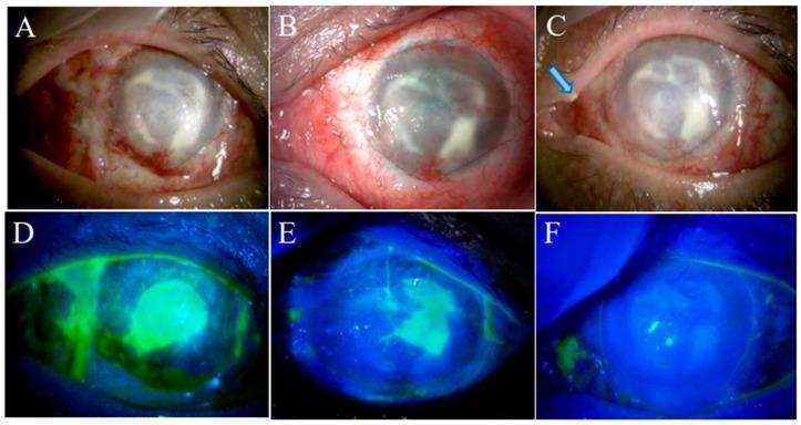
Six-panel clinical image series demonstrating the management of a persistent corneal epithelial defect in a patient following Cultivated Oral Mucosal Epithelial Transplantation (COMET). Panels A–C show slit-lamp clinical photographs, while D–F show corresponding cobalt blue light images with fluorescein staining. (A, D) Two weeks post-COMET, a large central corneal epithelial defect is present, evidenced by intense green fluorescein pooling (D) and surrounding conjunctival injection. (B, E) At one month, the defect remains persistent despite standard care. (C, F) Two weeks after the insertion of a lacrimal punctal plug (indicated by the blue arrow in C), the fluorescein staining is almost entirely absent (F), indicating successful epithelialization and resolution of the defect. The series illustrates the clinical utility of punctal plugs in managing persistent epithelial defects by increasing the residence time of natural tears or therapeutic drops on the ocular surface, particularly in cases of severe limbal stem cell deficiency (LSCD).
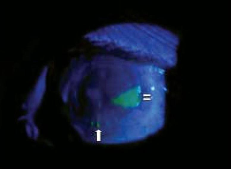
A clinical diagnostic image showing a slit-lamp examination of the human eye under cobalt blue light illumination with fluorescein staining. The image captures the anterior segment post-corneal or limbal grafting. A significant area of bright green fluorescein uptake is visible at the 3 o’clock position (indicated by double white arrows), representing a residual corneal epithelial defect during the healing process. At the 6 o’clock position (indicated by a single white arrow), two loose surgical sutures are visible on the ocular surface, also showing minor fluorescein pooling. The surrounding corneal surface shows approximately 90% re-epithelialization. This visual aid is intended for ophthalmology education to demonstrate postoperative monitoring, the assessment of corneal epithelial healing, and the identification of suture-related complications such as loose sutures.
Ophthalmic Dyes: MS Ophthalmology Theory Notes
How to write the answer
1. Definition
- Ocular-surface assessment
- Lacrimal drainage testing
- Retinal and choroidal angiography
- Cataract surgery
- Corneal transplantation
- Vitreoretinal surgery
2. Classification of Ophthalmic Dyes
| Group | Dyes | Main use |
|---|---|---|
| Ocular-surface vital dyes | Fluorescein, rose bengal, lissamine green | Corneal/conjunctival staining, dry eye assessment |
| Angiographic dyes | Sodium fluorescein, indocyanine green | FFA and ICGA |
| Anterior-segment surgical dyes | Trypan blue, fluorescein | Anterior capsule, corneal wound/Descemet membrane visualization |
| Vitreoretinal vital dyes | Indocyanine green, brilliant blue G, trypan blue, triamcinolone | ILM, ERM, posterior hyaloid and vitreous visualization |
| Miscellaneous diagnostic dyes | Methylene blue, gentian violet | Selected surgical marking applications, not routine intraocular use |
3. Basic Concept: Vital Staining
- Fluorescein mainly demonstrates epithelial defects and spaces between damaged epithelial cells.
- Rose bengal and lissamine green stain devitalized or damaged epithelial cells and mucus.
- Surgical dyes improve contrast between transparent tissue layers.
4. Fluorescein
Properties
- Water-soluble, orange dye
- Appears bright green under cobalt-blue illumination
- Excitation peak approximately 490 nm
- Emits yellow-green fluorescence around 530 nm
- Available as impregnated paper strips, topical solution, and intravenous sodium fluorescein for angiography
Mechanism of corneal staining
- Loss of epithelium
- Disruption of epithelial tight junctions
- Intercellular spaces
- Pools of tear fluid over an epithelial defect
Uses
A. Ocular-surface examination
- Corneal abrasion
- Corneal ulcer and epithelial defect
- Superficial punctate keratitis
- Dry eye disease
- Exposure keratopathy
- Neurotrophic keratopathy
- Contact-lens-related epithelial damage
- Recurrent corneal erosion
- Herpetic epithelial keratitis
B. Tear-film assessment
Fluorescein tear break-up time, TBUT
- Instill a small amount of fluorescein.
- Ask patient to blink naturally.
- Observe under cobalt-blue light.
- Time from last blink to the first dark break in fluorescent tear film is recorded.
C. Seidel test
- Corneal perforation
- Traumatic wound
- Postoperative cataract wound
- Filtering bleb leak
D. Contact-lens fitting
- Assessment of rigid gas-permeable lens fit
- Detection of corneal bearing, pooling and edge lift
- Identifying contact-lens-related epithelial injury
E. Fluorescein disappearance test, FDT
F. Fundus fluorescein angiography
Fluorescein staining patterns: viva table
| Pattern | Likely implication |
|---|---|
| Diffuse interpalpebral punctate staining | Dry eye, exposure, toxicity |
| Inferior corneal staining | Exposure, lagophthalmos, meibomian-gland dysfunction |
| Superior staining | Contact lens, superior limbic keratoconjunctivitis, foreign body under upper lid |
| Dendritic ulcer with terminal bulbs | HSV epithelial keratitis |
| Geographic epithelial ulcer | HSV, toxic keratopathy, severe epithelial disease |
| Pooling | Epithelial depression or defect |
| Negative staining | Elevated lesion, for example epithelial basement membrane abnormality, where dye surrounds but does not stain lesion |
| Seidel-positive stream | Wound leak or corneal perforation |
Technique
- Use a sterile fluorescein strip moistened with non-bacteriostatic saline.
- Touch the strip to inferior palpebral conjunctiva, not directly to cornea.
- Ask patient to blink.
- Examine with cobalt-blue filter and preferably yellow barrier filter.
Precautions
- Remove contact lenses first. Fluorescein may permanently stain soft contact lenses.
- Do not directly scrape the cornea with a dye strip, because this may cause artifactual linear staining.
- In suspected open globe injury, do not apply pressure to the eye.
Adverse effects
- Transient stinging
- Yellow discoloration of soft contact lens
- Rare hypersensitivity
5. Rose Bengal
Properties
- Red dye
- Usually supplied as impregnated strips
- Stains devitalized epithelial cells, mucus and cells inadequately protected by mucin
- Best seen under white light or red-free illumination
Mechanism
- Damaged or devitalized epithelial cells
- Mucin-deficient epithelial surfaces
- Areas not adequately protected by tear-film mucin
Uses
- Dry-eye disease, especially aqueous-deficient dry eye
- Sjögren syndrome
- Keratoconjunctivitis sicca
- Ocular cicatricial pemphigoid
- Stevens-Johnson syndrome
- Exposure keratopathy
- Superior limbic keratoconjunctivitis
- Assessment of conjunctival involvement in ocular-surface disease
Advantages
- Sensitive in identifying abnormal conjunctival epithelium
- Useful in ocular-surface staining scores
- Can reveal more extensive conjunctival involvement than fluorescein
Disadvantages
- Causes considerable burning, irritation and tearing
- Can itself be toxic to epithelium at higher concentration or prolonged contact
- Less comfortable for patients
- Routine use has declined in favor of lissamine green
6. Lissamine Green
Properties
- Green vital dye
- Stains devitalized or damaged epithelial cells and mucus
- Similar ocular-surface staining pattern to rose bengal
- Much better tolerated by patients
Indications
- Dry eye evaluation
- Sjögren syndrome
- Conjunctival staining assessment
- Ocular cicatricial disorders
- Exposure disease
- Ocular-surface disease scoring
Advantages
- Minimal ocular irritation
- Better patient comfort than rose bengal
- Especially useful for conjunctival staining
- Useful in dry-eye clinical trials and Sjögren evaluation
Limitation
7. Fluorescein vs Rose Bengal vs Lissamine Green
| Feature | Fluorescein | Rose Bengal | Lissamine Green |
|---|---|---|---|
| Main target | Epithelial defect/intercellular disruption | Damaged/devitalized cells and mucus | Damaged/devitalized cells and mucus |
| Best illumination | Cobalt-blue light | White or red-free light | White light |
| Main clinical use | Corneal epithelial defects, TBUT, Seidel test | Dry eye and conjunctival surface disease | Dry eye and conjunctival surface disease |
| Corneal staining | Excellent | Can stain cornea | Less useful for epithelial defects |
| Conjunctival staining | Less sensitive | Good | Good |
| Patient discomfort | Minimal | Significant | Minimal |
| Toxicity | Low | Higher | Lower |
| Routine modern preference | Very common | Declining use | Often preferred over rose bengal |
One-line viva answer
8. Sodium Fluorescein for Fundus Fluorescein Angiography, FFA
Definition
Important properties
- Water soluble
- About 70%-80% protein bound in circulation
- Fluoresces under blue excitation light
- Excreted through kidneys, causing yellow-green urine for about 24-36 hours
Phases
- Choroidal flush
- Arterial phase
- Arteriovenous/capillary phase
- Early venous phase
- Late venous phase
- Recirculation phase
Hyperfluorescence patterns
| Pattern | Meaning |
|---|---|
| Window defect | RPE atrophy permits increased choroidal fluorescence |
| Leakage | Increasing area and intensity with blurred margins |
| Pooling | Dye accumulates in anatomical spaces, such as subretinal fluid or PED |
| Staining | Late dye retention in scar, drusen, optic disc or vessel wall |
Hypofluorescence patterns
| Pattern | Meaning |
|---|---|
| Blocked fluorescence | Blood, pigment or exudate blocks background fluorescence |
| Filling defect | Nonperfusion or absent vascular filling |
Indications
- Diabetic retinopathy
- Retinal vein occlusion
- Retinal vasculitis
- Cystoid macular edema
- Neovascular AMD
- Central serous chorioretinopathy
- Macular ischemia
- Choroiditis
- Retinal neovascularization
Adverse effects
- Nausea/vomiting
- Yellow skin discoloration
- Yellow urine
- Extravasation injury
- Urticaria
- Bronchospasm
- Anaphylaxis, rare but serious
9. Indocyanine Green, ICG
Properties
- Water-soluble tricarbocyanine dye
- Binds strongly to plasma proteins
- Absorbs and emits in the near-infrared spectrum
- Near-infrared light penetrates pigment, fluid and blood better than visible light
Main role: Indocyanine Green Angiography, ICGA
Best clinical applications
- Polypoidal choroidal vasculopathy, PCV
- Type 1 macular neovascularization
- Occult choroidal neovascularization
- Central serous chorioretinopathy
- Choroidal inflammatory disorders
- Choroidal hemangioma
- Evaluation of choroidal circulation
FFA versus ICGA
| Feature | FFA | ICGA |
|---|---|---|
| Dye | Sodium fluorescein | Indocyanine green |
| Light | Visible blue-green spectrum | Near infrared |
| Best circulation viewed | Retina | Choroid |
| Penetration through blood/pigment | Limited | Better |
| Key uses | Leakage, DR, RVO, CME | PCV, type 1 MNV, choroidal disease |
ICG in vitreoretinal surgery
- Macular hole
- Epiretinal membrane surgery
- Myopic traction maculopathy
- Selected diabetic macular edema surgery
Disadvantages and safety concerns
- Potential retinal pigment epithelium and retinal toxicity
- Risk increases with high concentration, prolonged exposure, direct macular contact, intense endoillumination and hypo-osmolar preparations
- Avoid unnecessary prolonged macular exposure
- Use minimal effective concentration and promptly remove dye
10. Trypan Blue
Properties
- Blue vital dye
- Stains collagen-rich or nonviable tissue and transparent capsules/membranes
- Common anterior-segment concentration: approximately 0.06% to 0.15%
- Generally used intraoperatively
Main uses
A. Cataract surgery
- White mature cataract
- Intumescent cataract
- Hypermature cataract
- Poor red reflex
- Corneal opacity
- Dense posterior subcapsular cataract
- Pediatric cataract
- Vitrectomized eye
- Intraoperative miosis with poor capsule visibility
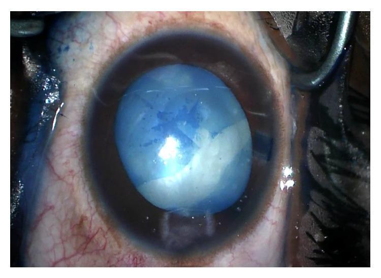
B. Corneal surgery
- Staining donor Descemet membrane in DMEK
- Visualization of Descemet membrane during endothelial keratoplasty
- Identification of retained Descemet membrane in selected procedures
C. Vitreoretinal surgery
- Epiretinal membrane staining
- Proliferative vitreoretinopathy membranes
- Occasionally used with other dyes in chromovitrectomy
Technique in cataract surgery
- Create side-port incision.
- Fill anterior chamber with air or OVD, depending on technique.
- Inject a small quantity of trypan blue onto anterior capsule.
- Allow brief contact.
- Irrigate/aspirate excess dye.
- Fill chamber with OVD.
- Perform CCC.
“Under-air” staining
Complications
- Endothelial toxicity with prolonged exposure or high concentration
- Inflammation, uncommon
- Inadvertent staining of intraocular tissues
- Theoretical retinal toxicity if posterior capsule is absent and dye reaches posterior segment
11. Brilliant Blue G, BBG
Properties
- Blue dye, often known as brilliant blue G or acid blue
- Preferentially stains the internal limiting membrane
- Better ILM selectivity and generally lower retinal toxicity concern than ICG
Uses
- ILM peeling in macular hole surgery
- ILM peeling in epiretinal membrane surgery
- Myopic foveoschisis and selected traction maculopathies
- Chromovitrectomy
Advantages
- Good ILM contrast
- Less affinity for retina/RPE than ICG
- Widely preferred for ILM staining
Limitations
- Less effective for epiretinal membrane alone than trypan blue in some circumstances
- Requires careful use near fovea
- Light exposure, concentration and exposure duration still matter
Important safety warning
12. Triamcinolone Acetonide
Is it truly a dye?
Uses
- Identifying posterior hyaloid during pars plana vitrectomy
- Detecting residual cortical vitreous
- Assisting membrane dissection
- Visualizing vitreous prolapse in anterior chamber during complicated cataract surgery
Advantages
- Excellent visualization of vitreous
- Allows more complete vitreous removal
- Familiar and inexpensive in many settings
Risks
- Steroid-induced IOP rise
- Inflammation
- Endophthalmitis risk from contamination if preparation is not preservative-free
- Retinal toxicity concerns from vehicle/preservatives
13. Other Surgical Stains
| Dye / agent | Principal use | Important note |
|---|---|---|
| ICG | ILM staining, ICGA | Potential macular/RPE toxicity |
| Brilliant blue G | ILM staining | Often preferred to ICG |
| Trypan blue | Anterior capsule, ERM, Descemet membrane | Essential in white cataract |
| Triamcinolone | Vitreous visualization | Not a true dye |
| Fluorescein | Seidel testing, corneal defects, DSAEK/DMEK-related visualization in selected contexts | Does not stain intact epithelium |
| Methylene blue | Surgical marking, rarely ocular surface | Not for routine intraocular use because of toxicity concerns |
| Gentian violet | Marking in some external/oculoplastic procedures | Avoid intraocular exposure |
| Infracyanine green | ILM staining | Iodine-free alternative to ICG in selected settings |
14. Dyes in Corneal Surgery
Fluorescein
- Detection of epithelial defects
- Seidel test
- Contact-lens fitting
- Tear-film evaluation
Trypan blue
- Donor Descemet membrane staining during DMEK
- Assists visualization of Descemet membrane
Rose bengal and lissamine green
- Evaluation of dry eye
- Ocular surface disease
- Conjunctival and mucin-deficient epithelial staining
ICG and BBG
15. Dyes in Cataract Surgery
Most important: Trypan blue
Indications
- White cataract
- Mature cataract
- Poor red reflex
- Corneal opacity
- Dense cataract
- Pediatric cataract
- Pseudoexfoliation with poor visualization
- Difficult capsulorhexis
Advantage
Related viva point
16. Dyes in Vitreoretinal Surgery: Chromovitrectomy
Definition
Targets and preferred stains
| Structure | Preferred stain / aid |
|---|---|
| Posterior hyaloid / cortical vitreous | Preservative-free triamcinolone |
| Internal limiting membrane | Brilliant blue G |
| Epiretinal membrane | Trypan blue |
| ILM and ERM combined visualization | Dual dyes or sequential staining protocols |
| Macular lesion/choroidal circulation | ICG angiography, not routine surgical stain alone |
Advantages
- Better visualization
- More complete membrane removal
- Reduced accidental retinal trauma
- Improved surgical precision
Risks
- Retinal toxicity
- RPE toxicity
- Phototoxicity from dye plus endoillumination
- Concentration-related damage
- Osmolarity and solvent-related toxicity
17. Dyes in Dry-Eye Assessment
Common ocular-surface staining systems
Oxford grading scale
National Eye Institute / NEI grading
Sjögren syndrome
- Corneal fluorescein staining
- Conjunctival lissamine green staining
18. High-Yield Comparisons
Trypan blue vs Brilliant Blue G
| Feature | Trypan blue | Brilliant blue G |
|---|---|---|
| Main target | Anterior capsule, ERM | ILM |
| Cataract surgery | Very useful | No routine role |
| Vitreoretinal use | ERM staining | ILM staining |
| Main surgical role | Capsulorhexis in white cataract | Macular-hole and ILM-peel surgery |
| Safety concern | High concentration/prolonged exposure | Phototoxicity risk with prolonged exposure and intense illumination |
ICG vs Brilliant Blue G
| Feature | ICG | Brilliant blue G |
|---|---|---|
| Main target | ILM | ILM |
| Contrast | Strong | Good |
| Safety | More concern for RPE/retinal toxicity | Usually regarded as safer |
| Current preference | Selected cases | Common choice for ILM staining |
Rose Bengal vs Lissamine Green
| Feature | Rose bengal | Lissamine green |
|---|---|---|
| Staining profile | Similar | Similar |
| Patient comfort | More irritating | Much better tolerated |
| Toxicity concern | Greater | Lower |
| Routine preference | Less common | More common |
FFA vs ICGA
| Feature | FFA | ICGA |
|---|---|---|
| Dye | Sodium fluorescein | Indocyanine green |
| Best examined circulation | Retinal | Choroidal |
| Leakage information | Excellent | Useful but choroid-focused |
| Penetration through blood/pigment | Poorer | Better |
| Best-known role | DR, RVO, macular leakage | PCV and occult/type 1 MNV |
19. Model Theory Answer: “Write a Short Note on Ophthalmic Dyes”
Teach me tissue adhesives and ophthalmic glues
cyanoacrylate fibrin glue corneal perforation
fibrin glue pterygium surgery
ophthalmic tissue adhesives cyanoacrylate fibrin glue corneal perforation pterygium review
cyanoacrylate tissue adhesive corneal perforation bandage contact lens image
-
Core concepts and classification
- Cyanoacrylate adhesives
- Fibrin sealants
- Other biologic and synthetic hydrogel adhesives
-
Corneal use
- Impending perforation, microperforation, descemetocele, and corneal melt
- Selection by defect size, location, cause, and tissue loss
- Application technique, bandage contact lens, and follow-up
-
Ocular-surface and anterior-segment surgery
- Pterygium conjunctival-autograft fixation
- Amniotic-membrane transplantation
- Conjunctival closure and lamellar corneal graft fixation
-
Comparison and safety
- Cyanoacrylate versus fibrin glue
- Strength, biodegradability, inflammation, infection considerations, cost
- Complications and indications for escalation to patch graft or keratoplasty
-
Evidence, recent materials, and examination preparation
- Current evidence and limits of available data
- PEG, collagen, gelatin-based, and other newer adhesives
- Model short note and viva questions
Which part of tissue adhesives should I teach in full detail next?
- Cataract and IOLs: FLACS, capsular tension ring (CTR), phakic IOLs, modern IOLs, accommodative IOLs
- Retina and vitreous: BRVO management, ARMD, OCT, ERG, ROP, retinal imaging, FFA, pars plana vitrectomy, Pascal laser, silicone oil
- Cornea and ocular surface: stem-cell therapy, ocular surface squamous neoplasia, amniotic membrane, collagen cross-linking/C3R, keratoprosthesis, lamellar keratoplasty, pterygium surgery, toxic contact lens reactions
- Glaucoma: neuroprotection, glaucoma implants, OCT in glaucoma
- Anterior segment / iris / diagnostic imaging: pupilloplasty, UBM
- General ophthalmic technology and materials: OVDs, lasers, dyes, tissue adhesives/glues
- Cataract and IOLs: FLACS, CTR, phakic IOLs, modern/accommodative IOLs
- Retina and vitreous: BRVO, ARMD, OCT, ERG, ROP, FFA, retinal imaging, vitrectomy, lasers, silicone oil
- Cornea and ocular surface: stem cells, amniotic membrane, OSSN, C3R, keratoplasty, KPro, pterygium, contact-lens toxicity
- Glaucoma: neuroprotection, drainage implants, OCT
- Imaging, lasers and materials: UBM, dyes, OVDs, tissue glues, lasers, pupilloplasty
- Femtosecond laser-assisted cataract surgery (FLACS)
- Capsular tension ring (CTR)
- Phakic intraocular lenses
- Modern pseudophakic IOLs, including toric, multifocal, EDOF and accommodating IOLs
- IOL selection, biometry and high-yield complications
1. Foundation: IOL basics
Definition
Parts
- Optic: central refracting portion
- Haptics: peripheral supporting elements
- Overall diameter: optic + haptic-to-haptic length
- Optic diameter: commonly 6 mm for standard adult posterior chamber IOLs
Ideal IOL properties
- Biocompatible and inert
- Stable fixation and centration
- Optically clear, scratch-resistant
- Minimal inflammation, posterior capsule opacification (PCO), dysphotopsia and glistenings
- Injectable through a small incision
- Predictable effective lens position (ELP)
Classification
| Basis | Categories |
|---|---|
| Site | Posterior chamber IOL (PCIOL), anterior chamber IOL (ACIOL), iris-fixated/iris-claw IOL, scleral-fixated IOL |
| Material | PMMA, silicone, hydrophobic acrylic, hydrophilic acrylic |
| Optic | Spherical, aspheric, monofocal, multifocal, extended-depth-of-focus (EDOF), toric, accommodating |
| Construction | One-piece or three-piece; rigid or foldable |
| Fixation | In-the-bag, sulcus, iris-claw, scleral fixation |
Materials: exam comparison
| Material | Advantages | Limitations |
|---|---|---|
| PMMA | Excellent optics, stable, inexpensive | Rigid, needs large incision |
| Silicone | Foldable, small incision | Silicone-oil adherence, therefore generally avoid if future retinal surgery with silicone oil is likely |
| Hydrophobic acrylic | Commonest modern material, foldable, low PCO with square edge, good capsular adhesion | Glistenings or surface light scatter may occur in some models |
| Hydrophilic acrylic | Flexible, good injector delivery | Greater calcification risk in some settings, including exposure to intraocular gas/air in susceptible lenses |
Design features that prevent PCO
- Sharp square posterior optic edge produces a capsular bend and contact inhibition of lens epithelial cell migration.
- In-the-bag placement and meticulous cortical clean-up further reduce PCO.
- PCO, if visually significant, is managed by Nd:YAG posterior capsulotomy.
2. FLACS: Femtosecond Laser-Assisted Cataract Surgery
Definition
- Corneal incisions
- Anterior capsulotomy
- Lens fragmentation/softening
- Arcuate or limbal-relaxing incisions for astigmatism
Principle
Essential components
- Docking interface: applanation or liquid-filled interface
- Image-guidance system: commonly OCT-based
- Laser delivery platform
- Patient interface and suction mechanism
- Integrated or adjacent phacoemulsification system
Steps of FLACS
- Pharmacological dilation and sterile preparation.
- Docking of the laser interface to the eye.
- Imaging and treatment planning.
- Laser creation of:
- Primary and side-port corneal incisions
- Precisely centered capsulotomy
- Lens fragmentation pattern
- Arcuate keratotomy if planned
- Transfer to operating microscope.
- Opening of laser capsulotomy and removal of free capsule disc.
- Hydrodissection, phacoaspiration and cortical clean-up.
- IOL implantation in the capsular bag.
Benefits
- Highly reproducible capsulotomy size, circularity and centration
- Precise corneal incisions
- Lens prefragmentation may reduce effective phaco time and cumulative dissipated energy
- Astigmatic arcuate incisions can be planned precisely
- Useful in selected challenging cases:
- Shallow anterior chamber
- Dense cataract
- Low endothelial reserve
- White/intumescent cataract, with careful case selection
- Premium IOL procedures where centration is especially important
Limitations
- High capital, consumable and maintenance costs
- Extra docking and workflow time
- Requires pupil dilation and adequate corneal clarity
- Not always feasible in deep-set eyes, marked kyphosis, severe tremor, poor cooperation, inability to lie flat, or significant conjunctival scarring
- Incomplete capsulotomy, capsular tags and incomplete fragmentation can occur
- Does not replace surgical judgement or conventional phaco skill
Complications
During docking
- Subconjunctival hemorrhage
- Corneal folds and imaging artefacts
- Transient rise in IOP from suction
- Loss of suction and incomplete treatment
During surgery
- Incomplete corneal incision
- Capsular tags, microadhesions or incomplete capsulotomy
- Anterior capsular tear if the capsule is pulled before identifying and releasing tags
- Incomplete nuclear fragmentation
- Miosis due to prostaglandin release
- Rare capsular block syndrome if hydrodissection is forceful in a gas-fragmented lens
FLACS versus conventional phaco: what to write as “recent evidence”
3. Capsular Tension Ring (CTR)
Definition
Design
- Usually made of PMMA
- Open ring with eyelets at both ends
- Inserted into the capsular bag after capsulorhexis and preferably after adequate hydrodissection
- Available in different diameters
Principle
Indications
Zonular weakness or dialysis
- Pseudoexfoliation syndrome
- Traumatic zonular dialysis
- High myopia
- Marfan syndrome and other ectopia lentis states
- Previous vitreoretinal surgery
- Mature/hypermature cataract with weak zonules
- Lens subluxation of limited extent
- Prior acute angle-closure attack with zonulopathy, in selected cases
Prevention of capsular contraction and IOL decentration
- Pseudoexfoliation
- Retinitis pigmentosa
- High myopia
- Uveitis, selectively
- Conditions predisposed to capsular phimosis
Types
| Device | Main use |
|---|---|
| Standard CTR | Mild-to-moderate diffuse zonular weakness or limited zonular dialysis |
| Modified CTR, Cionni ring | Significant or progressive zonular loss; has one or two fixation eyelets for scleral suturing |
| Capsular tension segment (CTS) | Localized zonular weakness; can be scleral fixated, often used with CTR in extensive dialysis |
| Ahmed capsular tension segment | Segmental capsular support and scleral fixation |
Surgical technique
- Create a well-centered continuous curvilinear capsulorhexis.
- Use dispersive OVD to maintain the bag and protect endothelium.
- Perform gentle hydrodissection, avoiding extension of zonular damage.
- Place capsular hooks or iris retractors, if severe focal dialysis.
- Insert the CTR slowly into the capsular bag using an injector or forceps.
- Ensure the leading eyelet does not engage or tear the capsulorhexis margin.
- Complete phacoemulsification with reduced stress on the weak zonular area.
- Implant IOL in the bag if support is adequate.
- In major dialysis, use a Cionni-modified device or CTS with scleral fixation.
Timing of insertion: common viva point
- Early insertion: improves early bag stability but may trap cortex behind the ring and make cortical aspiration difficult.
- Late insertion: allows easier cortical clean-up but may allow further zonular stress during phaco.
Advantages
- Stabilizes capsular bag during surgery
- Reduces capsular folds and equatorial bag collapse
- Improves IOL centration
- May improve rotational stability of toric IOLs
- Reduces risk of late decentration in selected cases, although it cannot eliminate progressive zonulopathy
Limitations and complications
- Cortex may become trapped between ring and capsule
- Capsular tear may extend if the ring is inserted forcefully
- Can worsen a pre-existing zonular dialysis if incorrectly inserted
- Standard CTR is inadequate when zonular loss is extensive or progressive
- Late in-the-bag IOL-CTR complex subluxation can still occur, especially in pseudoexfoliation
- A CTR should not be used as a substitute for scleral fixation when support is grossly insufficient
Recent evidence
4. Phakic Intraocular Lenses (pIOLs)
Definition
Place in refractive surgery
- Refractive error is too high for safe corneal laser ablation
- Cornea is thin or topographically unsuitable for laser refractive surgery
- Accommodation should be preserved
- The patient is a young adult with stable refraction
Classification
| Type | Position | Examples / features |
|---|---|---|
| Angle-supported anterior-chamber pIOL | Anterior chamber angle | Largely historical due to endothelial and angle complications |
| Iris-fixated pIOL | Clipped to mid-peripheral iris | Artisan/Verisyse-type designs, anterior or retropupillary fixation |
| Posterior-chamber pIOL | Between posterior iris and anterior crystalline lens, supported in ciliary sulcus | ICL and related implantable phakic contact lens designs |
Posterior chamber phakic IOL / ICL
Central-port ICL
Indications
- Stable refraction, generally for at least one year
- Moderate-to-high myopia, with or without astigmatism
- Hyperopia in selected cases
- Age typically 21 years or older, according to device approvals and local practice
- Adequate anterior chamber depth
- Adequate endothelial cell density
- Healthy cornea, crystalline lens, retina and optic nerve
- Strong desire to preserve accommodation
Contraindications
- Progressive refraction or unstable keratoconus
- Shallow anterior chamber
- Low endothelial cell count
- Narrow/occludable angle
- Cataract or significant lens opacity
- Active uveitis
- Uncontrolled glaucoma
- Corneal endothelial disease
- Significant retinal pathology requiring treatment first
- Unrealistic expectations or inability to comply with follow-up
Preoperative work-up
- Manifest and cycloplegic refraction
- Corneal tomography and pachymetry
- Anterior chamber depth, measured from endothelium to anterior lens surface
- White-to-white and/or sulcus-to-sulcus measurement
- Endothelial cell count
- Gonioscopy
- Dilated retinal examination, especially in high myopia
- IOP, optic-nerve assessment and macular OCT if indicated
- Lens-vault prediction and sizing assessment using ultrasound biomicroscopy, anterior-segment OCT or device-specific nomograms
Vault
- Low vault: risk of anterior subcapsular cataract
- Excessive vault: angle crowding, pigment dispersion and secondary angle closure
- Ideal target varies by device and imaging method, but an adequate central vault with open angles is the goal.
Complications
| Complication | Mechanism / prevention |
|---|---|
| Cataract, especially anterior subcapsular | Low vault, lens touch, older designs; careful sizing and follow-up |
| Pupillary block | More relevant to older non-central-port designs; prevented by PI or central-port design |
| Raised IOP | Retained OVD, steroid response, pigment dispersion, angle crowding, pupillary block |
| Endothelial cell loss | More important with anterior chamber pIOLs; monitor ECD |
| Uveitis | Surgical trauma, pigment dispersion, malposition |
| Pupillary ovalization | Mainly iris-claw lenses |
| Toric pIOL rotation | Causes residual astigmatism; may require repositioning |
| Retinal tear/detachment | Related partly to high-myopia phenotype; do meticulous peripheral retinal evaluation |
| Glare, halos, dysphotopsia | Optical effects, residual refractive error |
Recent advances
- Central-port posterior chamber ICLs
- Toric pIOLs
- Improved vault prediction using AS-OCT and UBM
- Larger optic zones and customized sizing
- Diffractive phakic lenses for carefully selected presbyopic patients, but evidence remains limited
5. Pseudophakic IOLs: Modern IOL Options
A. Monofocal IOL
Advantages
- Best contrast sensitivity
- Lowest rate of halos and glare
- Broadest suitability in eyes with retinal disease, glaucoma, corneal irregularity or uncertain visual potential
- Predictable and economical
Disadvantage
- Near spectacles are usually needed.
Aspheric monofocal IOL
B. Toric IOL
Principle
Indications
- Regular corneal astigmatism
- Cataract patient seeking reduced spectacle dependence
- Adequate capsular support
- Reliable keratometry and stable ocular surface
Contraindications / caution
- Irregular astigmatism, unstable keratoconus, corneal scar or severe dry eye
- Poor capsular support
- Unreliable biometry
- Marked zonulopathy unless support is secured
Key points
- Accurate biometry and posterior corneal astigmatism consideration are essential.
- Marking can be manual, image-guided or digitally guided.
- Rotation matters: each degree of toric IOL rotation causes roughly 3.3% loss of cylindrical correction. At 30 degrees, the intended astigmatic correction is effectively lost.
Complications
- Misalignment/rotation
- Residual refractive astigmatism
- IOL tilt or decentration
- Need for early surgical repositioning if substantial rotation occurs
C. Multifocal IOL
Principle
- Diffractive optics
- Refractive zones
- Hybrid designs
Advantages
- Greater spectacle independence at distance and near
Limitations
- Halos and glare
- Reduced contrast sensitivity, especially in dim illumination
- Neuroadaptation required
- Residual refractive error, dry eye, decentration or PCO can cause major dissatisfaction
Avoid or use cautiously in
- Significant ocular surface disease
- Corneal irregularity
- Macular disease, diabetic maculopathy, epiretinal membrane
- Advanced glaucoma or impaired contrast sensitivity
- Optic neuropathy
- Unrealistic patient expectations
- Occupations requiring excellent night contrast, depending on individual needs
D. Trifocal IOL
Advantages
- Stronger near and intermediate spectacle independence than many bifocal designs
Disadvantages
- Dysphotopsia and contrast trade-off remain
- Needs precise centration and refractive targeting
- Not ideal for eyes with retinal or optic-nerve disease
E. Extended Depth-of-Focus (EDOF) IOL
Principle
Advantages
- Often fewer halos than traditional multifocal lenses
- Good distance and intermediate vision
- Useful for computer users
Limitations
- Fine near tasks may still require spectacles
- Dysphotopsia and contrast effects can still occur
- Terminology and optical mechanisms vary between platforms
Important viva distinction
- Multifocal IOL: several discrete foci.
- EDOF IOL: elongated focus, usually better intermediate performance and often less severe dysphotopsia.
- Monofocal-plus/enhanced monofocal: improved intermediate range but not a true EDOF by all definitions.
F. Accommodating IOL
Definition
Mechanisms
- Single-optic movement: anterior movement of the optic during attempted accommodation.
- Dual-optic systems: relative movement of two optics changes overall power.
- Fluid-based or shape-changing designs: aim to alter curvature or refractive power.
Theoretical advantage
Limitations
- True, sustained objective accommodation has been difficult to demonstrate consistently.
- Capsular fibrosis and haptic restriction may reduce movement over time.
- Near outcomes may be partly due to depth of focus, pupil miosis or residual myopia rather than true accommodation.
- Current use is less widespread than monofocal, toric, multifocal, trifocal and EDOF designs.
Exam conclusion
6. IOL Power Calculation and Modern Biometry
Essential formula concept
- Axial length
- Corneal power
- Anterior chamber depth / lens position predictors
- Lens thickness
- White-to-white in some formulas
- Desired postoperative refraction
- Effective lens position (ELP)
Common formula evolution
| Generation | Examples | Main idea |
|---|---|---|
| First | SRK I | Regression-based |
| Second | SRK II | Axial-length correction |
| Third | SRK/T, Holladay 1, Hoffer Q | Uses predicted ELP |
| Fourth | Haigis, Holladay 2 | Multiple biometric variables |
| Modern theoretical/AI-assisted | Barrett Universal II, Kane, EVO, Hill-RBF, Olsen | More variables, ray tracing, large datasets or AI elements |
High-yield choices
- Short eyes: modern formulas such as Barrett Universal II, Kane, Hoffer QST and Holladay 2 may be useful.
- Long/high-myopic eyes: use modern formulas and consider axial-length adjustment where appropriate.
- Post-refractive surgery eyes: use no-history methods, tomography-derived corneal data and dedicated calculators where available.
- Toric IOLs: include posterior corneal astigmatism and surgically induced astigmatism.
Causes of refractive surprise
- Keratometry error, especially dry eye
- Incorrect axial length, especially posterior staphyloma
- Incorrect IOL constant
- Wrong IOL selection or implantation
- Unpredicted ELP
- Corneal edema or irregular astigmatism
- Prior refractive surgery
- IOL tilt, decentration or rotation
7. How to Select an IOL: Clinical Algorithm
Step 1: Determine visual potential
- Cornea and ocular surface
- Macula: OCT if indicated
- Optic nerve and glaucoma status
- Diabetic retinopathy
- Amblyopia and previous retinal surgery
Step 2: Determine refractive objective
- Distance target with reading glasses
- Monovision
- Distance + intermediate
- Maximum spectacle independence
Step 3: Assess corneal astigmatism
- Regular and significant: consider toric IOL
- Irregular: treat ocular surface/corneal condition first; avoid routine toric or multifocal decisions until measurements are reliable
Step 4: Assess suitability for presbyopia correction
Step 5: Assess capsular support
- Good support: in-the-bag PCIOL
- Mild zonulopathy: CTR + PCIOL
- Major zonulopathy: modified CTR/CTS with scleral fixation, or consider secondary fixation strategy
- No capsular support: scleral-fixated, iris-fixated or selected ACIOL depending on anatomy and surgeon expertise
8. Common Long-Answer Questions and Model Opening Lines
“Write a short note on FLACS.”
“Describe capsular tension ring.”
“Discuss phakic IOL.”
“Write a note on accommodating IOL.”
9. Rapid Revision Pearls
- FLACS: more precise capsulotomy and incisions, but no proven overall long-term superiority or cost-effectiveness over standard phaco.
- CTR: supports the bag, not the weak zonules themselves. Use a Cionni ring or CTS with scleral fixation if zonular loss is major.
- Standard CTR: can trap cortex. Do not insert forcefully.
- Phakic IOL: preserves accommodation; assess ACD, ECD, angle, retina and vault.
- Low pIOL vault: cataract risk.
- High pIOL vault: angle crowding/pigment/IOP risk.
- Toric IOL: 1 degree of rotation loses about 3.3% cylinder effect.
- Multifocal/trifocal IOL: better spectacle independence, but halos and lower contrast sensitivity.
- EDOF: distance-to-intermediate range with less near performance than trifocal in many designs.
- Accommodating IOL: variable evidence for sustained true accommodation.
- Hydrophobic acrylic square-edge IOL: standard modern choice for low PCO risk.
- Avoid silicone IOL when silicone-oil tamponade is likely in future retinal surgery.
- Branch retinal vein occlusion (BRVO)
- Age-related macular degeneration (AMD/ARMD)
- OCT and OCT angiography
- Electroretinography (ERG)
- Retinopathy of prematurity (ROP)
- Retinal lasers, including Pascal
- Pars plana vitrectomy (PPV)
- Silicone oil and other endotamponades
- Retinal imaging modalities
- Fundus fluorescein angiography (FFA)
1. Branch Retinal Vein Occlusion (BRVO)
Definition
Pathogenesis
- Venous narrowing
- Turbulent blood flow
- Endothelial injury
- Thrombus formation
- Venous obstruction
- Raised intraluminal venous pressure
- Capillary leakage causing macular edema
- Retinal hemorrhage
- Capillary nonperfusion and ischemia
- Increased VEGF production, causing macular edema and neovascularization
Risk factors
Ocular
- Hypertension-related arteriosclerosis
- Primary open-angle glaucoma
- Raised IOP
- Short axial length, reported in some populations
Systemic
- Hypertension
- Diabetes mellitus
- Dyslipidemia
- Smoking
- Obesity
- Renal disease
- Hyperhomocysteinemia
- Myeloproliferative disorders
- Thrombophilia, especially in young, bilateral, recurrent, or atypical RVO
Classification
By site
- Major BRVO: first-order branch vein occlusion, commonly superotemporal
- Macular BRVO: smaller macular venous branch involved
- Hemispheric retinal vein occlusion: one hemiretina involved, often considered intermediate between BRVO and CRVO
By perfusion
- Perfused BRVO
- Ischemic BRVO: extensive capillary nonperfusion, higher risk of retinal/disc neovascularization
Clinical features
Symptoms
- Painless unilateral decrease in vision
- Metamorphopsia
- Central or paracentral scotoma
- Often incidentally detected if macula is spared
Signs
- Dilated, tortuous vein
- Flame-shaped and dot-blot retinal hemorrhages
- Cotton-wool spots
- Retinal edema
- Macular edema if macular circulation is affected
- Collateral vessels later
- Neovascularization of retina or disc in ischemic disease
- Vitreous hemorrhage as a late complication
Investigations
| Investigation | Role |
|---|---|
| Visual acuity, IOP, slit-lamp and dilated fundus exam | Baseline assessment |
| OCT macula | Detects and follows macular edema, subretinal fluid, DRIL, outer retinal damage |
| OCT-A | Demonstrates superficial/deep plexus nonperfusion and collateral circulation, but does not show leakage |
| FFA | Defines ischemia, macular leakage, macular perfusion, neovascularization and capillary nonperfusion |
| Widefield FFA | Better for peripheral ischemia and targeted laser planning |
| Systemic work-up | Detects cardiovascular/metabolic risk factors |
Management
A. Treat systemic risk factors
- BP control
- Diabetes control
- Lipid management
- Smoking cessation
- Assessment for glaucoma
- Selective hematologic work-up where indicated
B. Macular edema
- Ranibizumab
- Aflibercept
- Bevacizumab, widely used off-label in many settings
- Faricimab, a bispecific antibody targeting VEGF-A and angiopoietin-2
- Initial loading followed by pro re nata regimen
- Treat-and-extend protocol
- Individualized OCT-guided treatment
C. Intravitreal corticosteroid
- In pseudophakia
- When anti-VEGF response is suboptimal
- If injection burden is difficult
- In anti-VEGF contraindication or selected inflammatory phenotypes
- IOP elevation
- Cataract, particularly in phakic eyes
- Need for repeat treatment
D. Laser photocoagulation
- Grid laser for macular edema: historical role, now usually secondary to anti-VEGF therapy.
- Sector scatter laser: indicated for retinal/disc neovascularization associated with nonperfusion, not prophylactically merely because ischemia exists.
Prognosis
- Baseline VA
- Duration/severity of macular edema
- Foveal ischemia
- Integrity of ellipsoid zone and external limiting membrane on OCT
- Disorganization of retinal inner layers, DRIL
- Development of neovascularization/vitreous hemorrhage
Recent advances
BRVO treatment is directed primarily at macular edema and neovascularization, while systemic evaluation reduces future ocular and cardiovascular risk.
2. Age-Related Macular Degeneration (AMD/ARMD)
Definition
Risk factors
Non-modifiable
- Increasing age
- Family history/genetic susceptibility
- White ethnicity
- Complement-pathway gene variants, including CFH and ARMS2/HTRA1 associations
Modifiable
- Smoking, the strongest modifiable risk factor
- Hypertension/cardiovascular risk factors
- Obesity
- Poor diet and low antioxidant intake
- Excess ultraviolet exposure is less clearly established
Classification
1. Early AMD
- Medium drusen
- Mild RPE pigmentary abnormalities
- Usually no visual symptoms
2. Intermediate AMD
- Large drusen, typically at least 125 micrometers
- Numerous medium drusen
- Noncentral geographic atrophy
3. Late AMD
A. Dry AMD
- Drusen and RPE dysfunction
- Geographic atrophy (GA): sharply demarcated RPE and photoreceptor loss
B. Neovascular or wet AMD
- Subretinal fluid
- Intraretinal fluid
- Pigment epithelial detachment
- Hemorrhage
- Fibrosis/disciform scar
Classification of macular neovascularization
| Type | Location | Typical imaging |
|---|---|---|
| Type 1 MNV | Sub-RPE | Irregular fibrovascular PED, sub-RPE flow on OCT-A |
| Type 2 MNV | Subretinal, above RPE | Subretinal hyperreflective material, classic leakage on FFA |
| Type 3 MNV | Intraretinal, formerly retinal angiomatous proliferation | Intraretinal cysts/hyperreflective foci, often with PED |
| Polypoidal choroidal vasculopathy | Aneurysmal type 1 neovascularization | Orange nodules, peaked PED, ICGA polypoidal lesions |
Clinical features
- Painless central visual loss
- Metamorphopsia
- Micropsia
- Difficulty recognizing faces and reading
- Central scotoma
- Reduced contrast and dark adaptation
Investigations
| Investigation | Key use |
|---|---|
| OCT | First-line diagnosis and monitoring of exudation |
| OCT-A | Noninvasive MNV visualization, no leakage information |
| FFA | Leakage pattern, classic/occult MNV, activity in difficult cases |
| ICGA | Particularly useful in PCV and type 1 MNV |
| Fundus autofluorescence | RPE health and geographic atrophy mapping |
| Color/widefield fundus photography | Baseline documentation and serial comparison |
Management
Dry AMD
- Smoking cessation
- Control cardiovascular risk factors
- Amsler monitoring and urgent review for new distortion/scotoma
- Low-vision support where necessary
- AREDS2 supplementation for selected intermediate AMD or advanced AMD in one eye
- Vitamin C
- Vitamin E
- Zinc
- Copper
- Lutein
- Zeaxanthin
Geographic atrophy: major advance
- Pegcetacoplan, a C3 inhibitor
- Avacincaptad pegol, a C5 inhibitor
Neovascular AMD
- Ranibizumab
- Aflibercept
- Bevacizumab, off-label in many regions
- Brolucizumab, used cautiously because of intraocular inflammation/retinal vasculitis concerns
- Faricimab: VEGF-A and Ang-2 inhibition
- Higher-dose aflibercept formulations in some regions for interval extension
- Fixed dosing
- Pro re nata
- Treat-and-extend, widely used in routine practice
Polypoidal choroidal vasculopathy
- Anti-VEGF is foundational.
- Photodynamic therapy may be added in selected cases, especially persistent polypoidal lesions or recurrent hemorrhage.
Recent advances
- OCT-guided treat-and-extend regimens
- Faricimab and high-dose aflibercept for reducing injection burden in selected patients
- Complement inhibition for geographic atrophy
- Home monitoring and AI-assisted fluid detection
- Long-acting delivery systems and gene therapy remain areas of active development rather than routine universal care
Dry AMD is not simply “benign.” It can progress to geographic atrophy or neovascular AMD. New metamorphopsia in a patient with dry AMD is an urgent symptom of possible conversion to MNV.
3. Optical Coherence Tomography (OCT)
Definition
Principle
- A reference beam
- A sample beam reflected from ocular tissues
Types
| Type | Main feature |
|---|---|
| Time-domain OCT | Older, slower, lower resolution |
| Spectral-domain OCT | Faster and higher resolution; common clinical platform |
| Swept-source OCT | Longer wavelength, deeper penetration through pigment, hemorrhage and media opacity; better choroid imaging |
| Enhanced-depth imaging OCT | Better choroidal visualization |
| OCT angiography | Flow-based visualization of retinal and choroidal vasculature without dye |
Retinal OCT applications
- Diabetic macular edema
- BRVO/CRVO macular edema
- Neovascular AMD
- Central serous chorioretinopathy
- Epiretinal membrane
- Vitreomacular traction
- Macular hole
- Myopic maculopathy
- Retinal dystrophies
- Hydroxychloroquine monitoring
- Optic nerve/RNFL and ganglion-cell analysis in glaucoma
Important OCT signs
| OCT finding | Clinical implication |
|---|---|
| Intraretinal cysts | Macular edema |
| Subretinal fluid | MNV, CSC, inflammatory choroidopathy, other causes |
| PED | AMD, PCV, CSC and other RPE disorders |
| Hyperreflective foci | RPE migration/inflammation, risk biomarker in some disorders |
| Subretinal hyperreflective material | Fibrovascular tissue, blood, MNV-associated material |
| Ellipsoid-zone disruption | Photoreceptor injury and poorer visual prognosis |
| DRIL | Associated with poorer VA in macular edema |
| VMT | Partial vitreous separation exerting foveal traction |
| Full-thickness macular hole | Defect from ILM to RPE with elevated margins |
OCT-Angiography
Principle
Advantages
- Noninvasive
- Rapid
- Layer-by-layer vascular segmentation
- Identifies nonexudative MNV
- Useful in diabetic retinopathy, AMD, retinal vein occlusion, macular telangiectasia and inherited retinal disease
Limitations
- Does not show leakage
- Motion artifact
- Projection artifact
- Segmentation errors, especially in edema/PED
- Poor images with media opacity or poor fixation
- Slow-flow lesions may be missed
FFA shows dynamic leakage and perfusion. OCT-A shows flow architecture but no leakage.
4. Fundus Fluorescein Angiography (FFA)
Definition
Principle
- Is a water-soluble orange dye
- Absorbs blue light near 490 nm
- Emits yellow-green fluorescence near 530 nm
- Is largely protein-bound intravascularly
- Is excreted by the kidneys
Phases of FFA
- Choroidal flush: patchy background choroidal fluorescence
- Arterial phase
- Arteriovenous/capillary phase
- Early venous phase: laminar flow
- Late venous phase
- Recirculation phase
Hyperfluorescence: causes
| Pattern | Meaning |
|---|---|
| Window defect | RPE atrophy allows increased choroidal fluorescence; early and stable intensity/size |
| Leakage | Increasing intensity and area with fuzzy margins |
| Pooling | Dye accumulation in an anatomical space, for example subretinal fluid/PED |
| Staining | Late fluorescence of tissue such as scar, drusen, disc or vessel wall |
Hypofluorescence: causes
| Pattern | Cause |
|---|---|
| Blocked fluorescence | Hemorrhage, pigment, exudate |
| Filling defect | Nonperfusion, arterial occlusion, choriocapillaris defect |
Indications
- Diabetic retinopathy and macular edema
- BRVO/CRVO evaluation
- Macular ischemia
- Neovascular AMD
- Central serous chorioretinopathy
- Retinal vasculitis
- Choroiditis
- Cystoid macular edema
- Retinal neovascularization
- Unexplained visual loss with suspected vascular/retinal pathology
Contraindications and adverse effects
Relative contraindications
- Previous severe fluorescein reaction
- Pregnancy, depending on risk-benefit analysis
- Severe asthma or major allergy history requires caution
Complications
- Nausea and vomiting, common minor effects
- Yellow discoloration of skin
- Bright yellow urine
- Extravasation pain/tissue irritation
- Urticaria
- Bronchospasm
- Anaphylaxis, rare but potentially fatal
FFA versus ICGA
| Feature | FFA | ICGA |
|---|---|---|
| Dye | Sodium fluorescein | Indocyanine green |
| Best visualized circulation | Retinal circulation | Choroidal circulation |
| Blocked by blood/pigment | More affected | Less affected because infrared light penetrates pigment/blood better |
| Major uses | DR, RVO, CME, leakage | PCV, occult/type 1 MNV, choroidal inflammatory disorders |
5. Electroretinography (ERG)
Definition
Principle
Components
| Wave | Main origin |
|---|---|
| a-wave | Photoreceptors, mainly photoreceptor hyperpolarization |
| b-wave | Bipolar cells and Müller-cell contribution |
| Oscillatory potentials | Inner retina, especially amacrine-cell activity |
| c-wave | RPE-photoreceptor complex, less often used clinically |
Types
Full-field ERG
- Retinitis pigmentosa
- Cone-rod dystrophy
- Congenital stationary night blindness
- Drug toxicity
- Widespread retinal dysfunction
- Unexplained reduced vision when a diffuse retinal dystrophy is suspected
Pattern ERG
Multifocal ERG
- Occult macular dystrophy
- Hydroxychloroquine toxicity
- Macular dysfunction with normal fundus
- Early regional retinal dysfunction
Electro-oculography
- Best vitelliform macular dystrophy
Dark-adapted versus light-adapted responses
| Condition | Dominant tested system |
|---|---|
| Dark-adapted/scotopic ERG | Rod pathway |
| Light-adapted/photopic ERG | Cone pathway |
Classic ERG patterns
| Disease | ERG finding |
|---|---|
| Retinitis pigmentosa | Reduced/extinguished rod responses early, later cone involvement |
| Cone dystrophy | Markedly reduced photopic response |
| Congenital stationary night blindness | Electronegative ERG, reduced b-wave relative to a-wave |
| X-linked juvenile retinoschisis | Electronegative ERG |
| Central retinal artery occlusion | Markedly reduced b-wave with relatively preserved a-wave, negative ERG |
| Birdshot chorioretinopathy | May show diffuse retinal dysfunction |
| Hydroxychloroquine toxicity | mfERG may detect localized parafoveal dysfunction |
A negative/electronegative ERG means the b-wave is smaller than the a-wave, suggesting post-photoreceptor inner retinal dysfunction.
6. Retinopathy of Prematurity (ROP)
Definition
Pathogenesis: two-phase model
Phase 1: Hyperoxia and vaso-obliteration
- Relative hyperoxia suppresses VEGF and IGF-1
- Normal retinal vascular growth is interrupted
- Peripheral retina remains avascular
Phase 2: Hypoxia-driven neovascularization
- Avascular retina becomes hypoxic
- VEGF rises
- Pathological neovascularization and fibrovascular proliferation develop
- Traction can cause retinal detachment
Risk factors
- Lower gestational age
- Lower birth weight
- Prolonged supplemental oxygen exposure
- Sepsis
- Apnea/respiratory distress
- Poor postnatal weight gain
- Anemia/transfusions
- Intraventricular hemorrhage
- Poor neonatal care and oxygen monitoring
International Classification of ROP
Zones
- Zone I: circle centered on optic disc, radius twice disc-fovea distance
- Zone II: from outer edge of zone I to nasal ora serrata and toward temporal equator
- Zone III: residual temporal crescent of peripheral retina
Stages
| Stage | Finding |
|---|---|
| 1 | Demarcation line |
| 2 | Ridge |
| 3 | Extraretinal fibrovascular proliferation |
| 4A | Partial tractional RD, macula spared |
| 4B | Partial tractional RD, macula involved |
| 5 | Total retinal detachment |
Plus disease
Aggressive ROP
Treatment indication: Type 1 ROP
- Zone I, any stage with plus disease
- Zone I, stage 3 without plus disease
- Zone II, stage 2 or 3 with plus disease
- Zone I, stage 1 or 2 without plus
- Zone II, stage 3 without plus
Treatment
A. Laser photocoagulation
- Well-established treatment
- Definitive peripheral ablation
- Less concern about prolonged systemic VEGF suppression compared with anti-VEGF
- Technically demanding in small infants
- More myopia
- May be difficult in posterior zone I disease
- Peripheral field is ablated
- Does not allow normal peripheral vascularization
B. Intravitreal anti-VEGF
- Bevacizumab
- Ranibizumab
- Aflibercept in some settings
- Very effective in zone I/posterior aggressive disease
- Rapid regression
- Preserves more peripheral retina
- May induce less myopia than laser
- Useful where media opacity or poor pupil dilation makes laser difficult
- Late recurrence/reactivation can occur, sometimes months later
- Requires long-term follow-up until peripheral vascularization is complete
- Systemic absorption and long-term neurodevelopmental/systemic safety remain important concerns
- Optimal agent and lowest effective dose remain unsettled
C. Surgery
- Lens-sparing vitrectomy for selected stage 4 disease
- Vitrectomy with or without lensectomy for advanced tractional detachment
- Anatomical success and visual outcome worsen markedly in stage 4B and stage 5 disease
Current evidence
Laser remains a standard definitive treatment, especially for zone II disease. Anti-VEGF is particularly valuable for zone I, posterior and aggressive ROP, but mandates prolonged follow-up for reactivation.
7. Retinal Lasers
Principle of retinal photocoagulation
- Wavelength
- Power
- Spot size
- Duration
- Retinal pigmentation
- Media clarity
- Lens used
Common laser wavelengths
| Laser | Wavelength | Major uses |
|---|---|---|
| Argon green | 514 nm | Historical/common retinal photocoagulation |
| Frequency-doubled Nd:YAG green | 532 nm | Common retinal laser |
| Yellow | 561-577 nm | Good hemoglobin absorption, macular applications |
| Diode infrared | 810 nm | Transscleral cyclophotocoagulation, some retinal uses |
| Krypton red | 647 nm | Better penetration through blood/pigment, largely less common now |
Types of retinal laser
| Procedure | Main purpose |
|---|---|
| Focal laser | Treat focal leakage/microaneurysms |
| Grid laser | Diffuse macular edema, now less frequently primary therapy |
| Panretinal photocoagulation, PRP | Regress neovascular drive in proliferative retinopathies |
| Barrier/barrage laser | Surround retinal breaks, lattice with holes or localized detachment |
| Sector scatter laser | Neovascularization due to sectoral ischemia, such as BRVO |
| Macular laser | Limited modern use due to anti-VEGF dominance |
| Micropulse/subthreshold laser | Tissue-sparing treatment in selected macular diseases |
PRP: indications
- Proliferative diabetic retinopathy
- Ischemic CRVO with neovascularization or high risk
- Ocular ischemic syndrome
- Proliferative sickle retinopathy
- Selected retinal vasculitis and Eales disease
Complications of retinal laser
- Pain
- Iatrogenic retinal break, rare
- Macular edema
- Reduced peripheral field after PRP
- Reduced night vision
- Reduced color/contrast sensitivity
- Choroidal effusion, exudative RD, rare
- Accidental foveal burn
- Bruch membrane rupture/secondary CNV, rare
8. Pascal Laser
Full form
Definition
Principle
- 2 × 2
- 3 × 3
- 4 × 4
- Arc
- Grid
- PRP arrays
Advantages
- Faster delivery of PRP
- More uniform spot pattern
- Less total procedure time
- Often better tolerated
- Potentially less collateral thermal diffusion with short pulses
- Useful in PRP, sector laser and pattern macular treatment
Limitations
- Shorter pulse duration requires higher power to achieve the intended burn
- Lesion intensity must be titrated carefully
- Less flexibility in irregular peripheral anatomy in some situations
- Cost and availability limitations
- A dense or excessively intense pattern can still produce significant field effects
Pascal versus conventional laser
9. Pars Plana Vitrectomy (PPV)
Definition
Anatomical basis
- Ora serrata anteriorly
- Ciliary body posteriorly
- Phakic adult eye: approximately 3.5-4 mm posterior to limbus
- Pseudophakic/aphakic eye: approximately 3-3.5 mm posterior to limbus
- In children: distance is adjusted according to age and globe size
Instrument systems
- 20 gauge: older, larger, sutured
- 23 gauge
- 25 gauge
- 27 gauge: very small, less flow, useful in selected fine maneuvers
- Infusion cannula
- Vitreous cutter
- Illumination probe
Indications
Vitreous hemorrhage
- Non-clearing diabetic vitreous hemorrhage
- Dense hemorrhage preventing retinal evaluation/treatment
- Vitreous hemorrhage associated with retinal tear/detachment
- Selected trauma
Retinal detachment
- Pseudophakic RRD
- Giant retinal tear
- Posterior breaks
- RRD with PVR
- Nonvisualized breaks due to hemorrhage/media opacity
- Complex or recurrent RD
Diabetic retinopathy
- Tractional RD threatening or involving macula
- Combined tractional-rhegmatogenous RD
- Non-clearing vitreous hemorrhage
- Dense premacular/subhyaloid hemorrhage, selected cases
- Severe fibrovascular traction
Macular disease
- Epiretinal membrane
- Full-thickness macular hole
- Vitreomacular traction
- Selected myopic traction maculopathy
Other
- Endophthalmitis, depending on visual acuity/severity
- Retained lens fragments
- Intraocular foreign body
- Diagnostic vitreous biopsy
- Dislocated IOL or lens material
- Severe posterior segment trauma
Goals in retinal detachment surgery
- Remove vitreoretinal traction
- Identify and treat all retinal breaks
- Flatten retina and drain subretinal fluid if required
- Create chorioretinal adhesion using laser/cryo
- Maintain retinal apposition with internal tamponade
Basic surgical steps
- Preoperative retinal mapping and consent
- Conjunctival displacement in transconjunctival systems
- Create pars plana ports
- Confirm infusion cannula position before opening infusion
- Core vitrectomy
- Induce posterior vitreous detachment, if not already present
- Peripheral vitreous shaving with scleral depression
- Remove membranes where indicated
- Identify all breaks
- Drain subretinal fluid, often through a break or drainage retinotomy
- Fluid-air exchange
- Endolaser retinopexy
- Gas or silicone-oil tamponade, if needed
- Close/suture leaking ports
Complications
Intraoperative
- Iatrogenic retinal break
- Lens touch
- Suprachoroidal hemorrhage
- Choroidal detachment
- Retinal incarceration at port
- Incomplete membrane removal
- Infusion misdirection
Postoperative
- Cataract progression, especially nuclear sclerosis in phakic patients
- Elevated IOP or hypotony
- Endophthalmitis
- Corneal edema
- Cystoid macular edema
- Recurrent RD/PVR
- Epiretinal membrane
- Retinal toxicity, rare
- Visual-field defects
- Need for reoperation
10. Silicone Oil
Definition
Mechanism
- Supporting the retina against the RPE
- Preventing fluid from entering retinal breaks
- Maintaining chorioretinal adhesion while laser scars mature
Types
- Conventional silicone oil: commonly 1,000 or 5,000 centistokes
- Heavy silicone oil: denser than water, designed to tamponade inferior pathology, but has limited long-term use due to complications/emulsification concerns
Indications
- Complex RRD with PVR
- Giant retinal tears
- Recurrent retinal detachment
- Severe diabetic tractional/combined RD
- Retinal detachment with proliferative vitreoretinopathy
- Ocular trauma
- Cases requiring long-term tamponade
- Patients unable to posture for gas tamponade
- Need for early air travel or situations where gas is unsuitable
Advantages compared with gas
- Long-term support
- No expansion with nitrous oxide/altitude in the same manner as gas
- No strict prolonged face-down positioning in some cases, although positioning can still be clinically important
- Fundus can be examined through oil
- Appropriate for complex disease
Disadvantages
- Usually requires a second surgery for removal
- Less favorable visual outcomes than gas in uncomplicated RRD, partly because oil is used in more complex eyes
- Emulsification and anterior-segment complications
Complications
| Complication | Mechanism |
|---|---|
| Cataract | Common in phakic eyes |
| Raised IOP/glaucoma | Pupillary block, emulsified oil in trabecular meshwork, inflammation, steroid response |
| Hypotony | Ciliary body dysfunction, PVR/traction |
| Keratopathy/band keratopathy | Oil in anterior chamber, endothelial toxicity |
| Corneal decompensation | Endothelial damage |
| Emulsification | More likely with longer retention, inflammation and lower-viscosity oil |
| Recurrent RD after removal | Persistent PVR or unsealed breaks |
| Retinal toxicity/inner retinal thinning | Multifactorial, particularly with long-term oil |
Pupillary block prevention
Silicone oil removal
- Retina is stable
- Adequate chorioretinal adhesion is established
- Oil-related complications develop
- Risk of redetachment is acceptable
Use silicone oil primarily for complex retinal detachment or where long-term tamponade is needed. It is not the routine preferred tamponade for uncomplicated RRD.
11. Other Vitreous Substitutes
| Agent | Features | Main cautions |
|---|---|---|
| Air | Short duration, useful in selected simple breaks/macular holes | Rapid absorption |
| SF6 gas | Expansile, intermediate duration | No air travel or nitrous oxide anesthesia until fully absorbed |
| C3F8 gas | More expansile and longer acting | Longer visual recovery, IOP rise, strict no-fly/no-N2O advice |
| Perfluorocarbon liquid | Heavy liquid, intraoperative retinal flattening, giant tear management | Must be removed, retinal toxicity if retained |
| Silicone oil | Long-term tamponade | Emulsification, glaucoma, cataract, keratopathy |
A patient with intraocular expansile gas must not fly or receive nitrous oxide anesthesia until the gas is fully absorbed, due to risk of acute dangerous IOP elevation.
12. Retinal Imaging Modalities: Exam Table
| Modality | Main information | Main strengths | Limitations |
|---|---|---|---|
| Color fundus photo | Surface retinal appearance | Documentation, screening, serial comparison | No depth or leakage information |
| Red-free photo | Nerve fiber layer, hemorrhage, vessels | Enhances retinal detail | Limited depth information |
| Ultra-widefield imaging | Peripheral retina | RVO, DR, uveitis, peripheral lesions | Peripheral distortion/artifact possible |
| OCT | Retinal microstructure | Macular fluid, traction, photoreceptor integrity | No direct leakage/flow information |
| OCT-A | Flow architecture | Noninvasive vascular mapping | No leakage, artifact-prone |
| FFA | Retinal perfusion and leakage | RVO, DR, vasculitis, MNV activity | Invasive dye test |
| ICGA | Choroidal circulation | PCV, type 1 MNV, choroiditis | Invasive, less available |
| FAF | RPE metabolic status | Geographic atrophy, inherited retinal disease | Interpretation needs clinical context |
| B-scan ultrasonography | Posterior segment through opaque media | RD, VH, mass, PVD | Lower retinal detail than OCT |
13. High-Yield Recent Advances
- Anti-VEGF treatment remains first-line for vision-threatening macular edema due to BRVO and for neovascular AMD.
- Faricimab targets VEGF-A and Ang-2 and may allow longer treatment intervals in some RVO and AMD patients, but superiority in durability over existing agents is not yet established.
- OCT-A is valuable for noninvasive vascular mapping, but FFA remains important when leakage and dynamic perfusion assessment are needed.
- Swept-source OCT gives deeper choroidal and vitreoretinal imaging.
- Geographic atrophy complement inhibitors can slow lesion enlargement but require counseling about limited functional recovery and risk of neovascular conversion.
- ROP anti-VEGF preserves peripheral retina and may reduce myopia, but requires prolonged surveillance for late reactivation and raises systemic-safety questions.
- Small-gauge vitrectomy with high-speed cutters has improved surgical efficiency and reduced tissue traction.
- Heads-up 3D vitreoretinal surgery, intraoperative OCT, digital visualization, robotic assistance and AI-supported imaging are evolving adjuncts.
- Pascal and micropulse lasers aim for more efficient or tissue-sparing laser delivery, but appropriate case selection and endpoint titration remain central.
14. One-Minute Viva Answers
What is the difference between FFA and OCT-A?
What is the first-line treatment for BRVO macular edema?
What are the major complications of silicone oil?
What does a negative ERG indicate?
What is plus disease in ROP?
What is the role of PPV in diabetic retinopathy?
Cornea and Ocular Surface: MS Ophthalmology Theory Revision
- Limbal stem-cell deficiency and stem-cell therapy
- Amniotic membrane transplantation
- Ocular surface squamous neoplasia
- Corneal collagen cross-linking / C3R
- Lamellar keratoplasty, especially DALK
- Keratoprosthesis
- Pterygium surgery
- Toxic contact-lens-related ocular surface disease
1. Limbal Stem Cells and Limbal Stem-Cell Deficiency (LSCD)
Normal limbal stem cells
Location
Functions
- Lifelong regeneration of corneal epithelium
- Maintenance of transparent, avascular corneal surface
- Healing after epithelial injury
- Prevention of conjunctivalization
Stem-cell niche
- Palisades of Vogt
- Limbal stromal fibroblasts
- Blood vessels and extracellular matrix
- Corneal nerves
- Melanocytes and immune cells
Limbal Stem-Cell Deficiency
Definition
Etiology
| Acquired, unilateral | Acquired, bilateral | Inherited/congenital |
|---|---|---|
| Chemical or thermal burns | Stevens-Johnson syndrome/toxic epidermal necrolysis | Aniridia |
| Contact-lens-related toxicity | Ocular cicatricial pemphigoid | Ectodermal dysplasia |
| Multiple limbal surgeries | Severe bilateral burns | PAX6-related disease |
| Cryotherapy, radiation | Severe atopy | Congenital erythropoietic porphyria |
| Mitomycin-C toxicity | Chronic topical drug toxicity | |
| Ocular surface tumors and their treatment | Graft-versus-host disease |
Clinical features
- Persistent or recurrent epithelial defects
- Reduced vision, photophobia, pain and redness
- Whorl-like or late fluorescein staining
- Loss of limbal palisades of Vogt
- Superficial corneal vascularization
- Conjunctivalization of cornea
- Fibrovascular pannus and scarring
- Recurrent erosions
- In advanced disease: keratinization and symblepharon
Diagnosis
- Fluorescein staining pattern
- Impression cytology showing conjunctival goblet cells on cornea
- In vivo confocal microscopy
- Anterior-segment OCT
- Corneal epithelial markers, where available
Staging concept
- Partial LSCD: a sector or portion of limbus affected
- Total LSCD: entire limbus affected
- Unilateral versus bilateral LSCD is critical because it determines the donor source.
Management of LSCD
Step 1: Restore the ocular surface
- Stop toxic topical medications and preservatives where possible
- Treat dry eye and lid disease
- Preservative-free lubricants
- Control inflammation: topical steroids, ciclosporin/tacrolimus in selected cases
- Manage exposure, lagophthalmos and trichiasis
- Treat infection and neurotrophic keratopathy if present
- Autologous serum tears or platelet-rich plasma in selected patients
- Scleral lens for surface protection and visual rehabilitation
Step 2: Stem-cell restoration
| Procedure | Best indication | Key issue |
|---|---|---|
| Conjunctival limbal autograft, CLAU | Unilateral total LSCD with healthy fellow eye | Larger limbal tissue harvest from fellow eye |
| Simple limbal epithelial transplantation, SLET | Unilateral LSCD | Small biopsy from fellow eye expanded in vivo on amniotic membrane |
| Cultivated limbal epithelial transplantation, CLET | Unilateral or selected bilateral disease | Ex vivo cell expansion, specialized facility |
| Living-related conjunctival limbal allograft, lr-CLAL | Bilateral LSCD | Systemic immunosuppression required |
| Keratolimbal allograft, KLAL | Severe bilateral LSCD | Cadaveric tissue and systemic immunosuppression |
| Cultivated oral mucosal epithelial transplantation, COMET | Bilateral LSCD when limbal autograft unavailable | Can provide epithelial stability, but optical quality may be limited |
SLET: high-yield answer
- Harvest a small limbal biopsy from healthy fellow eye.
- Secure cryopreserved or fresh amniotic membrane over recipient cornea.
- Divide donor biopsy into small explants.
- Place explants on membrane.
- Cover with bandage contact lens.
- Cells expand across the membrane and repopulate the corneal surface.
Important principle
2. Amniotic Membrane Transplantation (AMT)
Definition
Biological properties
- Promotes epithelial migration and adhesion
- Provides a basement-membrane scaffold
- Reduces inflammation and inflammatory-cell infiltration
- Reduces fibrosis and myofibroblast activation
- Inhibits angiogenesis
- Reduces pain by covering exposed corneal nerves
- Has antimicrobial and anti-scarring effects
Types
| Type | Method | Use |
|---|---|---|
| Cryopreserved AM | Preserves more biologically active components | Ocular-surface reconstruction, inflammatory conditions |
| Dehydrated AM | Shelf-stable | Office-based or surgical use |
| Fresh AM | Limited practical use because of infection/transmission concerns | Research or selected settings |
| Sutureless device, for example AM ring device | Self-retained | Persistent epithelial defect, dry eye, moderate surface inflammation |
| Sutured AM | Graft or patch | Burns, LSCD, tumor excision, perforation support |
Orientation: commonI’ve stopped researching. The cornea and ocular surface module was paused during amniotic membrane transplantation.
- AMT orientation, techniques, indications and complications
- OSSN
- C3R/corneal cross-linking
- DALK and keratoprosthesis
- Pterygium surgery
- Contact-lens toxicity# Cornea and Ocular Surface: MS Ophthalmology Theory Notes
Topics
- Limbal stem-cell deficiency and stem-cell therapy
- Amniotic membrane transplantation
- Ocular surface squamous neoplasia
- Corneal collagen cross-linking, CXL/C3R
- Lamellar keratoplasty, especially DALK
- Keratoprosthesis
- Pterygium surgery
- Toxic contact-lens-related ocular surface disease
1. Limbal Stem-Cell Deficiency and Stem-Cell Therapy
Normal limbus and stem-cell function
Functions
- Continuous renewal of corneal epithelium
- Healing after epithelial trauma
- Maintenance of a transparent, avascular corneal surface
- Barrier function preventing conjunctival epithelial migration onto cornea
Limbal stem-cell deficiency (LSCD)
Definition
Causes
| Category | Examples |
|---|---|
| Chemical/thermal injury | Alkali burns, acid burns, thermal burns |
| Inflammatory/cicatrizing disease | Stevens-Johnson syndrome, toxic epidermal necrolysis, ocular cicatricial pemphigoid, graft-versus-host disease |
| Iatrogenic/toxic | Repeated ocular surgery, cryotherapy, mitomycin-C, radiation, chronic preserved topical medication |
| Contact lens related | Chronic soft contact-lens overwear, solution toxicity |
| Genetic | Aniridia, PAX6 abnormalities, ectodermal dysplasia |
| Neoplastic | Extensive ocular-surface squamous neoplasia or its treatment |
| Others | Severe atopy, neurotrophic disease, chronic ocular surface inflammation |
Clinical features
- Persistent/recurrent epithelial defect
- Photophobia, irritation, pain, redness
- Reduced vision
- Late fluorescein staining in a whorl or vortex pattern
- Loss of palisades of Vogt
- Conjunctivalization of cornea
- Superficial corneal vascularization and fibrovascular pannus
- Recurrent erosions, scarring, calcification
- In severe disease: keratinization, symblepharon and dry eye
Diagnosis
- Fluorescein staining
- Slit-lamp evaluation of limbus and palisades
- Impression cytology: conjunctival goblet cells on the cornea strongly support LSCD
- In vivo confocal microscopy
- Anterior segment OCT
- Corneal epithelial phenotype markers, in specialist centers
Management of LSCD
Principle
Conservative management
- Stop toxic medication and minimize preservatives
- Preservative-free lubricants
- Treat blepharitis, meibomian-gland dysfunction and dry eye
- Control inflammation with carefully supervised topical steroid
- Topical ciclosporin or tacrolimus in selected inflammatory disease
- Autologous serum tears or platelet-rich plasma tears
- Punctal occlusion, tarsorrhaphy, epilation of trichiasis
- Scleral lenses for surface protection and optical rehabilitation
- Treat exposure, infection and neurotrophic keratopathy
Surgical restoration of limbal stem cells
| Procedure | Best use | Major limitation |
|---|---|---|
| Conjunctival limbal autograft, CLAU | Unilateral total LSCD | Requires relatively large limbal harvest from fellow eye |
| Simple limbal epithelial transplantation, SLET | Unilateral LSCD | Depends on healthy fellow-eye limbus |
| Cultivated limbal epithelial transplantation, CLET | Unilateral LSCD, selected bilateral cases | Laboratory infrastructure and cost |
| Living-related conjunctival limbal allograft, lr-CLAL | Bilateral LSCD | Systemic immunosuppression |
| Keratolimbal allograft, KLAL | Severe bilateral LSCD | Rejection and immunosuppression burden |
| Cultivated oral mucosal epithelial transplantation, COMET | Severe bilateral LSCD with no limbal donor | Surface may remain less optically clear than corneal epithelium |
A. Conjunctival limbal autograft
- Tissue is harvested from the healthy contralateral eye.
- Transplanted to affected eye after removing conjunctivalized corneal tissue.
- Suitable for unilateral complete LSCD.
- Donor-site damage is possible if excessive limbus is harvested.
B. SLET: Simple limbal epithelial transplantation
Steps
- Excise fibrovascular pannus and abnormal epithelium from recipient cornea.
- Place cryopreserved amniotic membrane over bare corneal surface.
- Harvest a small limbal biopsy from healthy contralateral eye.
- Divide biopsy into multiple small explants.
- Arrange explants over amniotic membrane.
- Secure with fibrin glue or sutures and apply a bandage contact lens.
Advantages
- Small donor biopsy
- Less risk to donor eye than CLAU
- Does not need a cell-culture laboratory
- Cost-effective
- Particularly practical for unilateral chemical-burn-related LSCD
C. CLET
D. Allograft procedures
- Living related donor limbal tissue, or
- Cadaveric keratolimbal allograft.
Emerging advances
- Cultivated epithelial sheets
- Oral-mucosal epithelial transplantation
- Induced pluripotent stem-cell approaches
- Mesenchymal stem-cell-derived exosomes
- Biomaterial scaffolds and 3D engineered limbal niches
2. Amniotic Membrane Transplantation (AMT)
Definition
Properties and mechanisms
- Promotes epithelial migration, adhesion and differentiation
- Provides basement-membrane substrate
- Suppresses inflammation
- Reduces fibroblast activation and scarring
- Reduces neovascularization
- Reduces pain by covering exposed corneal nerves
- Has anti-protease and anti-microbial properties
Types
- Cryopreserved amniotic membrane
- Dehydrated amniotic membrane
- Sutured graft
- Fibrin-glue-assisted graft
- Sutureless/self-retained membrane device
- Multilayer membrane for deep ulcers or perforation risk
Orientation: viva question
- For a graft/inlay, place the epithelial/basement-membrane side up, facing the regenerating corneal epithelium.
- The stromal side is placed against the host tissue.
- In an overlay/patch technique, the membrane acts mainly as a biological dressing, although standard orientation is still generally maintained.
Surgical techniques
1. Inlay or graft technique
- Persistent epithelial defect
- Corneal ulcer
- Stromal thinning
- Post-pterygium or post-tumor excision defect
- Partial LSCD
2. Overlay or patch technique
- Acute chemical injury
- Acute Stevens-Johnson syndrome
- Severe ocular-surface inflammation
- Extensive epithelial defect
3. Multilayer technique
- Corneal melt
- Descemetocele
- Small corneal perforation, often with tissue adhesive or bandage lens
Indications
| Category | Examples |
|---|---|
| Persistent epithelial defects | Neurotrophic keratopathy, post-infectious defect, exposure |
| Corneal ulcer/melt | Sterile melts, descemetocele, selected infectious ulcers after control |
| Acute burns | Moderate chemical/thermal burns |
| Acute SJS/TEN | Reduce inflammation, lid-margin and conjunctival cicatrization |
| LSCD | Partial LSCD, adjunct to epithelial debridement or SLET |
| Ocular-surface reconstruction | Post-OSSN excision, symblepharon release, fornix reconstruction |
| Corneal surgery | Adjunct in pterygium surgery, lamellar graft fixation, surface defects |
AMT in acute ocular burns
Complications
- Membrane displacement, folding or dissolution
- Infection, uncommon
- Pyogenic granuloma
- Incomplete epithelialization
- Recurrence of underlying inflammation
- Transmission risk is extremely low with screened, processed tissue but must be discussed
3. Ocular Surface Squamous Neoplasia (OSSN)
Definition
Histological spectrum
- Squamous epithelial dysplasia
- Conjunctival intraepithelial neoplasia, CIN
- Carcinoma in situ
- Invasive squamous cell carcinoma
Risk factors
- Ultraviolet-B exposure and outdoor work
- Older age
- Male sex in many populations
- HIV infection and immunosuppression
- HPV association, though causality and subtype contribution vary
- Xeroderma pigmentosum
- Chronic ocular-surface inflammation
- Smoking
- Prior irradiation
- Vitamin A deficiency in some settings
Clinical features
- Usually unilateral, interpalpebral, nasal limbal lesion
- Gelatinous, papilliform, leukoplakic or nodular mass
- Prominent feeder vessels
- Corneal epithelial extension may appear as a gray, translucent, elevated lesion
- May mimic pterygium, pinguecula, papilloma, actinic keratosis or amelanotic melanoma
Investigations
- Slit-lamp photography and lesion mapping
- High-resolution anterior-segment OCT:
- Thickened hyperreflective epithelium
- Abrupt transition between normal and abnormal epithelium
- Helpful to distinguish OSSN from pterygium
- Ultrasound biomicroscopy if intraocular extension suspected
- Impression cytology, selected cases
- Excision biopsy for histopathology where diagnosis is uncertain or lesion requires removal
- Orbital imaging if deep invasion is suspected
Management
A. Surgical excision: “no-touch technique”
Principles
- Avoid directly grasping tumor to prevent seeding.
- Wide conjunctival margins, commonly 3-4 mm of clinically normal tissue where feasible.
- Alcohol-assisted epitheliectomy for corneal component.
- Excise lesion with involved Tenon tissue if needed.
- Apply double freeze-thaw cryotherapy to conjunctival margins.
- Send specimen for histopathology.
- Reconstruct surface with conjunctival autograft or amniotic membrane if required.
B. Topical chemotherapy or immunotherapy
| Drug | Main strengths | Major limitations |
|---|---|---|
| Interferon alpha-2b | Generally well tolerated, useful topical/subconjunctivally | Longer treatment duration, cost/availability |
| 5-fluorouracil | Effective and relatively accessible | Epithelial toxicity, pain, hyperemia |
| Mitomycin-C | Effective for refractory/extensive disease | More surface toxicity, punctal stenosis, LSCD risk |
Choosing surgery versus medical treatment
- Histological diagnosis is needed
- Invasion is suspected
- Lesion is focal and easily excisable
- Patient may not comply with prolonged topical therapy
- Resources are limited
- Isolated corneal lesion requires diagnostic clarification
Follow-up
Major complication of treatment
- Large lesions
- More than 6 clock hours of limbal involvement
- Recurrent lesions
- Corneal involvement
- Repeated surgery or topical mitomycin-C
4. Corneal Collagen Cross-Linking: CXL / C3R
Definition
Main indications
- Documented progressive keratoconus
- Progressive post-LASIK or post-PRK ectasia
- Pellucid marginal degeneration, selected cases
- Keratoglobus or other ectasias, selected cases
- PACK-CXL: photoactivated chromophore for infectious keratitis, as adjunctive treatment in selected refractory infections
Evidence of progression
- Increase in Kmax
- Increase in manifest cylinder or myopia
- Progressive thinning
- Worsening corrected vision
- Change in posterior corneal curvature/elevation
- Serial topographic/tomographic worsening
Dresden protocol: conventional epithelium-off CXL
- Remove central 8-9 mm corneal epithelium.
- Instill 0.1% riboflavin in dextran solution for approximately 30 minutes.
- Confirm stromal saturation and adequate corneal thickness.
- Expose cornea to UVA at 370 nm, 3 mW/cm² for 30 minutes.
- Total radiant exposure is 5.4 J/cm².
- Apply antibiotic, bandage contact lens and postoperative anti-inflammatory regimen.
Safety criterion
Mechanism
Types
| Technique | Advantages | Limitations |
|---|---|---|
| Conventional epi-off CXL | Strongest evidence and deeper stromal effect | Pain, epithelial defect, infection risk, slower recovery |
| Accelerated CXL | Shorter procedure | Biological equivalence to conventional protocol is variable |
| Transepithelial/epi-on CXL | Less pain, faster healing | Riboflavin penetration and efficacy may be lower |
| Iontophoresis-assisted epi-on CXL | Improves riboflavin penetration | Long-term equivalence still uncertain |
| Contact-lens-assisted CXL | For thin cornea | Altered oxygen/UVA dynamics |
| Hypo-osmolar riboflavin protocol | Can swell thin corneas | Careful safety assessment required |
| Customized/topography-guided CXL | Targets cone region | Evolving evidence |
Complications
- Severe pain in early postoperative days
- Delayed epithelial healing
- Sterile infiltrates
- Infectious keratitis
- Corneal haze/scarring
- Endothelial damage in excessively thin cornea
- Herpes simplex keratitis reactivation
- Rare loss of corrected vision
Recent evidence
5. Lamellar Keratoplasty and DALK
Classification of corneal transplantation
| Procedure | Tissue replaced | Main indication |
|---|---|---|
| Penetrating keratoplasty, PK | Full-thickness cornea | Full-thickness scar, perforation, extensive disease involving endothelium |
| Superficial anterior lamellar keratoplasty, SALK | Anterior stroma | Superficial scar/dystrophy |
| DALK | Epithelium and stroma, preserves host Descemet membrane and endothelium | Keratoconus, stromal scar with healthy endothelium |
| DSAEK/DSEK | Posterior stroma, Descemet membrane and endothelium | Endothelial failure |
| DMEK | Descemet membrane and endothelium only | Endothelial disease, especially Fuchs dystrophy |
Deep anterior lamellar keratoplasty (DALK)
Definition
Indications
- Keratoconus
- Stromal corneal scars with healthy endothelium
- Stromal dystrophy
- Postinfectious stromal opacity after infection is controlled
- Some cases of corneal ectasia
Contraindications
- Endothelial dysfunction
- Significant Descemet membrane scarring
- Deep stromal scar adherent to Descemet membrane, relative contraindication
- Acute hydrops with severe Descemet membrane disruption, depending on case
Big-bubble technique
- Partial-depth trephination.
- Insert needle deeply into stroma.
- Inject air to create a cleavage plane between posterior stroma and Descemet membrane.
- Remove anterior stroma.
- Open and remove residual posterior stromal tissue.
- Place donor graft with donor Descemet membrane removed.
- Suture graft.
Advantages over PK
- Preserves host endothelium
- Very low risk of endothelial rejection
- Better long-term endothelial survival
- Reduced risk of catastrophic open-sky complications
- Stronger wound architecture
- Useful in young keratoconus patients
Disadvantages and complications
- Technically demanding
- Descemet membrane perforation
- Conversion to PK may be required
- Double anterior chamber if Descemet membrane detaches
- Interface haze
- Residual stromal bed can reduce optical quality
- Suture-related astigmatism and infection
- Recurrence of disease in graft, uncommon depending on disease
6. Keratoprosthesis
Definition
Main types
- Boston Type I keratoprosthesis
- Boston Type II keratoprosthesis
- Osteo-odonto-keratoprosthesis, OOKP
- Other specialist devices, including tibial osteokeratoprosthesis
Boston Type I KPro
Indications
- Multiple failed corneal grafts
- Severe bilateral corneal opacity with poor graft prognosis
- Chemical injury, selected cases
- Aniridia
- Severe herpetic disease, selected cases
- Some eyes with autoimmune ocular-surface disease, although outcomes are more guarded
Contraindications or poor-prognosis factors
- No light perception or poor optic-nerve/macular potential
- Uncontrolled glaucoma
- Active ocular inflammation
- Severe dry keratinized surface without adequate reconstruction
- Inability to comply with lifelong follow-up
Complications
- Glaucoma progression
- Retroprosthetic membrane
- Sterile keratolysis
- Infectious keratitis/endophthalmitis
- Retinal detachment
- Device extrusion
- Vitreous hemorrhage
- Need for lifelong antimicrobial prophylaxis and bandage contact lens in typical Boston Type I KPro care
7. Pterygium and Pterygium Surgery
Definition
Risk factors
- Chronic ultraviolet exposure
- Outdoor work
- Dust, wind and dry climate
- Chronic ocular-surface irritation
- Geographic “pterygium belt” exposure
Indications for surgery
- Progressive corneal encroachment threatening visual axis
- Induced irregular astigmatism or reduced vision
- Persistent inflammation/irritation despite conservative care
- Restricted motility or diplopia, uncommon
- Cosmetic concern after informed discussion
- Suspicion of dysplasia/OSSN, especially atypical, nodular, leukoplakic, rapidly growing or unusually vascular lesions
Surgical options
| Technique | Recurrence risk | Comments |
|---|---|---|
| Bare sclera excision | High | Avoid as routine modern technique |
| Primary conjunctival closure | Moderate | Limited role |
| Conjunctival autograft, CAG | Low | Preferred standard in many primary cases |
| Limbal conjunctival autograft | Low | Adds limbal barrier function |
| Amniotic membrane graft | Useful when conjunctiva must be preserved | Higher recurrence than CAG in many comparisons |
| Mitomycin-C adjunct | Reduces recurrence | Risk of scleral melt and delayed healing |
Conjunctival autograft technique
- Excise pterygium head from cornea.
- Remove fibrovascular body and Tenon tissue carefully.
- Polish residual corneal tissue as needed.
- Harvest superior bulbar conjunctival graft, often including limbal tissue.
- Place graft over bare sclera with limbal edge oriented toward limbus.
- Secure with sutures or fibrin glue.
Fibrin glue versus sutures
- Glue shortens operative time and improves comfort.
- Sutures are inexpensive and secure, but cause more postoperative inflammation and foreign-body sensation.
Mitomycin-C
- Delayed epithelial healing
- Scleral thinning or melt
- Necrotizing scleritis
- Secondary infection
- Corneal edema
- Cataract or glaucoma, rarely due to intraocular toxicity
8. Toxic Contact-Lens-Related Ocular Surface Disease
Definition
Major mechanisms
- Hypoxia: reduced oxygen transmission, especially with overnight wear
- Mechanical injury: tight lens, poor fit, edge trauma, lens deposits
- Solution toxicity: preservatives, hydrogen peroxide not neutralized, surfactants
- Inflammatory/hypersensitivity reaction
- Microbial infection: particularly Pseudomonas in contact-lens-associated keratitis
Clinical syndromes
| Condition | Features | Management principle |
|---|---|---|
| Contact-lens overwear syndrome | Diffuse SPK, edema, pain, photophobia | Stop lens wear, lubricate, review fit |
| Solution toxicity | Diffuse punctate keratitis, redness, burning | Stop product, preservative-free tears, change system |
| Hydrogen peroxide injury | Acute severe burning, epithelial defect if not neutralized | Immediate irrigation, stop lens use, treat epithelial injury |
| Superior epithelial arcuate lesion, SEAL | Arcuate superior epithelial lesion, tight/silicone hydrogel lens | Modify fit/lens, temporary cessation |
| Contact lens-induced acute red eye, CLARE | Acute unilateral red eye after overnight wear, infiltrates | Discontinue lens, exclude microbial keratitis |
| Contact-lens peripheral ulcer, CLPU | Peripheral infiltrate/ulcer, often with closed-eye wear | Stop lens, antibiotic where epithelial break |
| Infiltrative keratitis | Small peripheral infiltrates | Stop lens, assess infection risk |
| Giant papillary conjunctivitis, GPC | Itch, mucus, giant upper tarsal papillae | Stop/reduce lens wear, replace lens more often, mast-cell stabilizer |
| Contact-lens-induced LSCD | Superior conjunctivalization, whorl staining | Stop lens, manage surface, consider LSCD pathway |
| Microbial keratitis | Pain, infiltrate, epithelial defect, AC reaction | Emergency culture/treatment pathway |
Toxic keratitis: high-yield point
Management approach
- Stop contact-lens wear immediately.
- Remove lens and retain it/case for culture if microbial keratitis is suspected.
- Assess epithelial defect, infiltrate size/location, AC reaction and vision.
- Use preservative-free lubrication.
- Treat lid disease, dry eye and fit problems.
- Change to daily disposable lens or a preservative-free peroxide system only after complete recovery.
- Avoid topical steroid until infection is excluded or controlled.
- Educate: no overnight wear, no water exposure, no swimming/showering with lenses, strict hand hygiene and lens-case replacement.
Red flags for microbial keratitis
- Moderate/severe pain
- Reduced vision
- Central/paracentral infiltrate
- Epithelial defect over infiltrate
- Anterior-chamber reaction/hypopyon
- Rapid progression
- Contact-lens wearer with red painful eye
Rapid Exam Revision
One-line definitions
- LSCD: failure of limbal stem-cell function causing conjunctivalization and epithelial instability of cornea.
- AMT: biological ocular-surface graft or dressing that promotes epithelialization and reduces inflammation, fibrosis and angiogenesis.
- OSSN: spectrum from conjunctival epithelial dysplasia to invasive squamous cell carcinoma.
- CXL: riboflavin-UVA photochemical strengthening of corneal stromal collagen to arrest ectatic progression.
- DALK: stromal corneal transplant preserving host Descemet membrane and endothelium.
- KPro: artificial cornea for repeated graft failure or very poor prognosis for conventional grafting.
- Pterygium: fibrovascular conjunctival growth crossing limbus onto cornea.
Most important viva distinctions
| Question | Answer |
|---|---|
| What is the hallmark of LSCD? | Conjunctivalization of cornea, supported by goblet cells on corneal impression cytology |
| Why avoid early PK in LSCD? | The graft will fail unless stem-cell and tear-film environment are restored first |
| What is the standard CXL protocol? | Epi-off riboflavin plus UVA, 3 mW/cm² for 30 minutes, total 5.4 J/cm² |
| What is the key advantage of DALK over PK? | Preserved host endothelium and lower endothelial rejection risk |
| What is the preferred modern pterygium operation? | Excision with conjunctival, often limbal-conjunctival, autograft |
| What is the most dangerous contact-lens complication? | Microbial keratitis |
| What is the key treatment principle in OSSN surgery? | No-touch excision with adequate margins and cryotherapy, plus histopathology |
Recent-advance pearls
- SLET offers practical in vivo expansion of limbal epithelial cells using a small biopsy from the fellow eye.
- High-resolution anterior-segment OCT helps diagnose and monitor OSSN noninvasively.
- Topical interferon alpha-2b, 5-FU and mitomycin-C have shifted OSSN care from surgery-only to individualized medical-surgical management.
- Conventional epi-off CXL remains the reference treatment for progressive keratoconus.
- Accelerated, transepithelial and iontophoresis-assisted CXL are evolving alternatives, but may not have identical biomechanical effect.
- Cryopreserved amniotic membrane is particularly important in acute SJS/TEN and selected moderate ocular chemical burns.
Glaucoma: MS Ophthalmology Theory Revision
Topics
- Basics of aqueous humor dynamics and IOP
- Classification of glaucoma
- Primary open-angle glaucoma (POAG)
- Primary angle-closure disease (PACD)
- Secondary glaucomas
- Glaucoma evaluation: gonioscopy, disc, fields and OCT
- Medical and laser treatment
- Trabeculectomy
- Glaucoma drainage devices / implants
- MIGS
- Neuroprotection in glaucoma
- Recent advances and rapid viva points
1. Fundamentals: Aqueous Humor and IOP
Aqueous humor production
Mechanisms
- Active secretion: major mechanism
- Ultrafiltration
- Diffusion
Functions
- Maintains IOP and globe shape
- Provides nutrition to avascular cornea and lens
- Removes metabolites
- Transports ascorbate and other substances
- Maintains optical clarity
Aqueous flow pathway
Conventional or trabecular pathway
Unconventional or uveoscleral pathway
- Ciliary muscle
- Supraciliary space
- Suprachoroidal space
- Sclera and venous circulation
Intraocular pressure
Goldmann equation
- F = aqueous formation rate
- C = outflow facility
- Pv = episcleral venous pressure
2. Definition of Glaucoma
- Retinal ganglion-cell death
- Retinal nerve fiber layer loss
- Characteristic optic-disc cupping
- Corresponding visual-field defects
3. Classification of Glaucoma
| Main group | Examples |
|---|---|
| Primary open-angle glaucoma | POAG, normal-tension glaucoma, ocular hypertension |
| Primary angle-closure disease | Primary angle-closure suspect, primary angle closure, primary angle-closure glaucoma |
| Congenital/developmental glaucoma | Primary congenital glaucoma, anterior-segment dysgenesis |
| Secondary open-angle glaucoma | Pseudoexfoliation, pigmentary, steroid-induced, traumatic angle recession, uveitic, lens-particle, ghost-cell, neovascular |
| Secondary angle-closure glaucoma with pupillary block | Phacomorphic, posterior synechiae, aphakic/pseudophakic block |
| Secondary angle-closure without pupillary block | Neovascular glaucoma, ICE syndrome, malignant glaucoma, plateau iris, ciliary-body tumor, choroidal effusion |
4. Primary Open-Angle Glaucoma (POAG)
Definition
Risk factors
| Risk factor | Importance |
|---|---|
| Raised IOP | Major modifiable risk factor |
| Increasing age | Strong association |
| Family history | Important genetic risk |
| African or Hispanic ancestry | Higher prevalence and often more severe disease |
| Thin central corneal thickness | Risk factor and may cause underestimation of IOP |
| Myopia | Especially moderate-to-high myopia |
| Diabetes, vascular factors | Association varies |
| Disc hemorrhage | Marker of progression risk |
| Low ocular perfusion pressure | Important in some patients |
| Steroid response | May reveal predisposition |
Pathogenesis
IOP-dependent mechanisms
- Increased resistance to aqueous outflow at trabecular meshwork
- Mechanical stress at lamina cribrosa
- Retinal ganglion-cell axonal compression
- Impaired axoplasmic flow
- Optic-nerve head ischemia
IOP-independent mechanisms
- Vascular dysregulation
- Low perfusion pressure
- Oxidative stress
- Mitochondrial dysfunction
- Glutamate excitotoxicity
- Neuroinflammation
- Genetic susceptibility
Clinical features
Symptoms
- Gradual peripheral-field loss
- Difficulty with dark adaptation
- Late tunnel vision
- Central vision affected only in advanced disease
Signs
- Raised IOP may be present
- Open angle on gonioscopy
- Optic-disc cupping
- Rim thinning/notching, especially inferotemporal and superotemporal
- Vertical cup enlargement
- RNFL wedge defects
- Disc hemorrhage
- Corresponding visual-field defects
Optic-disc changes
ISNT rule
Glaucomatous disc signs
- Progressive cup enlargement
- Vertical cup-to-disc asymmetry greater than about 0.2
- Focal rim notch
- Laminar-dot sign
- Bayonetting of vessels
- Nasal displacement of vessels
- Peripapillary atrophy
- Disc hemorrhage
5. Visual Field Defects in Glaucoma
Early defects
- Increased pattern standard deviation
- Paracentral scotoma
- Nasal step of Roenne
- Seidel scotoma
Established defects
- Arcuate scotoma of Bjerrum
- Double arcuate scotoma
- Temporal wedge defect
Advanced disease
- Central island
- Temporal island
- Tubular field or tunnel vision
Important rule
6. Diagnosis and Work-up of Glaucoma
- Visual acuity and refraction
- Slit-lamp examination
- Goldmann applanation tonometry
- Pachymetry
- Gonioscopy
- Dilated optic-disc assessment
- Disc photographs
- Visual-field testing
- OCT RNFL and macular ganglion-cell analysis
- Assessment of systemic risk, medications and family history
Gonioscopy
Why it is essential
- Open
- Narrow/occludable
- Closed
- Synechially closed
- Abnormally pigmented
- Neovascularized
- Recessed after trauma
Angle structures from anterior to posterior
Shaffer grading
| Grade | Angle width | Interpretation |
|---|---|---|
| 4 | 35-45 degrees | Wide open |
| 3 | 25-35 degrees | Open |
| 2 | About 20 degrees | Narrow, possible closure |
| 1 | About 10 degrees | Very narrow |
| 0 | Closed | No angle structures visible |
7. OCT in Glaucoma
Role
Main OCT parameters
| Parameter | Clinical use |
|---|---|
| Peripapillary RNFL thickness | Detects axonal loss around optic nerve |
| Ganglion-cell complex, GCC | Macular ganglion-cell and inner plexiform layer analysis |
| Ganglion-cell inner plexiform layer, GCIPL | Early central glaucomatous damage |
| Optic-nerve head parameters | Rim area, cup volume, BMO-MRW |
| Progression analysis | Event and trend analysis over serial scans |
| Anterior-segment OCT | Angle configuration, iris-lens relationship, post-LPI assessment |
RNFL pattern
- Superior peak
- Inferior peak
- Thinner nasal and temporal sectors
OCT interpretation: limitations
- Do not diagnose glaucoma from a color code alone.
- “Red disease” means false-positive abnormal classification.
- “Green disease” means falsely reassuring normal classification.
- Myopia, tilted disc, peripapillary atrophy, poor signal strength, segmentation error, retinal disease and media opacity can mislead.
- Always correlate OCT with disc appearance and visual field.
8. Medical Treatment of Glaucoma
Therapeutic goal
- Baseline IOP
- Severity of damage
- Rate of progression
- Age and life expectancy
- Fellow-eye status
- Corneal thickness
- Risk factors such as disc hemorrhage or low perfusion pressure
- Mild disease: 20%-30% reduction
- Moderate disease: 30%-40% reduction
- Severe/progressive disease: often 40%-50% or more
Topical anti-glaucoma drugs
| Drug group | Examples | Mechanism | Major adverse effects |
|---|---|---|---|
| Prostaglandin analogues | Latanoprost, travoprost, bimatoprost, tafluprost | Increase uveoscleral outflow | Hyperemia, iris darkening, periocular fat atrophy, eyelash growth, uveitis/CME risk |
| Beta blockers | Timolol, betaxolol | Reduce aqueous production | Bradycardia, bronchospasm, hypotension, fatigue |
| Alpha-2 agonists | Brimonidine | Reduces production and increases uveoscleral outflow | Allergy, fatigue, dry mouth; avoid in infants |
| Carbonic anhydrase inhibitors | Dorzolamide, brinzolamide; oral acetazolamide | Reduce aqueous formation | Topical burning; systemic paresthesia, acidosis, renal stones, sulfa-related cautions |
| Cholinergics | Pilocarpine | Increases trabecular outflow by ciliary-muscle contraction | Brow ache, miosis, induced myopia, retinal-detachment risk |
| Rho-kinase inhibitors | Netarsudil, ripasudil in some regions | Increases trabecular outflow, reduces episcleral venous pressure | Hyperemia, corneal verticillata, conjunctival hemorrhage |
First-line medical choice
- Strong efficacy
- Once-daily dosing
- Limited systemic effects
9. Laser Treatment
A. Selective Laser Trabeculoplasty (SLT)
Principle
Indications
- POAG
- Ocular hypertension
- Pseudoexfoliative glaucoma
- Pigmentary glaucoma, cautiously
- Alternative to first-line drops
- Poor adherence or intolerance to drops
- Add-on treatment
Advantages
- Outpatient procedure
- Repeatable in many cases
- Reduces dependence on drops
- Avoids preservative toxicity
Complications
- Transient IOP spike
- Mild anterior uveitis
- Peripheral anterior synechiae, rare
- Corneal edema, rare
- Limited response in heavily scarred or advanced angle disease
B. Argon Laser Trabeculoplasty
C. Laser Peripheral Iridotomy (LPI)
Indications
- Acute angle closure after initial IOP control
- Primary angle closure
- Occludable/narrow angle at risk of pupillary block
- Fellow eye of acute angle-closure attack
- Iris bombe from posterior synechiae
Mechanism
Complications
- IOP spike
- Inflammation
- Corneal burn
- Hyphema
- Dysphotopsia
- Closure of iridotomy
D. Laser Peripheral Iridoplasty
- Plateau iris
- Persistent appositional angle closure after LPI
- Some acute angle-closure settings when LPI cannot be done immediately
10. Primary Angle-Closure Disease
Classification
| Condition | Definition |
|---|---|
| Primary angle-closure suspect, PACS | Occludable angle, but no raised IOP, PAS or glaucomatous optic neuropathy |
| Primary angle closure, PAC | Occludable angle with raised IOP and/or PAS, but no glaucomatous damage |
| Primary angle-closure glaucoma, PACG | PAC plus glaucomatous optic neuropathy and visual-field loss |
Mechanisms
- Relative pupillary block
- Plateau iris configuration
- Thick/anterior lens
- Short axial length
- Hypermetropia
- Ciliary-body rotation
- Lens enlargement with age
Acute angle closure
Symptoms
- Severe ocular pain
- Headache
- Halos around lights
- Blurred vision
- Nausea and vomiting
Signs
- Markedly raised IOP
- Ciliary injection
- Corneal edema
- Shallow anterior chamber
- Mid-dilated fixed pupil
- Closed angle
Emergency management
- Analgesic and antiemetic
- Topical aqueous suppressants
- Systemic acetazolamide unless contraindicated
- Hyperosmotic agent such as mannitol if severe and medically suitable
- Topical steroid
- Pilocarpine once IOP has fallen enough for iris sphincter to respond
- Definitive LPI when cornea clears
- Prophylactic LPI in fellow eye, if indicated
11. Trabeculectomy
Definition
Indications
- Progressive glaucoma despite maximum tolerated medical therapy and/or laser
- Advanced glaucoma requiring very low target IOP
- Poor adherence to medical therapy
- Rapid progression
- Inadequate response to MIGS or prior treatment
- Selected pediatric or secondary glaucomas
Basic steps
- Conjunctival peritomy
- Hemostasis
- Mitomycin-C or 5-FU application where indicated
- Partial-thickness scleral flap
- Deep scleral block and internal ostium/sclerostomy
- Peripheral iridectomy
- Adjustable/releasable scleral flap sutures
- Conjunctival watertight closure
- Formation of diffuse posterior bleb
Antimetabolites
- Mitomycin-C: stronger anti-fibrotic effect
- 5-Fluorouracil: intraoperative or postoperative use
Complications
Early
- Hypotony
- Shallow/flat anterior chamber
- Choroidal detachment
- Hyphema
- Bleb leak
- Malignant glaucoma
- Suprachoroidal hemorrhage
- Bleb failure due to fibrosis
Late
- Bleb leak
- Bleb-related infection or blebitis
- Endophthalmitis
- Hypotony maculopathy
- Cataract
- Dysesthesia
- Encapsulated bleb
- Ptosis
12. Glaucoma Drainage Devices (GDDs)
Definition
Components
- Tube
- Plate/end plate
- Tube-covering graft: sclera, cornea, pericardium or synthetic material
- Conjunctival covering
Classification
| Type | Examples | Principle |
|---|---|---|
| Valved | Ahmed valve, Krupin valve | Valve provides early flow resistance and lowers hypotony risk |
| Non-valved | Baerveldt, Molteno, ClearPath | Tube is ligated initially until capsule forms; often lower long-term IOP |
Indications
- Previous failed trabeculectomy
- Extensive conjunctival scarring
- Neovascular glaucoma
- Uveitic glaucoma
- Post-keratoplasty glaucoma
- Aphakic/pseudophakic glaucoma
- Iridocorneal endothelial syndrome
- Epithelial ingrowth
- Complex pediatric glaucoma
- Traumatic glaucoma
- Refractory glaucoma after multiple surgery
Surgical principles
- Select quadrant, commonly superotemporal.
- Secure plate posteriorly beneath rectus muscles.
- Create a scleral tunnel.
- Insert tube into anterior chamber, ciliary sulcus, or pars plana.
- Cover tube with patch graft.
- Ensure watertight conjunctival closure.
Non-valved implant
Complications
| Early | Late |
|---|---|
| Hypotony and choroidal detachment | Tube erosion/exposure |
| Shallow AC | Endophthalmitis |
| Hyphema | Corneal endothelial loss/decompensation |
| Tube blockage by iris, vitreous or blood | Diplopia/strabismus |
| Hypertensive phase, especially Ahmed valve | Tube migration or retraction |
| Suprachoroidal hemorrhage | Encapsulated plate/capsular fibrosis |
| Malposition of tube | Persistent IOP elevation or hypotony |
Tube position
- Anterior chamber placement is standard in many eyes.
- Sulcus placement may reduce endothelial risk in pseudophakic eyes.
- Pars plana placement requires prior or concurrent adequate vitrectomy.
Ahmed versus Baerveldt: classic comparison
| Feature | Ahmed valve | Baerveldt implant |
|---|---|---|
| Valve | Yes | No |
| Early hypotony | Less common | Higher risk without ligature |
| Early IOP reduction | Faster | Delayed until ligature opens |
| Long-term IOP | May be slightly higher | Often lower in suitable eyes |
| Hypertensive phase | More common | Can occur but less typical |
| Use | Eyes where early hypotony avoidance matters | Need for lower long-term IOP in selected refractory eyes |
13. MIGS: Minimally Invasive Glaucoma Surgery
Definition
Main categories
| Target | Examples | Mechanism |
|---|---|---|
| Trabecular meshwork / Schlemm canal | iStent, Hydrus, Trabectome, Kahook Dual Blade, goniotomy | Bypass or remove trabecular resistance |
| Suprachoroidal space | Selected devices, evolving availability | Increase uveoscleral outflow |
| Subconjunctival pathway | XEN gel stent, PreserFlo MicroShunt | Create controlled bleb-forming outflow |
| Ciliary processes | Endocyclophotocoagulation | Reduces aqueous production |
Indications
- Mild-to-moderate open-angle glaucoma
- Cataract surgery combined with IOP-lowering intervention
- Medication intolerance or poor adherence
- Target IOP not extremely low
- Open angle with accessible trabecular meshwork
Limitations
- Conventional trabecular MIGS cannot lower IOP below episcleral venous pressure.
- Not generally suitable as sole therapy for severe rapidly progressive glaucoma needing very low IOP.
- Bleb-forming “MIGS” may achieve lower pressures but have bleb-related risks.
14. Neuroprotection in Glaucoma
Definition
Why it is needed
- Normal-tension glaucoma
- Advanced glaucoma
- Eyes with vascular dysregulation
- Eyes with disc hemorrhage or low ocular perfusion pressure
Proposed mechanisms of ganglion-cell damage
- Mechanical laminar stress
- Ischemia and reperfusion injury
- Oxidative stress
- Mitochondrial dysfunction
- Excitotoxicity from glutamate
- Calcium influx and apoptosis
- Neuroinflammation
- Reduced neurotrophic-factor support
Potential neuroprotective strategies
| Strategy | Rationale | Current status |
|---|---|---|
| IOP reduction | Reduces mechanical/ischemic injury | Only proven standard neuroprotective intervention |
| Brimonidine | Alpha-2 agonist, possible anti-apoptotic effects | Suggested benefit, not definitive independent proof |
| Calcium-channel blockers | Improve vascular dysregulation theoretically | Not standard glaucoma treatment |
| Memantine | NMDA antagonism, reduces excitotoxicity | Major trials did not establish routine clinical use |
| Citicoline | Mitochondrial/neurotransmitter support | Limited evidence, adjunct only |
| Nicotinamide, vitamin B3 | Supports NAD metabolism and mitochondrial resilience | Research stage, safety concerns at high dose |
| Coenzyme Q10 | Antioxidant/mitochondrial support | Insufficient evidence for routine use |
| Ginkgo biloba | Antioxidant/vascular effects | Inconsistent evidence and bleeding interactions |
| Gene/cell therapy | Retinal ganglion-cell survival/regeneration | Experimental |
Exam conclusion on neuroprotection
15. High-Yield Secondary Glaucomas
| Type | Key clue | Management principle |
|---|---|---|
| Pseudoexfoliative glaucoma | Pseudoexfoliative material, poor dilation, high fluctuating IOP | Often aggressive, laser may help temporarily, surgery often needed |
| Pigmentary glaucoma | Krukenberg spindle, mid-peripheral iris transillumination, heavy TM pigment | Treat IOP; LPI has limited established role |
| Steroid-induced glaucoma | Raised IOP after steroid use | Stop/reduce steroid if possible; treat IOP |
| Uveitic glaucoma | Inflammation plus steroid response | Control inflammation and IOP; avoid miotics; GDD often useful in refractory cases |
| Neovascular glaucoma | Rubeosis iridis, NVA, ischemic retina | Treat cause with PRP and anti-VEGF plus IOP control; often needs GDD/cyclodestruction |
| Traumatic angle recession | Broad ciliary-body band, history of trauma | Long-term monitoring; medical treatment then surgery as needed |
| Phacomorphic glaucoma | Intumescent lens, shallow AC | Control IOP then lens extraction |
| Phacolytic glaucoma | Hypermature cataract, macrophages in AC | Control inflammation/IOP then cataract extraction |
| Malignant glaucoma | Shallow AC despite patent PI, high or normal IOP after surgery | Cycloplegia, aqueous suppression, YAG hyaloidotomy or vitrectomy |
16. Recent Advances in Glaucoma
- Selective laser trabeculoplasty is increasingly used as first-line or early treatment for open-angle glaucoma.
- Preservative-free fixed combinations reduce drop burden and ocular-surface toxicity.
- Rho-kinase inhibitors offer a new pharmacological mechanism by improving trabecular outflow.
- Sustained-release delivery systems and drug-eluting implants are evolving, with availability varying by country.
- MIGS has expanded options for mild-to-moderate open-angle glaucoma, especially with cataract surgery.
- Bleb-forming microshunts occupy an intermediate space between trabeculectomy and traditional MIGS.
- Swept-source OCT, BMO-MRW, macular GCIPL and OCT angiography improve structural assessment, but do not replace optic-disc examination and perimetry.
- Artificial intelligence is being developed for disc-photo, OCT and visual-field interpretation, but needs clinician oversight and validation across populations.
- Neuroprotection through mitochondrial/metabolic pathways, including nicotinamide, remains investigational.
Rapid Viva Answers
What is the most important modifiable risk factor in glaucoma?
What is the definitive test to classify angle as open or closed?
What is the first-line treatment for most POAG patients?
What is the hallmark of glaucoma on OCT?
Why is a thin cornea important?
What is a trabeculectomy?
When do you prefer a tube shunt?
What is the only proven neuroprotective treatment?
What is the difference between PAC and PACG?
5. Anterior segment / iris / diagnostic imaging
-
Pupilloplasty
- Indications: traumatic mydriasis, iris defects, dysphotopsia, surgical iris loss
- Techniques: McCannel, Siepser slipknot, single-pass four-throw, cerclage
- Complications and selection of technique
-
Ultrasound biomicroscopy (UBM)
- Principle, probe frequencies and image interpretation
- Indications: angle-closure mechanism, plateau iris, ciliary body tumors, iris lesions, IOL position, trauma
- UBM versus AS-OCT
6. General ophthalmic technology and materials
-
Ophthalmic viscosurgical devices (OVDs)
- Cohesive, dispersive, viscoadaptive and viscous-dispersive types
- Soft-shell technique
- Uses in cataract, glaucoma and endothelial protection
- Postoperative IOP rise and other complications
-
Ophthalmic lasers
- Basic laser physics: wavelength, coherence, collimation, tissue interaction
- Argon/532 nm, diode, Nd:YAG, femtosecond, excimer
- Retinal photocoagulation, YAG capsulotomy/iridotomy, SLT, cyclophotocoagulation
-
Ophthalmic dyes
- Fluorescein, rose bengal, lissamine green, trypan blue, indocyanine green, brilliant blue G
- Mechanism, indications, interpretation, toxicities
-
Tissue adhesives / glues
- Cyanoacrylate, fibrin glue and newer biomaterials
- Corneal perforation, conjunctival autograft, amniotic membrane and lamellar graft fixation
- Technique, contraindications and complications
Big-picture overview
- UBM versus AS-OCT
- Cohesive versus dispersive OVD
- Nd:YAG versus argon laser
- Fluorescein versus rose bengal versus lissamine green
- Cyanoacrylate versus fibrin glue
- Siepser versus McCannel pupilloplasty
- Pupilloplasty
- UBM
- OVDs
- Lasers
- Dyes
- Tissue glues# Pupilloplasty: MS Ophthalmology Theory Notes
Definition
Why pupil reconstruction matters
- Regulates retinal illumination
- Reduces higher-order optical aberrations
- Improves depth of focus
- Reduces glare and photophobia
- Gives a regular central aperture for quality vision
- Provides a cosmetic central black aperture
- Glare
- Halos
- Photophobia
- Monocular diplopia
- Reduced contrast sensitivity
- Decreased quality of vision
- Cosmetic disfigurement
Indications
1. Traumatic iris damage
- Traumatic mydriasis due to sphincter tear
- Irregular pupil after blunt trauma
- Radial iris tears
- Iridodialysis, usually combined with iris root repair
- Partial aniridia after penetrating injury
2. Iatrogenic iris defects
- Intraoperative iris trauma
- Iris prolapse with tissue loss
- Complicated cataract surgery
- IFIS-related sphincter damage
- Previous iridectomy or iridotomy-related dysphotopsia
- Postoperative Urrets-Zavalia syndrome with fixed dilated pupil
3. Functional indications
- Symptomatic traumatic or atonic mydriasis
- Pupillary distortion producing glare or monocular diplopia
- Decentered pupil in a pseudophakic eye
- Edge glare from IOL optic
- Reduction of excessive retinal light exposure in partial aniridia
- Improvement of quality of vision with multifocal IOL in selected eyes
4. Corneal and graft-related indications
- Floppy/atrophic iris threatening peripheral anterior synechiae after keratoplasty
- Iris defects contributing to glare after corneal surgery
- Adjunct to endothelial or penetrating keratoplasty in selected cases
5. Cosmetic indication
Contraindications and cautions
Absolute or major relative contraindications
- Active severe anterior uveitis
- Uncontrolled infection or endophthalmitis
- Inadequate residual iris tissue for suturing
- Severe progressive iris atrophy
- Uncontrolled glaucoma where a smaller pupil might worsen angle problems
- Major posterior segment pathology with poor visual potential, unless the goal is cosmetic or photophobia relief
Preoperative assessment
- Document visual acuity, refraction, glare symptoms and diplopia.
- Assess iris tissue: focal sphincter tear, diffuse atrophy, sector loss or complete absence.
- Examine IOL type, position and capsular support.
- Check for zonular weakness, vitreous in anterior chamber and prior vitrectomy.
- Measure IOP and perform gonioscopy where trauma or angle-recession glaucoma is possible.
- Examine retina, especially after blunt or penetrating trauma.
- Exclude active inflammation.
- Counsel that the reconstructed pupil is generally nonreactive or only partially reactive.
Principles of Pupilloplasty
- Centered
- Round or near-round
- Regular
- Functionally sized
- Non-obstructive pupil
- Reduced retinal illumination
- Difficulty in future retinal examination or treatment
- Difficulty with future cataract/IOL procedures
- Risk of postoperative angle crowding in susceptible eyes
Suture Material and Instruments
Common materials
- 10-0 polypropylene (Prolene): traditional iris suture material
- 9-0 polypropylene: sometimes used for greater durability or handling
- 10-0 nylon: less commonly used in some approaches
Instruments
- Microforceps
- Iris hooks/retractors where necessary
- 27G or 30G needle
- 9-0/10-0 polypropylene suture with long curved needle
- Paracentesis blade
- OVD, usually cohesive or dispersive according to need
- Anterior vitrectomy setup if vitreous is present
Classification of Pupilloplasty Techniques
| Technique | Best suited for | Main feature |
|---|---|---|
| McCannel suture | Focal iris defect or local sphincter tear | Externalized suture retrieval through corneal incision |
| Modified McCannel | Localized iris repair | Controlled external knot placement |
| Siepser slipknot | Small focal defects, irregular pupil, traumatic mydriasis | Intracameral sliding knot |
| Single-pass four-throw, SFT | Focal defects and sphincter repair | Self-retaining, self-locking configuration |
| Cerclage pupilloplasty | Diffuse traumatic mydriasis, atonic pupil | Purse-string reduction of entire pupil |
| Iris root repair | Iridodialysis | Refixation of peripheral iris to sclera |
| Artificial iris / iris prosthesis | Large sectoral loss or near-total aniridia | Substitute where native iris is inadequate |
1. McCannel Pupilloplasty
Principle
Basic technique
- Create paracentesis opposite the iris defect.
- Fill anterior chamber with OVD.
- Pass a long needle carrying 10-0 polypropylene through one iris edge and then the opposing iris edge.
- Exit through peripheral cornea or a corneal paracentesis.
- Retrieve and tie the suture externally.
- Bury or trim knot appropriately.
Advantages
- Reliable for focal defects
- Familiar technique
- Strong approximation
Limitations
- Requires externalization
- Extra corneal wounds may be needed
- More endothelial manipulation
- Less convenient for multiple sutures
- Risk of iris tissue cheese-wiring if excessive tension is applied
2. Siepser Slipknot Pupilloplasty
Principle
- Focal sphincter tears
- Sectoral iris defects
- Mild-to-moderate traumatic mydriasis
- Eccentric pupil
- Iris repair combined with secondary IOL surgery
Basic steps
- Create two small paracenteses.
- Form the chamber with OVD.
- Pass 10-0 polypropylene through one iris edge and then the opposing edge.
- Retrieve the suture through a paracentesis, creating a loop outside the wound.
- Pass the free end through the loop, generally with a double throw.
- Slide the knot intraocularly by pulling the suture ends.
- Adjust tension until the pupil is round and centered.
- Cut suture ends short.
Advantages
- Small-incision surgery
- Knot remains intraocular
- Good control over tension
- Less corneal manipulation than McCannel
- Suitable for repeat sutures
- Can be combined with phaco or IOL fixation
Limitations
- Technically demanding
- Knot can loosen if improperly constructed
- Repeated manipulation risks endothelial trauma
- Not ideal if iris tissue is extremely friable or absent
3. Single-Pass Four-Throw Technique
Principle
Basic steps
- Pass polypropylene once through the two iris margins.
- Retrieve a loop through paracentesis.
- Pass the free end through the loop four times.
- Draw the knot down to approximate iris tissue.
- Adjust pupil size and centration.
- Trim ends.
Advantages
- Only one pass through iris tissue
- Secure, self-retaining configuration
- Less intraocular manipulation
- Can be quick once mastered
- Useful for a focal iris defect
Limitations
- The four throws can create a bulky knot
- Requires good visualization and careful control of tension
- May not suit diffuse or extensive iris loss
4. Cerclage Pupilloplasty
Definition
Indications
- Diffuse traumatic mydriasis
- Atonic pupil
- Urrets-Zavalia syndrome
- Large irregular pupil with reasonably intact circumferential iris rim
- Partial aniridia with sufficient remaining iris
Basic concept
Advantages
- Produces a round, central aperture
- Best for diffuse rather than focal sphincter damage
- Markedly reduces photophobia and glare
- Useful where a simple sectoral repair would leave an irregular aperture
Limitations
- Excessive tightening may create a pinhole pupil
- Multiple passes increase surgical time and iris trauma
- Risk of postoperative inflammation and pigment dispersion
- May complicate future retinal visualization
5. Iris Root Repair for Iridodialysis
Definition
Clinical clue
Management
Methods
- Open-loop scleral fixation
- Closed-chamber ab interno scleral fixation
- Mattress suture techniques
- Needle-guided externalization of iris sutures
Basic principle
6. Laser Pupilloplasty
Potential uses
- Selected decentered pupils
- Some eyes with multifocal IOL dysphotopsia due to pupillary decentration
- Selected iris configuration abnormalities
Limitations
- Limited ability to reconstruct tissue defects
- Thermal damage and inflammation possible
- Not suitable for major traumatic iris loss
- Suture pupilloplasty remains the main reconstructive strategy
Pupilloplasty in Traumatic Mydriasis
Clinical problem
- Hyphema
- Angle recession
- Iridodialysis
- Lens subluxation
- Traumatic cataract
- Zonular dialysis
- Vitreous hemorrhage
- Retinal tear/detachment
- Traumatic optic neuropathy
Management algorithm
- Treat acute trauma and rule out open globe.
- Control inflammation and IOP.
- Assess for associated lens, angle and retinal injury.
- Observe initially if sphincter function may recover and symptoms are limited.
- If persistent symptomatic mydriasis:
- Focal tear: Siepser or SFT technique
- Diffuse sphincter dysfunction: cerclage pupilloplasty
- Major tissue loss: artificial iris, sometimes combined with secondary IOL fixation
Pupilloplasty with IOL Surgery
Combined indications
- Aphakia with traumatic mydriasis
- Dislocated IOL plus iris defect
- Cataract with traumatic iris defect
- Pseudophakia with dysphotopsia from a large eccentric pupil
- IOL edge visible through iris defect
Points to remember
- Secure the IOL before final pupil centration.
- Assess whether the IOL is centered in relation to the visual axis.
- Ensure no vitreous is incarcerated at pupil or wound.
- In a pseudophakic eye, sulcus or scleral-fixated IOL position affects iris configuration.
- Do not make the pupil too small over a multifocal or extended-depth-of-focus IOL without considering optical consequences.
Complications
| Complication | Prevention / management |
|---|---|
| Iris bleeding / hyphema | Gentle handling, adequate OVD, maintain IOP |
| Iris atrophy or cheese-wiring | Avoid excessive tension and fragile tissue |
| Postoperative uveitis | Steroid and cycloplegic as indicated |
| IOP elevation | Remove OVD thoroughly, treat inflammation |
| Pupil decentration | Symmetric bites and gradual tension adjustment |
| Overcorrection / pinhole pupil | Reconstruct a functional pupil, do not overtighten |
| Residual glare or photophobia | Assess for iris tissue loss, IOL edge issues, retinal disease |
| Suture loosening or breakage | Secure knot construction, long-term follow-up |
| Endothelial damage | Use OVD, minimize intraocular manipulation |
| Cystoid macular edema | Reduce iris trauma and manage inflammation |
| Peripheral anterior synechiae | Avoid excessive peripheral iris traction |
| Dysphotopsia | Ensure centration and appropriate pupil size |
Technique Selection: High-Yield Table
| Clinical situation | Best approach |
|---|---|
| Small focal sphincter tear | Siepser slipknot or SFT |
| Moderate sectoral iris defect | Multiple Siepser/SFT sutures |
| Diffuse traumatic mydriasis with intact iris rim | Cerclage pupilloplasty |
| Iridodialysis | Iris root scleral fixation |
| Large sectoral iris loss | Iris prosthesis, possibly with limited pupilloplasty |
| Near-total aniridia | Artificial iris implant, if suitable |
| Pupil decentration after multifocal IOL | Carefully selected laser or suture centration procedure |
| Floppy iris after keratoplasty | Pupilloplasty to reduce synechial risk in selected cases |
Recent Advances
- Single-pass four-throw techniques have simplified intracameral iris repair by creating a self-locking knot with less repeated suture manipulation.
- Double sliding-knot and modified Siepser techniques improve centration and knot stability.
- Custom silicone artificial iris implants may be combined with IOL fixation in severe traumatic aniridia.
- Small-gauge instrumentation and microincision approaches reduce surgical trauma.
- Anterior-segment OCT and UBM help assess iris defects, angle anatomy, IOL position and surgical planning in complex trauma.
- Laser pupilloplasty remains selective rather than routine.
Model Theory Answer: “Write a Short Note on Pupilloplasty”
Viva Questions
What is the commonest indication for pupilloplasty?
Which technique is commonly used for a focal iris defect through a small incision?
Which technique is best for diffuse traumatic mydriasis?
What is the suture commonly used?
What pupil size should be aimed for?
What is the difference between pupilloplasty and iridodialysis repair?
What should you always look for in traumatic mydriasis?
How to write a long answer
1. Definition
- Coagulate tissue
- Cut or ablate tissue
- Create photodisruption
- Produce selective cellular effects
- Activate photosensitizers
2. Basic Laser Physics
Essential properties
| Property | Meaning | Clinical significance |
|---|---|---|
| Monochromaticity | Single or narrow wavelength | Selective absorption by target chromophore |
| Coherence | Waves are in phase spatially and temporally | Focused, controlled energy delivery |
| Collimation | Beam has minimal divergence | Accurate delivery over distance |
| High energy density | Energy concentrated in a small spot | Allows tissue effect with limited surrounding damage |
Components of a laser
- Active medium: material generating laser light
- Energy source/pump: electrical current, flash lamp, diode source
- Optical resonator: two mirrors surrounding active medium
- Output coupler: partially transmitting mirror through which laser beam exits
- Delivery system: slit lamp, indirect ophthalmoscope, endoprobe, microscope or fiber optic cable
3. Laser-Tissue Interactions
- Wavelength
- Power
- Spot size
- Exposure duration
- Pigmentation and chromophore content
- Tissue thickness
- Media clarity
- Location of treatment
A. Photocoagulation
- Retinal photocoagulation
- Peripheral iridoplasty
- Argon laser trabeculoplasty
- Cyclophotocoagulation
B. Photodisruption
- Nd:YAG capsulotomy
- Nd:YAG peripheral iridotomy
- Laser vitreolysis, selected cases
C. Photoablation
- Excimer laser PRK
- LASIK stromal ablation
- PTK
D. Photochemical reaction
- Photodynamic therapy with verteporfin
- Corneal collagen cross-linking: riboflavin plus UVA
E. Selective photothermolysis
- Selective laser trabeculoplasty, SLT
- Laser targeting pigmented trabecular meshwork cells
4. Chromophores Relevant to Ophthalmology
| Chromophore | Wavelengths absorbed | Clinical relevance |
|---|---|---|
| Melanin | Broad absorption, especially green to near-infrared | RPE, choroid, iris, ciliary body |
| Hemoglobin | Blue-green-yellow range | Retinal vessels, neovascular tissue |
| Xanthophyll | Blue light | Foveal pigment, hence blue lasers are avoided near fovea |
| Water | Infrared wavelengths | Tissue vaporization and cutting with some lasers |
| Riboflavin | UVA around 370 nm | Corneal collagen cross-linking |
5. Classification of Ophthalmic Lasers
By active medium
| Laser | Active medium | Wavelength | Main ophthalmic use |
|---|---|---|---|
| Argon blue-green | Ionized argon gas | 488 nm, 514 nm | Historical retinal laser, ALT |
| Frequency-doubled Nd:YAG | Solid-state Nd:YAG, frequency doubled | 532 nm green | Retinal photocoagulation, iridoplasty, LPI |
| Krypton red | Krypton gas | 647 nm | Retinal photocoagulation, penetrates blood/pigment |
| Yellow laser | Solid-state or dye laser | 561-577 nm | Retinal treatment, vascular lesions |
| Diode laser | Semiconductor | 810 nm infrared | Cyclophotocoagulation, retinal photocoagulation, ROP |
| Nd:YAG laser | Neodymium:YAG crystal | 1064 nm infrared | Posterior capsulotomy, iridotomy, membranectomy |
| Excimer laser | Argon-fluoride gas | 193 nm ultraviolet | PRK, LASIK, PTK |
| Femtosecond laser | Near-infrared | About 1053 nm | LASIK flap, SMILE, FLACS, corneal incisions |
| CO₂ laser | Carbon dioxide gas | 10,600 nm | Mainly oculoplastic, not routine intraocular surgery |
| Holmium:YAG | Solid-state | 2100 nm | Historical laser thermokeratoplasty |
6. Retinal Photocoagulation Lasers
Commonly used retinal lasers
| Laser | Wavelength | Strengths | Limitations |
|---|---|---|---|
| 532 nm green | Green | Widely available, absorbed by melanin and hemoglobin | More blocked by dense blood/pigment |
| 577 nm yellow | Yellow | High hemoglobin absorption, relatively lower xanthophyll absorption | Device availability |
| 647 nm krypton red | Red | Better penetration through blood and pigment | Less commonly used now |
| 810 nm diode | Infrared | Deep penetration, transscleral use, ROP and CPC | Less precise visible endpoint in some cases |
Principles of retinal photocoagulation
Variables controlling burns
- Power: higher power increases intensity
- Duration: longer duration increases thermal spread
- Spot size: larger spot requires higher power but treats larger area
- Pigmentation: darker fundus needs less power
- Media opacity: cataract, corneal edema or vitreous hemorrhage may require adjustment
A. Focal laser photocoagulation
Indications
- Selected focal diabetic macular edema due to leaking microaneurysms
- Selected focal retinal vascular leakage
- Selected extrafoveal lesions
Principle
Complications
- Paracentral scotoma
- Foveal burn
- Choroidal neovascularization
- Reduced color vision
- Scar enlargement over time
B. Grid laser photocoagulation
Indications
- Diffuse diabetic macular edema, historically
- Chronic macular edema in selected non-center-involving situations
Technique
C. Panretinal photocoagulation (PRP)
Definition
Indications
- Proliferative diabetic retinopathy
- High-risk PDR
- Neovascular glaucoma due to retinal ischemia
- Ischemic CRVO with neovascularization
- Proliferative sickle cell retinopathy
- Eales disease
- Selected retinal vasculitis
Mechanism
- Reduces metabolic oxygen demand
- Increases oxygen diffusion from choroid to inner retina
- Reduces hypoxia-induced VEGF production
- Causes regression of retinal and iris neovascularization
Technique
- Usually delivered in 1-3 sessions
- Spots placed from outside vascular arcades to near peripheral retina
- Avoid long posterior ciliary nerves and vessels
- Avoid direct treatment of macula and optic disc
- Conventional burns are moderate intensity, gray-white, not intense white burns
Complications
- Pain
- Reduced peripheral visual field
- Reduced night vision
- Reduced color and contrast sensitivity
- Macular edema
- Exudative retinal detachment, rare
- Choroidal effusion
- Accidental foveal burn
- Pupillary dysfunction, rare
- Worsening of pre-existing macular edema
D. Sectoral scatter photocoagulation
Indication
- Retinal or disc neovascularization due to sectoral ischemia in BRVO.
E. Barrage or barrier laser photocoagulation
Indications
- Symptomatic retinal tear
- Retinal hole with subretinal fluid
- Selected lattice degeneration with holes
- Localized retinal detachment in selected cases
Principle
Complications
- Inadequate treatment leading to retinal detachment
- Excessive burns causing inflammation or scotoma
- New retinal break
- Rare choroidal neovascularization
7. Pattern Scanning Laser: Pascal
Full form
Principle
Uses
- PRP
- Sectoral photocoagulation
- Macular grid treatment
- Retinal tears
- Diabetic retinopathy
Advantages
- Rapid delivery
- Uniform spot placement
- Shorter treatment time
- Often less painful than conventional laser
- Reduced thermal spread with short pulses
- Helpful for large PRP sessions
Limitations
- Higher power may be needed because pulse duration is shorter
- Dense patterns may still produce significant tissue damage
- Not a substitute for careful titration and correct retinal placement
8. Subthreshold and Micropulse Laser
Principle
Uses
- Chronic central serous chorioretinopathy
- Selected diabetic macular edema
- Macular edema in retinal vein occlusion, selected cases
- Some macular telangiectasia and other retinal disorders
Advantages
- Minimal visible retinal scar
- Less damage to photoreceptors and RPE
- May be repeatable near macula
Limitations
- No visible endpoint makes titration difficult
- Evidence and protocols vary
- Not appropriate for proliferative retinal disease needing destructive PRP
9. Argon Laser Trabeculoplasty (ALT)
Principle
Indications
- Primary open-angle glaucoma
- Pseudoexfoliative glaucoma
- Pigmentary glaucoma
Limitations
- Causes structural thermal damage
- Less repeatable than SLT
- Less commonly used now
10. Selective Laser Trabeculoplasty (SLT)
Principle
Indications
- Primary open-angle glaucoma
- Ocular hypertension
- Pseudoexfoliative glaucoma
- Pigmentary glaucoma, with caution
- First-line treatment in suitable patients
- Add-on treatment when drops are inadequate
- Medication intolerance, nonadherence, or ocular-surface toxicity
Advantages over ALT
- Less thermal tissue destruction
- Usually repeatable
- Outpatient procedure
- Reduces topical medication burden
- Useful as initial treatment in selected open-angle glaucoma
Technique
- Gonioscopy lens is used.
- Laser spots are placed over 180 or 360 degrees of trabecular meshwork.
- Mild bubble formation is used as a treatment endpoint.
- IOP is monitored after treatment, especially in high-risk eyes.
Complications
- Transient IOP elevation
- Mild anterior uveitis
- Headache or discomfort
- Peripheral anterior synechiae, uncommon
- Corneal edema, rare
- Variable or diminishing response over time
11. Laser Peripheral Iridotomy (LPI)
Definition
Indications
- Acute primary angle closure, after initial medical IOP lowering
- Primary angle closure
- Primary angle-closure glaucoma
- Occludable angles where pupillary block is likely
- Fellow eye after acute angle-closure attack
- Iris bombe due to posterior synechiae
- Aphakic/pseudophakic pupillary block
Laser types
- Nd:YAG laser: commonly used because it causes photodisruption
- Argon laser: can pre-treat thick/dark iris, often followed by Nd:YAG
- Combined argon-Nd:YAG approach: useful in thick, heavily pigmented irides
Site
- Superior peripheral iris, commonly under the upper lid
- Choose an iris crypt where possible
- Avoid visible horizontal meridian to reduce dysphotopsia
- Avoid large blood vessels
Complications
- IOP spike
- Anterior uveitis
- Hyphema
- Corneal endothelial injury
- Lens pitting
- Iris burn
- Dysphotopsia
- Closure of iridotomy
- Rare retinal injury
Key examination point
- Plateau iris
- Peripheral anterior synechiae
- Lens-related crowding
- Ciliary body rotation
- Neovascular membrane
12. Argon Laser Peripheral Iridoplasty (ALPI)
Principle
Indications
- Plateau iris syndrome after a patent LPI
- Persistent appositional closure after LPI
- Acute angle closure when LPI cannot be immediately performed
- Some cases of phacomorphic angle closure as temporizing therapy
Complications
- Anterior uveitis
- IOP rise
- Iris atrophy
- Peripheral anterior synechiae
- Corneal burn
- Dysphotopsia, uncommon
13. Nd:YAG Posterior Capsulotomy
Definition
Indication
- Reduced visual acuity
- Glare
- Reduced contrast
- Difficulty viewing/treating retina
- Functional complaints with corresponding central PCO
Principle
Technique
- Dilate pupil where appropriate.
- Use a YAG capsulotomy lens.
- Focus slightly posterior to capsule to reduce IOL pitting.
- Make central opening large enough for visual axis, but avoid excessive size.
- Use lowest effective energy.
Complications
- IOP spike
- IOL pitting
- Anterior uveitis
- Cystoid macular edema
- Retinal tear or detachment, especially in high myopia
- Damage to anterior hyaloid
- Vitreous prolapse
- Re-opacification from residual capsule, uncommon
14. Nd:YAG Laser for Peripheral Iridotomy
Principle
Advantages
- Effective in a single session in many eyes
- Precise
- No incision
- Useful in pigmented irides when adequate energy and focusing are used
Precautions
- Use a contact lens to improve focusing and protect cornea.
- Avoid treating through corneal edema if possible.
- Pretreat high-risk eyes for IOP spike as per local protocol.
- Confirm patency by retroillumination or transillumination.
15. Nd:YAG Laser Membranectomy
Uses
- Pupillary membranes
- Fibrin membranes after surgery or uveitis, selected cases
- Anterior capsular phimosis, selected cases
- Vitreous strands causing pupillary block or wound traction, selected cases
Risks
- Inflammation
- IOP elevation
- IOL damage
- Retinal complications if excessive energy is used
16. Cyclophotocoagulation
Definition
Types
- Transscleral diode cyclophotocoagulation, continuous wave
- Micropulse transscleral cyclophotocoagulation
- Endoscopic cyclophotocoagulation, ECP
A. Continuous-wave transscleral diode CPC
Indications
- Neovascular glaucoma
- Painful blind eye with uncontrolled IOP
- Multiple failed glaucoma surgeries
- Severe uveitic or congenital glaucoma in selected cases
- Poor visual potential
Principle
Complications
- Severe inflammation
- Hypotony
- Phthisis bulbi
- Vision loss
- Chronic pain
- Cystoid macular edema
- Sympathetic ophthalmia, extremely rare
B. Micropulse transscleral CPC
Principle
Advantages
- Less destructive than continuous-wave CPC
- Can be considered earlier in selected glaucoma eyes with useful vision
- Less inflammation and hypotony risk, though risks remain
Limitation
C. Endoscopic cyclophotocoagulation
Principle
Uses
- Combined cataract surgery and glaucoma treatment
- Refractory glaucoma
- Selected pediatric glaucoma
17. Excimer Laser
Principle
Uses
| Procedure | Main use |
|---|---|
| PRK | Surface refractive correction |
| LASIK | Stromal refractive ablation beneath flap |
| PTK | Superficial corneal opacity, recurrent erosion, dystrophy |
| Topography-guided ablation | Irregular astigmatism, selected corneal disorders |
| Transepithelial PRK | Surface ablation with epithelial removal by laser |
Complications
- Corneal haze
- Regression
- Dry eye
- Overcorrection/undercorrection
- Ectasia
- Infection
- Irregular astigmatism
- Glare and halos
18. Femtosecond Laser
Principle
Uses
- LASIK flap creation
- SMILE lenticule creation
- Corneal tunnels for intracorneal ring segments
- Femtosecond laser-assisted cataract surgery:
- Corneal incisions
- Capsulotomy
- Lens fragmentation
- Arcuate incisions
- Lamellar keratoplasty preparation
- Astigmatic keratotomy
Advantages
- High precision
- Predictable flap dimensions
- Minimal collateral thermal damage
- Customizable depth and geometry
Complications
- Suction loss
- Incomplete flap or capsulotomy
- Interface bubbles
- Transient IOP rise during docking
- Miosis during FLACS
- Higher cost
19. Photodynamic Therapy
Principle
Uses
- Polypoidal choroidal vasculopathy, often combined with anti-VEGF
- Chronic central serous chorioretinopathy, using reduced-fluence or reduced-dose protocols in selected settings
- Selected choroidal hemangioma
- Historical role in neovascular AMD before anti-VEGF era
Complications
- Transient visual reduction
- RPE changes
- Choroidal ischemia
- Photosensitivity reaction
- Infusion-site reactions
20. Corneal Collagen Cross-Linking
Principle
Main indications
- Progressive keratoconus
- Post-refractive surgery ectasia
- Selected pellucid marginal degeneration
- PACK-CXL as adjunct in selected resistant infectious keratitis
Standard conventional protocol
- Epithelium removed
- Riboflavin saturation
- UVA 3 mW/cm² for 30 minutes
- Total energy 5.4 J/cm²
Main complications
- Pain
- Delayed epithelial healing
- Haze
- Sterile infiltrates
- Infectious keratitis
- Endothelial damage in thin cornea
- Herpetic reactivation
21. Laser Safety
Patient safety
- Correct eye and correct indication
- Informed consent, including visual risks
- Appropriate wavelength-specific protective eyewear
- Proper focusing and titration
- Avoid treatment over fovea unless specifically indicated
- Check IOP after procedures with known spike risk
- Follow-up for inflammation, retinal complications and pressure rise
Staff safety
- Wavelength-specific protective goggles
- Warning signs outside laser room
- Door safety controls
- Avoid reflective instruments
- Smoke evacuation where tissue plume occurs
- Trained personnel only
22. Important Comparisons
Nd:YAG versus Argon Laser
| Feature | Nd:YAG | Argon / green laser |
|---|---|---|
| Mechanism | Photodisruption | Photocoagulation |
| Tissue effect | Mechanical tissue disruption | Thermal coagulation |
| Common uses | Capsulotomy, iridotomy | Retinal laser, iridoplasty, ALT |
| Wavelength | 1064 nm | 488/514 nm or 532 nm |
| Major complication | IOL pitting, IOP spike, retinal risk | Thermal burn, inflammation, scarring |
ALT versus SLT
| Feature | ALT | SLT |
|---|---|---|
| Mechanism | Thermal coagulation | Selective photothermolysis |
| Target | Trabecular meshwork | Pigmented TM cells |
| Tissue damage | More structural | Less structural |
| Repeatability | Limited | More repeatable |
| Current role | Less common | Common first-line/add-on option |
PRP versus focal laser
| Feature | PRP | Focal laser |
|---|---|---|
| Area treated | Peripheral retina | Specific leaking lesion |
| Main aim | Reduce neovascular drive | Reduce focal leakage |
| Typical indication | PDR, ischemic neovascularization | Selected focal edema/microaneurysm |
| Major adverse effect | Field and night-vision loss | Focal scotoma/foveal injury |
LPI versus ALPI
| Feature | LPI | ALPI |
|---|---|---|
| Main mechanism | Bypasses pupillary block | Contracts peripheral iris |
| Main indication | Pupillary block angle closure | Plateau iris or persistent appositional closure |
| Laser mechanism | Usually Nd:YAG photodisruption | Argon photocoagulation |
| Effect | Creates iris hole | Pulls iris away from angle |
23. Rapid Viva Questions
What are the four basic laser-tissue interactions?
Which laser is used for posterior capsulotomy?
Which laser is used for PRK?
Which laser is used for SLT?
Which laser is commonly used for transscleral cyclophotocoagulation?
What is the main mechanism of PRP?
What is the main complication of PRP?
What is the difference between Nd:YAG iridotomy and argon iridoplasty?
What is the main endpoint in retinal photocoagulation?
What is the major danger of intraocular expansile gas after retinal laser or surgery?
How to write the answer
1. Definition
- Ocular-surface assessment
- Lacrimal drainage testing
- Retinal and choroidal angiography
- Cataract surgery
- Corneal transplantation
- Vitreoretinal surgery
2. Classification of Ophthalmic Dyes
| Group | Dyes | Main use |
|---|---|---|
| Ocular-surface vital dyes | Fluorescein, rose bengal, lissamine green | Corneal/conjunctival staining, dry eye assessment |
| Angiographic dyes | Sodium fluorescein, indocyanine green | FFA and ICGA |
| Anterior-segment surgical dyes | Trypan blue, fluorescein | Anterior capsule, corneal wound/Descemet membrane visualization |
| Vitreoretinal vital dyes | Indocyanine green, brilliant blue G, trypan blue, triamcinolone | ILM, ERM, posterior hyaloid and vitreous visualization |
| Miscellaneous diagnostic dyes | Methylene blue, gentian violet | Selected surgical marking applications, not routine intraocular use |
3. Basic Concept: Vital Staining
- Fluorescein mainly demonstrates epithelial defects and spaces between damaged epithelial cells.
- Rose bengal and lissamine green stain devitalized or damaged epithelial cells and mucus.
- Surgical dyes improve contrast between transparent tissue layers.
4. Fluorescein
Properties
- Water-soluble, orange dye
- Appears bright green under cobalt-blue illumination
- Excitation peak approximately 490 nm
- Emits yellow-green fluorescence around 530 nm
- Available as impregnated paper strips, topical solution, and intravenous sodium fluorescein for angiography
Mechanism of corneal staining
- Loss of epithelium
- Disruption of epithelial tight junctions
- Intercellular spaces
- Pools of tear fluid over an epithelial defect
Uses
A. Ocular-surface examination
- Corneal abrasion
- Corneal ulcer and epithelial defect
- Superficial punctate keratitis
- Dry eye disease
- Exposure keratopathy
- Neurotrophic keratopathy
- Contact-lens-related epithelial damage
- Recurrent corneal erosion
- Herpetic epithelial keratitis
B. Tear-film assessment
Fluorescein tear break-up time, TBUT
- Instill a small amount of fluorescein.
- Ask patient to blink naturally.
- Observe under cobalt-blue light.
- Time from last blink to the first dark break in fluorescent tear film is recorded.
C. Seidel test
- Corneal perforation
- Traumatic wound
- Postoperative cataract wound
- Filtering bleb leak
D. Contact-lens fitting
- Assessment of rigid gas-permeable lens fit
- Detection of corneal bearing, pooling and edge lift
- Identifying contact-lens-related epithelial injury
E. Fluorescein disappearance test, FDT
F. Fundus fluorescein angiography
Fluorescein staining patterns: viva table
| Pattern | Likely implication |
|---|---|
| Diffuse interpalpebral punctate staining | Dry eye, exposure, toxicity |
| Inferior corneal staining | Exposure, lagophthalmos, meibomian-gland dysfunction |
| Superior staining | Contact lens, superior limbic keratoconjunctivitis, foreign body under upper lid |
| Dendritic ulcer with terminal bulbs | HSV epithelial keratitis |
| Geographic epithelial ulcer | HSV, toxic keratopathy, severe epithelial disease |
| Pooling | Epithelial depression or defect |
| Negative staining | Elevated lesion, for example epithelial basement membrane abnormality, where dye surrounds but does not stain lesion |
| Seidel-positive stream | Wound leak or corneal perforation |
Technique
- Use a sterile fluorescein strip moistened with non-bacteriostatic saline.
- Touch the strip to inferior palpebral conjunctiva, not directly to cornea.
- Ask patient to blink.
- Examine with cobalt-blue filter and preferably yellow barrier filter.
Precautions
- Remove contact lenses first. Fluorescein may permanently stain soft contact lenses.
- Do not directly scrape the cornea with a dye strip, because this may cause artifactual linear staining.
- In suspected open globe injury, do not apply pressure to the eye.
Adverse effects
- Transient stinging
- Yellow discoloration of soft contact lens
- Rare hypersensitivity
5. Rose Bengal
Properties
- Red dye
- Usually supplied as impregnated strips
- Stains devitalized epithelial cells, mucus and cells inadequately protected by mucin
- Best seen under white light or red-free illumination
Mechanism
- Damaged or devitalized epithelial cells
- Mucin-deficient epithelial surfaces
- Areas not adequately protected by tear-film mucin
Uses
- Dry-eye disease, especially aqueous-deficient dry eye
- Sjögren syndrome
- Keratoconjunctivitis sicca
- Ocular cicatricial pemphigoid
- Stevens-Johnson syndrome
- Exposure keratopathy
- Superior limbic keratoconjunctivitis
- Assessment of conjunctival involvement in ocular-surface disease
Advantages
- Sensitive in identifying abnormal conjunctival epithelium
- Useful in ocular-surface staining scores
- Can reveal more extensive conjunctival involvement than fluorescein
Disadvantages
- Causes considerable burning, irritation and tearing
- Can itself be toxic to epithelium at higher concentration or prolonged contact
- Less comfortable for patients
- Routine use has declined in favor of lissamine green
6. Lissamine Green
Properties
- Green vital dye
- Stains devitalized or damaged epithelial cells and mucus
- Similar ocular-surface staining pattern to rose bengal
- Much better tolerated by patients
Indications
- Dry eye evaluation
- Sjögren syndrome
- Conjunctival staining assessment
- Ocular cicatricial disorders
- Exposure disease
- Ocular-surface disease scoring
Advantages
- Minimal ocular irritation
- Better patient comfort than rose bengal
- Especially useful for conjunctival staining
- Useful in dry-eye clinical trials and Sjögren evaluation
Limitation
7. Fluorescein vs Rose Bengal vs Lissamine Green
| Feature | Fluorescein | Rose Bengal | Lissamine Green |
|---|---|---|---|
| Main target | Epithelial defect/intercellular disruption | Damaged/devitalized cells and mucus | Damaged/devitalized cells and mucus |
| Best illumination | Cobalt-blue light | White or red-free light | White light |
| Main clinical use | Corneal epithelial defects, TBUT, Seidel test | Dry eye and conjunctival surface disease | Dry eye and conjunctival surface disease |
| Corneal staining | Excellent | Can stain cornea | Less useful for epithelial defects |
| Conjunctival staining | Less sensitive | Good | Good |
| Patient discomfort | Minimal | Significant | Minimal |
| Toxicity | Low | Higher | Lower |
| Routine modern preference | Very common | Declining use | Often preferred over rose bengal |
One-line viva answer
8. Sodium Fluorescein for Fundus Fluorescein Angiography, FFA
Definition
Important properties
- Water soluble
- About 70%-80% protein bound in circulation
- Fluoresces under blue excitation light
- Excreted through kidneys, causing yellow-green urine for about 24-36 hours
Phases
- Choroidal flush
- Arterial phase
- Arteriovenous/capillary phase
- Early venous phase
- Late venous phase
- Recirculation phase
Hyperfluorescence patterns
| Pattern | Meaning |
|---|---|
| Window defect | RPE atrophy permits increased choroidal fluorescence |
| Leakage | Increasing area and intensity with blurred margins |
| Pooling | Dye accumulates in anatomical spaces, such as subretinal fluid or PED |
| Staining | Late dye retention in scar, drusen, optic disc or vessel wall |
Hypofluorescence patterns
| Pattern | Meaning |
|---|---|
| Blocked fluorescence | Blood, pigment or exudate blocks background fluorescence |
| Filling defect | Nonperfusion or absent vascular filling |
Indications
- Diabetic retinopathy
- Retinal vein occlusion
- Retinal vasculitis
- Cystoid macular edema
- Neovascular AMD
- Central serous chorioretinopathy
- Macular ischemia
- Choroiditis
- Retinal neovascularization
Adverse effects
- Nausea/vomiting
- Yellow skin discoloration
- Yellow urine
- Extravasation injury
- Urticaria
- Bronchospasm
- Anaphylaxis, rare but serious
9. Indocyanine Green, ICG
Properties
- Water-soluble tricarbocyanine dye
- Binds strongly to plasma proteins
- Absorbs and emits in the near-infrared spectrum
- Near-infrared light penetrates pigment, fluid and blood better than visible light
Main role: Indocyanine Green Angiography, ICGA
Best clinical applications
- Polypoidal choroidal vasculopathy, PCV
- Type 1 macular neovascularization
- Occult choroidal neovascularization
- Central serous chorioretinopathy
- Choroidal inflammatory disorders
- Choroidal hemangioma
- Evaluation of choroidal circulation
FFA versus ICGA
| Feature | FFA | ICGA |
|---|---|---|
| Dye | Sodium fluorescein | Indocyanine green |
| Light | Visible blue-green spectrum | Near infrared |
| Best circulation viewed | Retina | Choroid |
| Penetration through blood/pigment | Limited | Better |
| Key uses | Leakage, DR, RVO, CME | PCV, type 1 MNV, choroidal disease |
ICG in vitreoretinal surgery
- Macular hole
- Epiretinal membrane surgery
- Myopic traction maculopathy
- Selected diabetic macular edema surgery
Disadvantages and safety concerns
- Potential retinal pigment epithelium and retinal toxicity
- Risk increases with high concentration, prolonged exposure, direct macular contact, intense endoillumination and hypo-osmolar preparations
- Avoid unnecessary prolonged macular exposure
- Use minimal effective concentration and promptly remove dye
10. Trypan Blue
Properties
- Blue vital dye
- Stains collagen-rich or nonviable tissue and transparent capsules/membranes
- Common anterior-segment concentration: approximately 0.06% to 0.15%
- Generally used intraoperatively
Main uses
A. Cataract surgery
- White mature cataract
- Intumescent cataract
- Hypermature cataract
- Poor red reflex
- Corneal opacity
- Dense posterior subcapsular cataract
- Pediatric cataract
- Vitrectomized eye
- Intraoperative miosis with poor capsule visibility
B. Corneal surgery
- Staining donor Descemet membrane in DMEK
- Visualization of Descemet membrane during endothelial keratoplasty
- Identification of retained Descemet membrane in selected procedures
C. Vitreoretinal surgery
- Epiretinal membrane staining
- Proliferative vitreoretinopathy membranes
- Occasionally used with other dyes in chromovitrectomy
Technique in cataract surgery
- Create side-port incision.
- Fill anterior chamber with air or OVD, depending on technique.
- Inject a small quantity of trypan blue onto anterior capsule.
- Allow brief contact.
- Irrigate/aspirate excess dye.
- Fill chamber with OVD.
- Perform CCC.
“Under-air” staining
Complications
- Endothelial toxicity with prolonged exposure or high concentration
- Inflammation, uncommon
- Inadvertent staining of intraocular tissues
- Theoretical retinal toxicity if posterior capsule is absent and dye reaches posterior segment
11. Brilliant Blue G, BBG
Properties
- Blue dye, often known as brilliant blue G or acid blue
- Preferentially stains the internal limiting membrane
- Better ILM selectivity and generally lower retinal toxicity concern than ICG
Uses
- ILM peeling in macular hole surgery
- ILM peeling in epiretinal membrane surgery
- Myopic foveoschisis and selected traction maculopathies
- Chromovitrectomy
Advantages
- Good ILM contrast
- Less affinity for retina/RPE than ICG
- Widely preferred for ILM staining
Limitations
- Less effective for epiretinal membrane alone than trypan blue in some circumstances
- Requires careful use near fovea
- Light exposure, concentration and exposure duration still matter
Important safety warning
12. Triamcinolone Acetonide
Is it truly a dye?
Uses
- Identifying posterior hyaloid during pars plana vitrectomy
- Detecting residual cortical vitreous
- Assisting membrane dissection
- Visualizing vitreous prolapse in anterior chamber during complicated cataract surgery
Advantages
- Excellent visualization of vitreous
- Allows more complete vitreous removal
- Familiar and inexpensive in many settings
Risks
- Steroid-induced IOP rise
- Inflammation
- Endophthalmitis risk from contamination if preparation is not preservative-free
- Retinal toxicity concerns from vehicle/preservatives
13. Other Surgical Stains
| Dye / agent | Principal use | Important note |
|---|---|---|
| ICG | ILM staining, ICGA | Potential macular/RPE toxicity |
| Brilliant blue G | ILM staining | Often preferred to ICG |
| Trypan blue | Anterior capsule, ERM, Descemet membrane | Essential in white cataract |
| Triamcinolone | Vitreous visualization | Not a true dye |
| Fluorescein | Seidel testing, corneal defects, DSAEK/DMEK-related visualization in selected contexts | Does not stain intact epithelium |
| Methylene blue | Surgical marking, rarely ocular surface | Not for routine intraocular use because of toxicity concerns |
| Gentian violet | Marking in some external/oculoplastic procedures | Avoid intraocular exposure |
| Infracyanine green | ILM staining | Iodine-free alternative to ICG in selected settings |
14. Dyes in Corneal Surgery
Fluorescein
- Detection of epithelial defects
- Seidel test
- Contact-lens fitting
- Tear-film evaluation
Trypan blue
- Donor Descemet membrane staining during DMEK
- Assists visualization of Descemet membrane
Rose bengal and lissamine green
- Evaluation of dry eye
- Ocular surface disease
- Conjunctival and mucin-deficient epithelial staining
ICG and BBG
15. Dyes in Cataract Surgery
Most important: Trypan blue
Indications
- White cataract
- Mature cataract
- Poor red reflex
- Corneal opacity
- Dense cataract
- Pediatric cataract
- Pseudoexfoliation with poor visualization
- Difficult capsulorhexis
Advantage
Related viva point
16. Dyes in Vitreoretinal Surgery: Chromovitrectomy
Definition
Targets and preferred stains
| Structure | Preferred stain / aid |
|---|---|
| Posterior hyaloid / cortical vitreous | Preservative-free triamcinolone |
| Internal limiting membrane | Brilliant blue G |
| Epiretinal membrane | Trypan blue |
| ILM and ERM combined visualization | Dual dyes or sequential staining protocols |
| Macular lesion/choroidal circulation | ICG angiography, not routine surgical stain alone |
Advantages
- Better visualization
- More complete membrane removal
- Reduced accidental retinal trauma
- Improved surgical precision
Risks
- Retinal toxicity
- RPE toxicity
- Phototoxicity from dye plus endoillumination
- Concentration-related damage
- Osmolarity and solvent-related toxicity
17. Dyes in Dry-Eye Assessment
Common ocular-surface staining systems
Oxford grading scale
National Eye Institute / NEI grading
Sjögren syndrome
- Corneal fluorescein staining
- Conjunctival lissamine green staining
18. High-Yield Comparisons
Trypan blue vs Brilliant Blue G
| Feature | Trypan blue | Brilliant blue G |
|---|---|---|
| Main target | Anterior capsule, ERM | ILM |
| Cataract surgery | Very useful | No routine role |
| Vitreoretinal use | ERM staining | ILM staining |
| Main surgical role | Capsulorhexis in white cataract | Macular-hole and ILM-peel surgery |
| Safety concern | High concentration/prolonged exposure | Phototoxicity risk with prolonged exposure and intense illumination |
ICG vs Brilliant Blue G
| Feature | ICG | Brilliant blue G |
|---|---|---|
| Main target | ILM | ILM |
| Contrast | Strong | Good |
| Safety | More concern for RPE/retinal toxicity | Usually regarded as safer |
| Current preference | Selected cases | Common choice for ILM staining |
Rose Bengal vs Lissamine Green
| Feature | Rose bengal | Lissamine green |
|---|---|---|
| Staining profile | Similar | Similar |
| Patient comfort | More irritating | Much better tolerated |
| Toxicity concern | Greater | Lower |
| Routine preference | Less common | More common |
FFA vs ICGA
| Feature | FFA | ICGA |
|---|---|---|
| Dye | Sodium fluorescein | Indocyanine green |
| Best examined circulation | Retinal | Choroidal |
| Leakage information | Excellent | Useful but choroid-focused |
| Penetration through blood/pigment | Poorer | Better |
| Best-known role | DR, RVO, macular leakage | PCV and occult/type 1 MNV |
19. Model Theory Answer: “Write a Short Note on Ophthalmic Dyes”
- Cyanoacrylate adhesives
- Fibrin sealants
- Other biologic and synthetic hydrogel adhesives
-
Corneal use
- Impending perforation, microperforation, descemetocele, and corneal melt
- Selection by defect size, location, cause, and tissue loss
- Application technique, bandage contact lens, and follow-up
-
Ocular-surface and anterior-segment surgery
- Pterygium conjunctival-autograft fixation
- Amniotic-membrane transplantation
- Conjunctival closure and lamellar corneal graft fixation
-
Comparison and safety
- Cyanoacrylate versus fibrin glue
- Strength, biodegradability, inflammation, infection considerations, cost
- Complications and indications for escalation to patch graft or keratoplasty
-
Evidence, recent materials, and examination preparation
- Current evidence and limits of available data
- PEG, collagen, gelatin-based, and other newer adhesives
- Model short note and viva questions
Teach me tissue adhesives and ophthalmic glues
corneal perforation tissue adhesive
AAO tissue adhesives repair corneal perforations cyanoacrylate fibrin glue
https://www.aao.org/education/current-insight/tissue-adhesiv…
pterygium fibrin glue sutures
Tissue Adhesives and Ophthalmic Glues
1. Definition
-
Tectonic sealing
Temporary closure of a small corneal perforation, wound leak, or impending perforation. -
Suture-sparing fixation
Fixation of conjunctival autografts, amniotic membrane, lamellar corneal grafts, and selected conjunctival wounds.
- Synthetic adhesives: cyanoacrylate
- Biological adhesives: fibrin glue/sealant
2. Classification
| Group | Examples | Main ophthalmic role |
|---|---|---|
| Synthetic adhesive | N-butyl-2-cyanoacrylate, 2-octyl cyanoacrylate | Strong, rapid tectonic seal for small corneal perforations |
| Biological adhesive | Fibrinogen + thrombin, with factor XIII/aprotinin in some formulations | Conjunctival autograft, AMT, lamellar graft fixation, selected corneal defects |
| Synthetic hydrogel sealants | Polyethylene glycol (PEG)-based hydrogel sealants, e.g., ReSure | Selected clear-corneal incision leaks after cataract surgery |
| Emerging biomaterial adhesives | Gelatin-based, collagen-based, methacrylated gelatin, chitosan, bioinspired hydrogels | Mostly preclinical, early clinical, or limited-availability applications |
3. Cyanoacrylate Adhesive
Chemistry and mechanism
- Seals the defect mechanically
- Prevents further aqueous leakage
- Provides temporary tectonic support
- Allows corneal stromal healing underneath
- May have some bacteriostatic activity, particularly against some Gram-positive organisms
Common forms
- N-butyl-2-cyanoacrylate
- Iso-butyl cyanoacrylate
- 2-octyl cyanoacrylate
Indications for cyanoacrylate
A. Corneal indications
-
Small corneal perforation
- Best for a small, focal perforation with apposed edges
- Common practical threshold: about < 2 mm
- Some series and reviews describe selected defects up to 3 mm, but success falls as size, tissue loss, and inflammation increase.
-
Impending perforation / descemetocele
- Severe focal thinning with only Descemet membrane remaining
- Used to prevent frank perforation.
-
Sterile corneal melt
- Peripheral ulcerative keratitis
- Rheumatoid arthritis-associated melt
- Mooren ulcer
- Neurotrophic keratopathy
- Exposure-related melt
- Postinfectious melt after infection is brought under control
-
Microbial keratitis with perforation
- Only alongside intensive, organism-directed antimicrobial treatment.
- Glue is not a substitute for culture, antimicrobial therapy, debridement, or control of infection.
-
Small postoperative or traumatic wound leak
- Selected focal corneal wound leaks.
- It may be used as a bridge to definitive repair.
B. Other occasional uses
- Temporary adhesive tarsorrhaphy in selected exposure/neurotrophic cases
- Small conjunctival wound support, although fibrin glue is generally preferred for surface work
When cyanoacrylate is unsuitable or insufficient
- Large perforation, especially >2-3 mm
- Extensive tissue loss or irregular defect with poor edge apposition
- Broad descemetocele with inadequate supporting stroma
- Significant uveal prolapse or iris incarceration
- Uncontrolled infectious keratitis
- Progressive autoimmune melt despite systemic control
- Posterior corneal involvement or an unstable anterior chamber
- Defect near limbus with poor adhesion or excessive conjunctival mobility
- Repeated glue failure
- Severe ocular trauma requiring exploration and suturing
- Multilayer amniotic membrane transplantation
- Conjunctival flap
- Corneal patch graft
- Lamellar keratoplasty
- Tectonic penetrating keratoplasty
- Scleral patch graft, depending on location
Cyanoacrylate is often a temporizing tectonic measure, not definitive treatment of the underlying disease.
4. Technique: Cyanoacrylate for a Corneal Perforation
Pre-procedure assessment
-
Confirm perforation or impending perforation:
- Seidel test
- Anterior chamber depth
- Iris prolapse/incarceration
- IOP, if safe to measure
- Extent and location of thinning
- Infectious versus sterile cause
-
Identify and treat the cause:
- Corneal scraping and microbiology if infection is suspected
- Antimicrobial therapy for microbial keratitis
- Systemic immunosuppression when required in autoimmune melt
- Lubrication, exposure management, and neurotrophic management where relevant
-
Assess whether glue is appropriate:
- Small, focal defect
- Adequate surrounding stromal support
- No large tissue deficit or major uveal prolapse
Steps
-
Anaesthesia and preparation
- Topical anaesthesia, with sterile preparation.
- Procedure can be performed at slit lamp in selected cooperative cases or in the operating theatre.
-
Reform the anterior chamber if needed
- In a shallow or flat chamber, a surgeon may use viscoelastic or air through a controlled approach to restore anatomy before sealing.
- Avoid excessive manipulation in fragile cornea.
-
Dry the application area
- Debride loose epithelium around the defect if necessary.
- Dry the corneal surface carefully with a cellulose sponge.
- A dry surface helps controlled polymerization and adherence.
-
Apply a very small amount
- Place a tiny drop of glue on a sterile applicator, such as a fine needle hub, plastic drape fragment, or suitable sterile instrument.
- Apply it directly over the focal defect.
- Avoid glue entering the anterior chamber.
-
Allow polymerization
- The adhesive rapidly hardens on contact with moisture.
-
Check seal
- Repeat Seidel test.
- Assess anterior chamber depth and pupil configuration.
-
Apply a bandage contact lens
- Essential in most cases because hardened cyanoacrylate has a rough surface.
- It improves comfort and reduces lid-related mechanical trauma.
-
Post-procedure treatment
- Topical antimicrobial prophylaxis or intensive antimicrobial therapy if infection is present.
- Cycloplegic where indicated.
- Treat the underlying disease aggressively.
- Shield the eye and arrange close follow-up.
Follow-up after cyanoacrylate
- Persistent Seidel positivity
- Recurrent leak after glue dislodgement
- Anterior chamber shallowing
- Infection beneath or adjacent to glue
- Corneal infiltrate
- IOP rise
- Synechiae
- Corneal vascularization
- Giant papillary conjunctivitis
- Progression of melt around the adhesive
5. Fibrin Glue
Composition and mechanism
- Fibrinogen component
- Thrombin component
- Often factor XIII and an antifibrinolytic component, such as aprotinin, depending on formulation
Properties
- Biocompatible
- Flexible
- Biodegradable
- Less inflammatory than cyanoacrylate
- Less rigid and less toxic to the ocular surface
- Lower tensile strength than cyanoacrylate
- More expensive
- Requires preparation and correct mixing
- Often derived from human plasma, so there is a theoretical transmission and hypersensitivity concern despite modern donor screening and viral inactivation procedures
Indications for fibrin glue
A. Ocular-surface surgery
-
Pterygium surgery
- Fixation of conjunctival autograft
- Fixation of limbal-conjunctival autograft
- Reduces operating time and postoperative discomfort compared with sutures
-
Amniotic membrane transplantation
- Fixation of single-layer or multilayer amniotic membrane
- Persistent epithelial defects
- Corneal ulceration
- Corneal melt
- Small perforations, often as part of a multilayer technique
-
Conjunctival closure
- Conjunctival wounds
- Selected strabismus or ocular-surface procedures
- Closure after excision of conjunctival lesions in selected settings
-
Lamellar corneal graft fixation
- Selected lamellar grafts or patch grafts
- Adjunct to sutures in selected cases
-
Glaucoma and oculoplastic procedures
- Selected conjunctival closure or graft fixation
- Use varies between surgeons and is not a replacement for sound wound construction.
B. Corneal perforation and thinning
6. Cyanoacrylate versus Fibrin Glue
| Feature | Cyanoacrylate | Fibrin glue |
|---|---|---|
| Nature | Synthetic | Biological |
| Mechanism | Moisture-triggered rapid polymerization | Fibrinogen-thrombin clot formation |
| Setting time | Very rapid | Relatively slower and more controllable |
| Tensile strength | High | Lower |
| Corneal perforation use | Preferred for focal small perforation and active leak | Selected small perforations, often adjunctive |
| Surface quality | Hard, rough, brittle | Smooth, flexible |
| Need for bandage contact lens | Usually yes | Often useful, but less mechanically necessary |
| Biodegradability | Poor, remains until dislodged or removed | Biodegradable |
| Inflammation | More inflammation and vascularization | Less inflammatory |
| Toxicity | Can be toxic to epithelium/stroma if excessive | Generally more biocompatible |
| Infection-related advantage | Some bacteriostatic activity | No comparable intrinsic antimicrobial action |
| Main surface-surgery role | Limited | Pterygium graft, AMT, conjunctiva |
| Cost/access | Usually cheaper, readily available | More expensive and requires preparation |
| Blood-borne transmission concern | No plasma-derived transmission risk | Theoretical concern with human plasma-derived product |
| Best exam answer | Small focal corneal perforation | Ocular-surface graft fixation and AMT |
Cyanoacrylate is stronger and longer lasting but more inflammatory; fibrin glue is biologically compatible and ideal for ocular-surface fixation but has lower tensile strength.
7. Fibrin Glue in Pterygium Surgery
Rationale
- Sutures
- Fibrin glue
- Autologous blood
- Sutureless and glue-free techniques
Basic technique
- Excise pterygium and prepare recipient scleral bed.
- Harvest a thin conjunctival or conjunctivo-limbal autograft.
- Preserve correct orientation:
- Limbal edge of graft toward limbus
- Epithelial surface upward
- Apply fibrinogen and thrombin components to recipient bed and/or graft undersurface.
- Position graft smoothly.
- Align edges and gently press for adherence.
- Remove excess glue and check for graft inversion, wrinkling, displacement, or retained Tenon tissue.
Advantages over sutures
- Shorter operative time
- Less postoperative pain and foreign-body sensation
- Less suture-related inflammation
- Faster postoperative comfort and rehabilitation
- No suture removal
Limitations and complications
- Cost
- Availability
- Graft displacement or retraction
- Graft edema or hematoma
- Granuloma
- Inclusion cyst
- Rare hypersensitivity
- Theoretical infection/transmission concern with plasma-derived products
- Recurrence still depends mainly on pterygium biology, surgical technique, graft size, Tenon removal, and postoperative inflammation control
8. Tissue Adhesives in Amniotic Membrane Transplantation
Uses
- Persistent epithelial defect
- Neurotrophic keratopathy
- Corneal ulcer
- Chemical injury
- Corneal melt
- Small perforation
- Conjunctival reconstruction
- Ocular surface reconstruction after lesion excision
Principle
Multilayer technique for corneal melt/perforation
- Debride necrotic tissue.
- Place small pieces of amniotic membrane into the stromal defect as an inlay.
- Cover with a larger membrane as an overlay.
- Secure with fibrin glue, sutures, or both.
- Add bandage contact lens if appropriate.
- Treat the underlying cause.
9. Other Adhesives and Sealants
A. PEG hydrogel sealants
Example
- ReSure Sealant is a PEG-based hydrogel product used in selected settings for intraoperative clear-corneal incision leakage.
Advantages
- Soft, transparent hydrogel
- Designed for ocular wound sealing
- Avoids the rough, hard surface of cyanoacrylate
- Can be useful for a leaking cataract incision when standard hydration is inadequate
Limitations
- Not a substitute for sutures in major wound instability
- Not intended for large corneal perforations or severe melts
- Cost and availability can limit routine use
- Evidence for universal routine use after cataract surgery is limited because clinically significant wound leaks and infection are uncommon. AAO discussion
B. Albumin-glutaraldehyde adhesives
C. Gelatin, collagen, chitosan, and bioinspired hydrogels
- Better transparency
- Greater elasticity
- Biodegradability
- Lower inflammation
- Better epithelial compatibility
- Controlled drug delivery
- Stronger adhesion in a wet environment
- Gelatin methacrylate-based sealants
- Collagen-based adhesives
- Chitosan-based adhesives
- Mussel-inspired catechol-containing hydrogels
- Light-activated hydrogels
- Stem-cell or drug-loaded bioadhesive matrices
10. Complications
A. Cyanoacrylate complications
| Complication | Mechanism / relevance |
|---|---|
| Glue dislodgement | Recurrent leak or need for reapplication |
| Persistent Seidel positivity | Inadequate seal or progressive tissue loss |
| Corneal toxicity | Excess glue, epithelial damage, stromal inflammation |
| Corneal neovascularization | Common with prolonged glue presence |
| Giant papillary conjunctivitis | Mechanical reaction to rough glue surface and contact lens |
| Secondary microbial keratitis | Infection may develop beneath glue or bandage contact lens |
| Secondary glaucoma | Inflammation, synechiae, or intraocular glue entry |
| Anterior chamber glue entry | Can cause endothelial polymerization, iridocorneal adhesions, pupillary block, synechiae |
| Irregular astigmatism/scarring | Particularly if central or prolonged |
| Delayed definitive management | A clinical error if glue is repeatedly used despite progressive melt |
B. Fibrin glue complications
- Lower strength, hence early failure or graft displacement
- Slower formation of a stable plug
- Cost and storage requirements
- Granuloma or local inflammatory response
- Rare hypersensitivity
- Theoretical blood-borne infection transmission risk with plasma-derived products
- Potentially less suitable than cyanoacrylate for a briskly leaking, focal corneal perforation
11. Surgical Decision-making Algorithm for Corneal Perforation
Step 1: Confirm severity
- Seidel test
- Location and size
- Anterior chamber depth
- Iris prolapse?
- Active infection?
- Extent of melt and stromal support?
Step 2: Treat the cause simultaneously
- Culture and antimicrobial treatment if infectious
- Systemic immunosuppression for autoimmune melt where indicated
- Lubrication, tarsorrhaphy, lid correction, serum tears, or neurotrophic therapy as appropriate
Step 3: Choose structural support
| Clinical situation | Preferred approach |
|---|---|
| Severe thinning, no leak | Medical therapy ± AMT; consider glue if imminent perforation |
| Focal microperforation, approximately <2 mm | Cyanoacrylate + bandage contact lens |
| Selected small perforation with better tissue apposition / surface reconstruction need | Fibrin glue ± multilayer AMT |
| Defect 2-3 mm | Individualized: glue may be attempted in selected cases, but low threshold for patch graft |
| Larger defect, uveal prolapse, uncontrolled melt, repeated glue failure | Tectonic patch graft or penetrating keratoplasty |
| Peripheral ulcerative keratitis | Glue may stabilize the globe, but systemic disease control is essential |
| Postoperative focal wound leak | Hydration, suture, glue, or sealant depending on wound architecture and severity |
12. Model Long-answer Opening
Tissue adhesives are materials used to approximate, fix, or seal ocular tissues without conventional sutures. In ophthalmology, they are principally used for temporary tectonic management of small corneal perforations and for sutureless fixation of ocular-surface grafts. Cyanoacrylate is a strong synthetic adhesive that polymerizes rapidly in the presence of moisture and is most useful for small focal corneal perforations. Fibrin glue is a biological, biodegradable sealant formed by the interaction of fibrinogen and thrombin; it is widely used for conjunctival autograft fixation in pterygium surgery, amniotic membrane transplantation, and selected lamellar graft procedures.
13. Ready-to-write Short Note: “Tissue Adhesives in Ophthalmology”
- Synthetic adhesives: cyanoacrylate
- Biological adhesives: fibrin glue
- Newer hydrogel sealants: PEG-based agents for selected corneal incision leaks.
14. Viva Pearls
-
What is the commonest glue for a small corneal perforation?
Cyanoacrylate tissue adhesive. -
What is the mechanism of cyanoacrylate?
Rapid moisture-induced anionic polymerization. -
What is the usual size of corneal perforation most suitable for cyanoacrylate?
A small focal perforation, commonly about <2 mm. Selected cases up to 3 mm may be attempted, but results are less predictable. -
Why is a bandage contact lens placed over cyanoacrylate?
Because the hardened glue has a rough surface and can cause lid irritation and giant papillary conjunctivitis. -
What are the two components of fibrin glue?
Fibrinogen and thrombin. -
Which glue is preferred for pterygium conjunctival autograft fixation?
Fibrin glue. -
Which is stronger: cyanoacrylate or fibrin glue?
Cyanoacrylate. -
Which produces less inflammation and is biodegradable?
Fibrin glue. -
Can glue alone treat infectious corneal perforation?
No. It is only structural support. Intensive antimicrobial treatment and microbiological evaluation are essential. -
When should one proceed to a tectonic graft rather than glue?
Large defect, poor tissue apposition, uveal prolapse, progressive uncontrolled melt, persistent leak, or repeated glue failure. -
What is the major drawback of fibrin glue in a leaking corneal perforation?
Lower tensile strength and potentially less durable seal. -
Name a newer sealant used for clear-corneal incision leaks after cataract surgery.
PEG hydrogel sealant, such as ReSure.
In corneal perforation, glue seals the hole; it does not cure the disease causing the hole.
Ubm Oct Slit lamp Ophthalmoscope Keratometer Iol calculation formulas
-
UBM
Principle, machine settings, normal anatomy, plateau iris, angle closure, ciliary-body lesions, trauma, IOL position, comparison with AS-OCT. -
OCT
Physics, retinal-layer interpretation, macular diseases, glaucoma OCT, OCT-A, artifacts, comparison with FFA. -
Slit-lamp biomicroscopy
Illumination techniques, examination sequence, cornea/anterior chamber/lens findings, grading cells and flare, contact lenses and gonioscopy attachments. -
Ophthalmoscopy
Direct versus indirect ophthalmoscopy, optics, technique, field of view, disc and retinal examination, common viva points. -
Keratometry
Principle, Javal-Schiotz keratometer, Bausch and Lomb keratometer, mires, astigmatism calculation, limitations, role in toric IOL planning. -
IOL calculation formulas
Biometry, formula generations, SRK I/II, SRK/T, Hoffer Q, Holladay, Haigis, Barrett Universal II, Kane, Hill-RBF, special eyes and sources of refractive surprise.
Which topic should I teach in full first?
- Cataract and IOLs: FLACS, capsular tension ring (CTR), phakic IOLs, modern IOLs, accommodative IOLs
- Retina and vitreous: BRVO management, ARMD, OCT, ERG, ROP, retinal imaging, FFA, pars plana vitrectomy, Pascal laser, silicone oil
- Cornea and ocular surface: stem-cell therapy, ocular surface squamous neoplasia, amniotic membrane, collagen cross-linking/C3R, keratoprosthesis, lamellar keratoplasty, pterygium surgery, toxic contact lens reactions
- Glaucoma: neuroprotection, glaucoma implants, OCT in glaucoma
- Anterior segment / iris / diagnostic imaging: pupilloplasty, UBM
- General ophthalmic technology and materials: OVDs, lasers, dyes, tissue adhesives/glues
- Cataract and IOLs: FLACS, CTR, phakic IOLs, modern/accommodative IOLs
- Retina and vitreous: BRVO, ARMD, OCT, ERG, ROP, FFA, retinal imaging, vitrectomy, lasers, silicone oil
- Cornea and ocular surface: stem cells, amniotic membrane, OSSN, C3R, keratoplasty, KPro, pterygium, contact-lens toxicity
- Glaucoma: neuroprotection, drainage implants, OCT
- Imaging, lasers and materials: UBM, dyes, OVDs, tissue glues, lasers, pupilloplasty
- Femtosecond laser-assisted cataract surgery (FLACS)
- Capsular tension ring (CTR)
- Phakic intraocular lenses
- Modern pseudophakic IOLs, including toric, multifocal, EDOF and accommodating IOLs
- IOL selection, biometry and high-yield complications
1. Foundation: IOL basics
Definition
Parts
- Optic: central refracting portion
- Haptics: peripheral supporting elements
- Overall diameter: optic + haptic-to-haptic length
- Optic diameter: commonly 6 mm for standard adult posterior chamber IOLs
Ideal IOL properties
- Biocompatible and inert
- Stable fixation and centration
- Optically clear, scratch-resistant
- Minimal inflammation, posterior capsule opacification (PCO), dysphotopsia and glistenings
- Injectable through a small incision
- Predictable effective lens position (ELP)
Classification
| Basis | Categories |
|---|---|
| Site | Posterior chamber IOL (PCIOL), anterior chamber IOL (ACIOL), iris-fixated/iris-claw IOL, scleral-fixated IOL |
| Material | PMMA, silicone, hydrophobic acrylic, hydrophilic acrylic |
| Optic | Spherical, aspheric, monofocal, multifocal, extended-depth-of-focus (EDOF), toric, accommodating |
| Construction | One-piece or three-piece; rigid or foldable |
| Fixation | In-the-bag, sulcus, iris-claw, scleral fixation |
Materials: exam comparison
| Material | Advantages | Limitations |
|---|---|---|
| PMMA | Excellent optics, stable, inexpensive | Rigid, needs large incision |
| Silicone | Foldable, small incision | Silicone-oil adherence, therefore generally avoid if future retinal surgery with silicone oil is likely |
| Hydrophobic acrylic | Commonest modern material, foldable, low PCO with square edge, good capsular adhesion | Glistenings or surface light scatter may occur in some models |
| Hydrophilic acrylic | Flexible, good injector delivery | Greater calcification risk in some settings, including exposure to intraocular gas/air in susceptible lenses |
Design features that prevent PCO
- Sharp square posterior optic edge produces a capsular bend and contact inhibition of lens epithelial cell migration.
- In-the-bag placement and meticulous cortical clean-up further reduce PCO.
- PCO, if visually significant, is managed by Nd:YAG posterior capsulotomy.
2. FLACS: Femtosecond Laser-Assisted Cataract Surgery
Definition
- Corneal incisions
- Anterior capsulotomy
- Lens fragmentation/softening
- Arcuate or limbal-relaxing incisions for astigmatism
Principle
Essential components
- Docking interface: applanation or liquid-filled interface
- Image-guidance system: commonly OCT-based
- Laser delivery platform
- Patient interface and suction mechanism
- Integrated or adjacent phacoemulsification system
Steps of FLACS
- Pharmacological dilation and sterile preparation.
- Docking of the laser interface to the eye.
- Imaging and treatment planning.
- Laser creation of:
- Primary and side-port corneal incisions
- Precisely centered capsulotomy
- Lens fragmentation pattern
- Arcuate keratotomy if planned
- Transfer to operating microscope.
- Opening of laser capsulotomy and removal of free capsule disc.
- Hydrodissection, phacoaspiration and cortical clean-up.
- IOL implantation in the capsular bag.
Benefits
- Highly reproducible capsulotomy size, circularity and centration
- Precise corneal incisions
- Lens prefragmentation may reduce effective phaco time and cumulative dissipated energy
- Astigmatic arcuate incisions can be planned precisely
- Useful in selected challenging cases:
- Shallow anterior chamber
- Dense cataract
- Low endothelial reserve
- White/intumescent cataract, with careful case selection
- Premium IOL procedures where centration is especially important
Limitations
- High capital, consumable and maintenance costs
- Extra docking and workflow time
- Requires pupil dilation and adequate corneal clarity
- Not always feasible in deep-set eyes, marked kyphosis, severe tremor, poor cooperation, inability to lie flat, or significant conjunctival scarring
- Incomplete capsulotomy, capsular tags and incomplete fragmentation can occur
- Does not replace surgical judgement or conventional phaco skill
Complications
During docking
- Subconjunctival hemorrhage
- Corneal folds and imaging artefacts
- Transient rise in IOP from suction
- Loss of suction and incomplete treatment
During surgery
- Incomplete corneal incision
- Capsular tags, microadhesions or incomplete capsulotomy
- Anterior capsular tear if the capsule is pulled before identifying and releasing tags
- Incomplete nuclear fragmentation
- Miosis due to prostaglandin release
- Rare capsular block syndrome if hydrodissection is forceful in a gas-fragmented lens
FLACS versus conventional phaco: what to write as “recent evidence”
3. Capsular Tension Ring (CTR)
Definition
Design
- Usually made of PMMA
- Open ring with eyelets at both ends
- Inserted into the capsular bag after capsulorhexis and preferably after adequate hydrodissection
- Available in different diameters
Principle
Indications
Zonular weakness or dialysis
- Pseudoexfoliation syndrome
- Traumatic zonular dialysis
- High myopia
- Marfan syndrome and other ectopia lentis states
- Previous vitreoretinal surgery
- Mature/hypermature cataract with weak zonules
- Lens subluxation of limited extent
- Prior acute angle-closure attack with zonulopathy, in selected cases
Prevention of capsular contraction and IOL decentration
- Pseudoexfoliation
- Retinitis pigmentosa
- High myopia
- Uveitis, selectively
- Conditions predisposed to capsular phimosis
Types
| Device | Main use |
|---|---|
| Standard CTR | Mild-to-moderate diffuse zonular weakness or limited zonular dialysis |
| Modified CTR, Cionni ring | Significant or progressive zonular loss; has one or two fixation eyelets for scleral suturing |
| Capsular tension segment (CTS) | Localized zonular weakness; can be scleral fixated, often used with CTR in extensive dialysis |
| Ahmed capsular tension segment | Segmental capsular support and scleral fixation |
Surgical technique
- Create a well-centered continuous curvilinear capsulorhexis.
- Use dispersive OVD to maintain the bag and protect endothelium.
- Perform gentle hydrodissection, avoiding extension of zonular damage.
- Place capsular hooks or iris retractors, if severe focal dialysis.
- Insert the CTR slowly into the capsular bag using an injector or forceps.
- Ensure the leading eyelet does not engage or tear the capsulorhexis margin.
- Complete phacoemulsification with reduced stress on the weak zonular area.
- Implant IOL in the bag if support is adequate.
- In major dialysis, use a Cionni-modified device or CTS with scleral fixation.
Timing of insertion: common viva point
- Early insertion: improves early bag stability but may trap cortex behind the ring and make cortical aspiration difficult.
- Late insertion: allows easier cortical clean-up but may allow further zonular stress during phaco.
Advantages
- Stabilizes capsular bag during surgery
- Reduces capsular folds and equatorial bag collapse
- Improves IOL centration
- May improve rotational stability of toric IOLs
- Reduces risk of late decentration in selected cases, although it cannot eliminate progressive zonulopathy
Limitations and complications
- Cortex may become trapped between ring and capsule
- Capsular tear may extend if the ring is inserted forcefully
- Can worsen a pre-existing zonular dialysis if incorrectly inserted
- Standard CTR is inadequate when zonular loss is extensive or progressive
- Late in-the-bag IOL-CTR complex subluxation can still occur, especially in pseudoexfoliation
- A CTR should not be used as a substitute for scleral fixation when support is grossly insufficient
Recent evidence
4. Phakic Intraocular Lenses (pIOLs)
Definition
Place in refractive surgery
- Refractive error is too high for safe corneal laser ablation
- Cornea is thin or topographically unsuitable for laser refractive surgery
- Accommodation should be preserved
- The patient is a young adult with stable refraction
Classification
| Type | Position | Examples / features |
|---|---|---|
| Angle-supported anterior-chamber pIOL | Anterior chamber angle | Largely historical due to endothelial and angle complications |
| Iris-fixated pIOL | Clipped to mid-peripheral iris | Artisan/Verisyse-type designs, anterior or retropupillary fixation |
| Posterior-chamber pIOL | Between posterior iris and anterior crystalline lens, supported in ciliary sulcus | ICL and related implantable phakic contact lens designs |
Posterior chamber phakic IOL / ICL
Central-port ICL
Indications
- Stable refraction, generally for at least one year
- Moderate-to-high myopia, with or without astigmatism
- Hyperopia in selected cases
- Age typically 21 years or older, according to device approvals and local practice
- Adequate anterior chamber depth
- Adequate endothelial cell density
- Healthy cornea, crystalline lens, retina and optic nerve
- Strong desire to preserve accommodation
Contraindications
- Progressive refraction or unstable keratoconus
- Shallow anterior chamber
- Low endothelial cell count
- Narrow/occludable angle
- Cataract or significant lens opacity
- Active uveitis
- Uncontrolled glaucoma
- Corneal endothelial disease
- Significant retinal pathology requiring treatment first
- Unrealistic expectations or inability to comply with follow-up
Preoperative work-up
- Manifest and cycloplegic refraction
- Corneal tomography and pachymetry
- Anterior chamber depth, measured from endothelium to anterior lens surface
- White-to-white and/or sulcus-to-sulcus measurement
- Endothelial cell count
- Gonioscopy
- Dilated retinal examination, especially in high myopia
- IOP, optic-nerve assessment and macular OCT if indicated
- Lens-vault prediction and sizing assessment using ultrasound biomicroscopy, anterior-segment OCT or device-specific nomograms
Vault
- Low vault: risk of anterior subcapsular cataract
- Excessive vault: angle crowding, pigment dispersion and secondary angle closure
- Ideal target varies by device and imaging method, but an adequate central vault with open angles is the goal.
Complications
| Complication | Mechanism / prevention |
|---|---|
| Cataract, especially anterior subcapsular | Low vault, lens touch, older designs; careful sizing and follow-up |
| Pupillary block | More relevant to older non-central-port designs; prevented by PI or central-port design |
| Raised IOP | Retained OVD, steroid response, pigment dispersion, angle crowding, pupillary block |
| Endothelial cell loss | More important with anterior chamber pIOLs; monitor ECD |
| Uveitis | Surgical trauma, pigment dispersion, malposition |
| Pupillary ovalization | Mainly iris-claw lenses |
| Toric pIOL rotation | Causes residual astigmatism; may require repositioning |
| Retinal tear/detachment | Related partly to high-myopia phenotype; do meticulous peripheral retinal evaluation |
| Glare, halos, dysphotopsia | Optical effects, residual refractive error |
Recent advances
- Central-port posterior chamber ICLs
- Toric pIOLs
- Improved vault prediction using AS-OCT and UBM
- Larger optic zones and customized sizing
- Diffractive phakic lenses for carefully selected presbyopic patients, but evidence remains limited
5. Pseudophakic IOLs: Modern IOL Options
A. Monofocal IOL
Advantages
- Best contrast sensitivity
- Lowest rate of halos and glare
- Broadest suitability in eyes with retinal disease, glaucoma, corneal irregularity or uncertain visual potential
- Predictable and economical
Disadvantage
- Near spectacles are usually needed.
Aspheric monofocal IOL
B. Toric IOL
Principle
Indications
- Regular corneal astigmatism
- Cataract patient seeking reduced spectacle dependence
- Adequate capsular support
- Reliable keratometry and stable ocular surface
Contraindications / caution
- Irregular astigmatism, unstable keratoconus, corneal scar or severe dry eye
- Poor capsular support
- Unreliable biometry
- Marked zonulopathy unless support is secured
Key points
- Accurate biometry and posterior corneal astigmatism consideration are essential.
- Marking can be manual, image-guided or digitally guided.
- Rotation matters: each degree of toric IOL rotation causes roughly 3.3% loss of cylindrical correction. At 30 degrees, the intended astigmatic correction is effectively lost.
Complications
- Misalignment/rotation
- Residual refractive astigmatism
- IOL tilt or decentration
- Need for early surgical repositioning if substantial rotation occurs
C. Multifocal IOL
Principle
- Diffractive optics
- Refractive zones
- Hybrid designs
Advantages
- Greater spectacle independence at distance and near
Limitations
- Halos and glare
- Reduced contrast sensitivity, especially in dim illumination
- Neuroadaptation required
- Residual refractive error, dry eye, decentration or PCO can cause major dissatisfaction
Avoid or use cautiously in
- Significant ocular surface disease
- Corneal irregularity
- Macular disease, diabetic maculopathy, epiretinal membrane
- Advanced glaucoma or impaired contrast sensitivity
- Optic neuropathy
- Unrealistic patient expectations
- Occupations requiring excellent night contrast, depending on individual needs
D. Trifocal IOL
Advantages
- Stronger near and intermediate spectacle independence than many bifocal designs
Disadvantages
- Dysphotopsia and contrast trade-off remain
- Needs precise centration and refractive targeting
- Not ideal for eyes with retinal or optic-nerve disease
E. Extended Depth-of-Focus (EDOF) IOL
Principle
Advantages
- Often fewer halos than traditional multifocal lenses
- Good distance and intermediate vision
- Useful for computer users
Limitations
- Fine near tasks may still require spectacles
- Dysphotopsia and contrast effects can still occur
- Terminology and optical mechanisms vary between platforms
Important viva distinction
- Multifocal IOL: several discrete foci.
- EDOF IOL: elongated focus, usually better intermediate performance and often less severe dysphotopsia.
- Monofocal-plus/enhanced monofocal: improved intermediate range but not a true EDOF by all definitions.
F. Accommodating IOL
Definition
Mechanisms
- Single-optic movement: anterior movement of the optic during attempted accommodation.
- Dual-optic systems: relative movement of two optics changes overall power.
- Fluid-based or shape-changing designs: aim to alter curvature or refractive power.
Theoretical advantage
Limitations
- True, sustained objective accommodation has been difficult to demonstrate consistently.
- Capsular fibrosis and haptic restriction may reduce movement over time.
- Near outcomes may be partly due to depth of focus, pupil miosis or residual myopia rather than true accommodation.
- Current use is less widespread than monofocal, toric, multifocal, trifocal and EDOF designs.
Exam conclusion
6. IOL Power Calculation and Modern Biometry
Essential formula concept
- Axial length
- Corneal power
- Anterior chamber depth / lens position predictors
- Lens thickness
- White-to-white in some formulas
- Desired postoperative refraction
- Effective lens position (ELP)
Common formula evolution
| Generation | Examples | Main idea |
|---|---|---|
| First | SRK I | Regression-based |
| Second | SRK II | Axial-length correction |
| Third | SRK/T, Holladay 1, Hoffer Q | Uses predicted ELP |
| Fourth | Haigis, Holladay 2 | Multiple biometric variables |
| Modern theoretical/AI-assisted | Barrett Universal II, Kane, EVO, Hill-RBF, Olsen | More variables, ray tracing, large datasets or AI elements |
High-yield choices
- Short eyes: modern formulas such as Barrett Universal II, Kane, Hoffer QST and Holladay 2 may be useful.
- Long/high-myopic eyes: use modern formulas and consider axial-length adjustment where appropriate.
- Post-refractive surgery eyes: use no-history methods, tomography-derived corneal data and dedicated calculators where available.
- Toric IOLs: include posterior corneal astigmatism and surgically induced astigmatism.
Causes of refractive surprise
- Keratometry error, especially dry eye
- Incorrect axial length, especially posterior staphyloma
- Incorrect IOL constant
- Wrong IOL selection or implantation
- Unpredicted ELP
- Corneal edema or irregular astigmatism
- Prior refractive surgery
- IOL tilt, decentration or rotation
7. How to Select an IOL: Clinical Algorithm
Step 1: Determine visual potential
- Cornea and ocular surface
- Macula: OCT if indicated
- Optic nerve and glaucoma status
- Diabetic retinopathy
- Amblyopia and previous retinal surgery
Step 2: Determine refractive objective
- Distance target with reading glasses
- Monovision
- Distance + intermediate
- Maximum spectacle independence
Step 3: Assess corneal astigmatism
- Regular and significant: consider toric IOL
- Irregular: treat ocular surface/corneal condition first; avoid routine toric or multifocal decisions until measurements are reliable
Step 4: Assess suitability for presbyopia correction
Step 5: Assess capsular support
- Good support: in-the-bag PCIOL
- Mild zonulopathy: CTR + PCIOL
- Major zonulopathy: modified CTR/CTS with scleral fixation, or consider secondary fixation strategy
- No capsular support: scleral-fixated, iris-fixated or selected ACIOL depending on anatomy and surgeon expertise
8. Common Long-Answer Questions and Model Opening Lines
“Write a short note on FLACS.”
“Describe capsular tension ring.”
“Discuss phakic IOL.”
“Write a note on accommodating IOL.”
9. Rapid Revision Pearls
- FLACS: more precise capsulotomy and incisions, but no proven overall long-term superiority or cost-effectiveness over standard phaco.
- CTR: supports the bag, not the weak zonules themselves. Use a Cionni ring or CTS with scleral fixation if zonular loss is major.
- Standard CTR: can trap cortex. Do not insert forcefully.
- Phakic IOL: preserves accommodation; assess ACD, ECD, angle, retina and vault.
- Low pIOL vault: cataract risk.
- High pIOL vault: angle crowding/pigment/IOP risk.
- Toric IOL: 1 degree of rotation loses about 3.3% cylinder effect.
- Multifocal/trifocal IOL: better spectacle independence, but halos and lower contrast sensitivity.
- EDOF: distance-to-intermediate range with less near performance than trifocal in many designs.
- Accommodating IOL: variable evidence for sustained true accommodation.
- Hydrophobic acrylic square-edge IOL: standard modern choice for low PCO risk.
- Avoid silicone IOL when silicone-oil tamponade is likely in future retinal surgery.
- Branch retinal vein occlusion (BRVO)
- Age-related macular degeneration (AMD/ARMD)
- OCT and OCT angiography
- Electroretinography (ERG)
- Retinopathy of prematurity (ROP)
- Retinal lasers, including Pascal
- Pars plana vitrectomy (PPV)
- Silicone oil and other endotamponades
- Retinal imaging modalities
- Fundus fluorescein angiography (FFA)
1. Branch Retinal Vein Occlusion (BRVO)
Definition
Pathogenesis
- Venous narrowing
- Turbulent blood flow
- Endothelial injury
- Thrombus formation
- Venous obstruction
- Raised intraluminal venous pressure
- Capillary leakage causing macular edema
- Retinal hemorrhage
- Capillary nonperfusion and ischemia
- Increased VEGF production, causing macular edema and neovascularization
Risk factors
Ocular
- Hypertension-related arteriosclerosis
- Primary open-angle glaucoma
- Raised IOP
- Short axial length, reported in some populations
Systemic
- Hypertension
- Diabetes mellitus
- Dyslipidemia
- Smoking
- Obesity
- Renal disease
- Hyperhomocysteinemia
- Myeloproliferative disorders
- Thrombophilia, especially in young, bilateral, recurrent, or atypical RVO
Classification
By site
- Major BRVO: first-order branch vein occlusion, commonly superotemporal
- Macular BRVO: smaller macular venous branch involved
- Hemispheric retinal vein occlusion: one hemiretina involved, often considered intermediate between BRVO and CRVO
By perfusion
- Perfused BRVO
- Ischemic BRVO: extensive capillary nonperfusion, higher risk of retinal/disc neovascularization
Clinical features
Symptoms
- Painless unilateral decrease in vision
- Metamorphopsia
- Central or paracentral scotoma
- Often incidentally detected if macula is spared
Signs
- Dilated, tortuous vein
- Flame-shaped and dot-blot retinal hemorrhages
- Cotton-wool spots
- Retinal edema
- Macular edema if macular circulation is affected
- Collateral vessels later
- Neovascularization of retina or disc in ischemic disease
- Vitreous hemorrhage as a late complication
Investigations
| Investigation | Role |
|---|---|
| Visual acuity, IOP, slit-lamp and dilated fundus exam | Baseline assessment |
| OCT macula | Detects and follows macular edema, subretinal fluid, DRIL, outer retinal damage |
| OCT-A | Demonstrates superficial/deep plexus nonperfusion and collateral circulation, but does not show leakage |
| FFA | Defines ischemia, macular leakage, macular perfusion, neovascularization and capillary nonperfusion |
| Widefield FFA | Better for peripheral ischemia and targeted laser planning |
| Systemic work-up | Detects cardiovascular/metabolic risk factors |
Management
A. Treat systemic risk factors
- BP control
- Diabetes control
- Lipid management
- Smoking cessation
- Assessment for glaucoma
- Selective hematologic work-up where indicated
B. Macular edema
- Ranibizumab
- Aflibercept
- Bevacizumab, widely used off-label in many settings
- Faricimab, a bispecific antibody targeting VEGF-A and angiopoietin-2
- Initial loading followed by pro re nata regimen
- Treat-and-extend protocol
- Individualized OCT-guided treatment
C. Intravitreal corticosteroid
- In pseudophakia
- When anti-VEGF response is suboptimal
- If injection burden is difficult
- In anti-VEGF contraindication or selected inflammatory phenotypes
- IOP elevation
- Cataract, particularly in phakic eyes
- Need for repeat treatment
D. Laser photocoagulation
- Grid laser for macular edema: historical role, now usually secondary to anti-VEGF therapy.
- Sector scatter laser: indicated for retinal/disc neovascularization associated with nonperfusion, not prophylactically merely because ischemia exists.
Prognosis
- Baseline VA
- Duration/severity of macular edema
- Foveal ischemia
- Integrity of ellipsoid zone and external limiting membrane on OCT
- Disorganization of retinal inner layers, DRIL
- Development of neovascularization/vitreous hemorrhage
Recent advances
BRVO treatment is directed primarily at macular edema and neovascularization, while systemic evaluation reduces future ocular and cardiovascular risk.
2. Age-Related Macular Degeneration (AMD/ARMD)
Definition
Risk factors
Non-modifiable
- Increasing age
- Family history/genetic susceptibility
- White ethnicity
- Complement-pathway gene variants, including CFH and ARMS2/HTRA1 associations
Modifiable
- Smoking, the strongest modifiable risk factor
- Hypertension/cardiovascular risk factors
- Obesity
- Poor diet and low antioxidant intake
- Excess ultraviolet exposure is less clearly established
Classification
1. Early AMD
- Medium drusen
- Mild RPE pigmentary abnormalities
- Usually no visual symptoms
2. Intermediate AMD
- Large drusen, typically at least 125 micrometers
- Numerous medium drusen
- Noncentral geographic atrophy
3. Late AMD
A. Dry AMD
- Drusen and RPE dysfunction
- Geographic atrophy (GA): sharply demarcated RPE and photoreceptor loss
B. Neovascular or wet AMD
- Subretinal fluid
- Intraretinal fluid
- Pigment epithelial detachment
- Hemorrhage
- Fibrosis/disciform scar
Classification of macular neovascularization
| Type | Location | Typical imaging |
|---|---|---|
| Type 1 MNV | Sub-RPE | Irregular fibrovascular PED, sub-RPE flow on OCT-A |
| Type 2 MNV | Subretinal, above RPE | Subretinal hyperreflective material, classic leakage on FFA |
| Type 3 MNV | Intraretinal, formerly retinal angiomatous proliferation | Intraretinal cysts/hyperreflective foci, often with PED |
| Polypoidal choroidal vasculopathy | Aneurysmal type 1 neovascularization | Orange nodules, peaked PED, ICGA polypoidal lesions |
Clinical features
- Painless central visual loss
- Metamorphopsia
- Micropsia
- Difficulty recognizing faces and reading
- Central scotoma
- Reduced contrast and dark adaptation
Investigations
| Investigation | Key use |
|---|---|
| OCT | First-line diagnosis and monitoring of exudation |
| OCT-A | Noninvasive MNV visualization, no leakage information |
| FFA | Leakage pattern, classic/occult MNV, activity in difficult cases |
| ICGA | Particularly useful in PCV and type 1 MNV |
| Fundus autofluorescence | RPE health and geographic atrophy mapping |
| Color/widefield fundus photography | Baseline documentation and serial comparison |
Management
Dry AMD
- Smoking cessation
- Control cardiovascular risk factors
- Amsler monitoring and urgent review for new distortion/scotoma
- Low-vision support where necessary
- AREDS2 supplementation for selected intermediate AMD or advanced AMD in one eye
- Vitamin C
- Vitamin E
- Zinc
- Copper
- Lutein
- Zeaxanthin
Geographic atrophy: major advance
- Pegcetacoplan, a C3 inhibitor
- Avacincaptad pegol, a C5 inhibitor
Neovascular AMD
- Ranibizumab
- Aflibercept
- Bevacizumab, off-label in many regions
- Brolucizumab, used cautiously because of intraocular inflammation/retinal vasculitis concerns
- Faricimab: VEGF-A and Ang-2 inhibition
- Higher-dose aflibercept formulations in some regions for interval extension
- Fixed dosing
- Pro re nata
- Treat-and-extend, widely used in routine practice
Polypoidal choroidal vasculopathy
- Anti-VEGF is foundational.
- Photodynamic therapy may be added in selected cases, especially persistent polypoidal lesions or recurrent hemorrhage.
Recent advances
- OCT-guided treat-and-extend regimens
- Faricimab and high-dose aflibercept for reducing injection burden in selected patients
- Complement inhibition for geographic atrophy
- Home monitoring and AI-assisted fluid detection
- Long-acting delivery systems and gene therapy remain areas of active development rather than routine universal care
Dry AMD is not simply “benign.” It can progress to geographic atrophy or neovascular AMD. New metamorphopsia in a patient with dry AMD is an urgent symptom of possible conversion to MNV.
3. Optical Coherence Tomography (OCT)
Definition
Principle
- A reference beam
- A sample beam reflected from ocular tissues
Types
| Type | Main feature |
|---|---|
| Time-domain OCT | Older, slower, lower resolution |
| Spectral-domain OCT | Faster and higher resolution; common clinical platform |
| Swept-source OCT | Longer wavelength, deeper penetration through pigment, hemorrhage and media opacity; better choroid imaging |
| Enhanced-depth imaging OCT | Better choroidal visualization |
| OCT angiography | Flow-based visualization of retinal and choroidal vasculature without dye |
Retinal OCT applications
- Diabetic macular edema
- BRVO/CRVO macular edema
- Neovascular AMD
- Central serous chorioretinopathy
- Epiretinal membrane
- Vitreomacular traction
- Macular hole
- Myopic maculopathy
- Retinal dystrophies
- Hydroxychloroquine monitoring
- Optic nerve/RNFL and ganglion-cell analysis in glaucoma
Important OCT signs
| OCT finding | Clinical implication |
|---|---|
| Intraretinal cysts | Macular edema |
| Subretinal fluid | MNV, CSC, inflammatory choroidopathy, other causes |
| PED | AMD, PCV, CSC and other RPE disorders |
| Hyperreflective foci | RPE migration/inflammation, risk biomarker in some disorders |
| Subretinal hyperreflective material | Fibrovascular tissue, blood, MNV-associated material |
| Ellipsoid-zone disruption | Photoreceptor injury and poorer visual prognosis |
| DRIL | Associated with poorer VA in macular edema |
| VMT | Partial vitreous separation exerting foveal traction |
| Full-thickness macular hole | Defect from ILM to RPE with elevated margins |
OCT-Angiography
Principle
Advantages
- Noninvasive
- Rapid
- Layer-by-layer vascular segmentation
- Identifies nonexudative MNV
- Useful in diabetic retinopathy, AMD, retinal vein occlusion, macular telangiectasia and inherited retinal disease
Limitations
- Does not show leakage
- Motion artifact
- Projection artifact
- Segmentation errors, especially in edema/PED
- Poor images with media opacity or poor fixation
- Slow-flow lesions may be missed
FFA shows dynamic leakage and perfusion. OCT-A shows flow architecture but no leakage.
4. Fundus Fluorescein Angiography (FFA)
Definition
Principle
- Is a water-soluble orange dye
- Absorbs blue light near 490 nm
- Emits yellow-green fluorescence near 530 nm
- Is largely protein-bound intravascularly
- Is excreted by the kidneys
Phases of FFA
- Choroidal flush: patchy background choroidal fluorescence
- Arterial phase
- Arteriovenous/capillary phase
- Early venous phase: laminar flow
- Late venous phase
- Recirculation phase
Hyperfluorescence: causes
| Pattern | Meaning |
|---|---|
| Window defect | RPE atrophy allows increased choroidal fluorescence; early and stable intensity/size |
| Leakage | Increasing intensity and area with fuzzy margins |
| Pooling | Dye accumulation in an anatomical space, for example subretinal fluid/PED |
| Staining | Late fluorescence of tissue such as scar, drusen, disc or vessel wall |
Hypofluorescence: causes
| Pattern | Cause |
|---|---|
| Blocked fluorescence | Hemorrhage, pigment, exudate |
| Filling defect | Nonperfusion, arterial occlusion, choriocapillaris defect |
Indications
- Diabetic retinopathy and macular edema
- BRVO/CRVO evaluation
- Macular ischemia
- Neovascular AMD
- Central serous chorioretinopathy
- Retinal vasculitis
- Choroiditis
- Cystoid macular edema
- Retinal neovascularization
- Unexplained visual loss with suspected vascular/retinal pathology
Contraindications and adverse effects
Relative contraindications
- Previous severe fluorescein reaction
- Pregnancy, depending on risk-benefit analysis
- Severe asthma or major allergy history requires caution
Complications
- Nausea and vomiting, common minor effects
- Yellow discoloration of skin
- Bright yellow urine
- Extravasation pain/tissue irritation
- Urticaria
- Bronchospasm
- Anaphylaxis, rare but potentially fatal
FFA versus ICGA
| Feature | FFA | ICGA |
|---|---|---|
| Dye | Sodium fluorescein | Indocyanine green |
| Best visualized circulation | Retinal circulation | Choroidal circulation |
| Blocked by blood/pigment | More affected | Less affected because infrared light penetrates pigment/blood better |
| Major uses | DR, RVO, CME, leakage | PCV, occult/type 1 MNV, choroidal inflammatory disorders |
5. Electroretinography (ERG)
Definition
Principle
Components
| Wave | Main origin |
|---|---|
| a-wave | Photoreceptors, mainly photoreceptor hyperpolarization |
| b-wave | Bipolar cells and Müller-cell contribution |
| Oscillatory potentials | Inner retina, especially amacrine-cell activity |
| c-wave | RPE-photoreceptor complex, less often used clinically |
Types
Full-field ERG
- Retinitis pigmentosa
- Cone-rod dystrophy
- Congenital stationary night blindness
- Drug toxicity
- Widespread retinal dysfunction
- Unexplained reduced vision when a diffuse retinal dystrophy is suspected
Pattern ERG
Multifocal ERG
- Occult macular dystrophy
- Hydroxychloroquine toxicity
- Macular dysfunction with normal fundus
- Early regional retinal dysfunction
Electro-oculography
- Best vitelliform macular dystrophy
Dark-adapted versus light-adapted responses
| Condition | Dominant tested system |
|---|---|
| Dark-adapted/scotopic ERG | Rod pathway |
| Light-adapted/photopic ERG | Cone pathway |
Classic ERG patterns
| Disease | ERG finding |
|---|---|
| Retinitis pigmentosa | Reduced/extinguished rod responses early, later cone involvement |
| Cone dystrophy | Markedly reduced photopic response |
| Congenital stationary night blindness | Electronegative ERG, reduced b-wave relative to a-wave |
| X-linked juvenile retinoschisis | Electronegative ERG |
| Central retinal artery occlusion | Markedly reduced b-wave with relatively preserved a-wave, negative ERG |
| Birdshot chorioretinopathy | May show diffuse retinal dysfunction |
| Hydroxychloroquine toxicity | mfERG may detect localized parafoveal dysfunction |
A negative/electronegative ERG means the b-wave is smaller than the a-wave, suggesting post-photoreceptor inner retinal dysfunction.
6. Retinopathy of Prematurity (ROP)
Definition
Pathogenesis: two-phase model
Phase 1: Hyperoxia and vaso-obliteration
- Relative hyperoxia suppresses VEGF and IGF-1
- Normal retinal vascular growth is interrupted
- Peripheral retina remains avascular
Phase 2: Hypoxia-driven neovascularization
- Avascular retina becomes hypoxic
- VEGF rises
- Pathological neovascularization and fibrovascular proliferation develop
- Traction can cause retinal detachment
Risk factors
- Lower gestational age
- Lower birth weight
- Prolonged supplemental oxygen exposure
- Sepsis
- Apnea/respiratory distress
- Poor postnatal weight gain
- Anemia/transfusions
- Intraventricular hemorrhage
- Poor neonatal care and oxygen monitoring
International Classification of ROP
Zones
- Zone I: circle centered on optic disc, radius twice disc-fovea distance
- Zone II: from outer edge of zone I to nasal ora serrata and toward temporal equator
- Zone III: residual temporal crescent of peripheral retina
Stages
| Stage | Finding |
|---|---|
| 1 | Demarcation line |
| 2 | Ridge |
| 3 | Extraretinal fibrovascular proliferation |
| 4A | Partial tractional RD, macula spared |
| 4B | Partial tractional RD, macula involved |
| 5 | Total retinal detachment |
Plus disease
Aggressive ROP
Treatment indication: Type 1 ROP
- Zone I, any stage with plus disease
- Zone I, stage 3 without plus disease
- Zone II, stage 2 or 3 with plus disease
- Zone I, stage 1 or 2 without plus
- Zone II, stage 3 without plus
Treatment
A. Laser photocoagulation
- Well-established treatment
- Definitive peripheral ablation
- Less concern about prolonged systemic VEGF suppression compared with anti-VEGF
- Technically demanding in small infants
- More myopia
- May be difficult in posterior zone I disease
- Peripheral field is ablated
- Does not allow normal peripheral vascularization
B. Intravitreal anti-VEGF
- Bevacizumab
- Ranibizumab
- Aflibercept in some settings
- Very effective in zone I/posterior aggressive disease
- Rapid regression
- Preserves more peripheral retina
- May induce less myopia than laser
- Useful where media opacity or poor pupil dilation makes laser difficult
- Late recurrence/reactivation can occur, sometimes months later
- Requires long-term follow-up until peripheral vascularization is complete
- Systemic absorption and long-term neurodevelopmental/systemic safety remain important concerns
- Optimal agent and lowest effective dose remain unsettled
C. Surgery
- Lens-sparing vitrectomy for selected stage 4 disease
- Vitrectomy with or without lensectomy for advanced tractional detachment
- Anatomical success and visual outcome worsen markedly in stage 4B and stage 5 disease
Current evidence
Laser remains a standard definitive treatment, especially for zone II disease. Anti-VEGF is particularly valuable for zone I, posterior and aggressive ROP, but mandates prolonged follow-up for reactivation.
7. Retinal Lasers
Principle of retinal photocoagulation
- Wavelength
- Power
- Spot size
- Duration
- Retinal pigmentation
- Media clarity
- Lens used
Common laser wavelengths
| Laser | Wavelength | Major uses |
|---|---|---|
| Argon green | 514 nm | Historical/common retinal photocoagulation |
| Frequency-doubled Nd:YAG green | 532 nm | Common retinal laser |
| Yellow | 561-577 nm | Good hemoglobin absorption, macular applications |
| Diode infrared | 810 nm | Transscleral cyclophotocoagulation, some retinal uses |
| Krypton red | 647 nm | Better penetration through blood/pigment, largely less common now |
Types of retinal laser
| Procedure | Main purpose |
|---|---|
| Focal laser | Treat focal leakage/microaneurysms |
| Grid laser | Diffuse macular edema, now less frequently primary therapy |
| Panretinal photocoagulation, PRP | Regress neovascular drive in proliferative retinopathies |
| Barrier/barrage laser | Surround retinal breaks, lattice with holes or localized detachment |
| Sector scatter laser | Neovascularization due to sectoral ischemia, such as BRVO |
| Macular laser | Limited modern use due to anti-VEGF dominance |
| Micropulse/subthreshold laser | Tissue-sparing treatment in selected macular diseases |
PRP: indications
- Proliferative diabetic retinopathy
- Ischemic CRVO with neovascularization or high risk
- Ocular ischemic syndrome
- Proliferative sickle retinopathy
- Selected retinal vasculitis and Eales disease
Complications of retinal laser
- Pain
- Iatrogenic retinal break, rare
- Macular edema
- Reduced peripheral field after PRP
- Reduced night vision
- Reduced color/contrast sensitivity
- Choroidal effusion, exudative RD, rare
- Accidental foveal burn
- Bruch membrane rupture/secondary CNV, rare
8. Pascal Laser
Full form
Definition
Principle
- 2 × 2
- 3 × 3
- 4 × 4
- Arc
- Grid
- PRP arrays
Advantages
- Faster delivery of PRP
- More uniform spot pattern
- Less total procedure time
- Often better tolerated
- Potentially less collateral thermal diffusion with short pulses
- Useful in PRP, sector laser and pattern macular treatment
Limitations
- Shorter pulse duration requires higher power to achieve the intended burn
- Lesion intensity must be titrated carefully
- Less flexibility in irregular peripheral anatomy in some situations
- Cost and availability limitations
- A dense or excessively intense pattern can still produce significant field effects
Pascal versus conventional laser
9. Pars Plana Vitrectomy (PPV)
Definition
Anatomical basis
- Ora serrata anteriorly
- Ciliary body posteriorly
- Phakic adult eye: approximately 3.5-4 mm posterior to limbus
- Pseudophakic/aphakic eye: approximately 3-3.5 mm posterior to limbus
- In children: distance is adjusted according to age and globe size
Instrument systems
- 20 gauge: older, larger, sutured
- 23 gauge
- 25 gauge
- 27 gauge: very small, less flow, useful in selected fine maneuvers
- Infusion cannula
- Vitreous cutter
- Illumination probe
Indications
Vitreous hemorrhage
- Non-clearing diabetic vitreous hemorrhage
- Dense hemorrhage preventing retinal evaluation/treatment
- Vitreous hemorrhage associated with retinal tear/detachment
- Selected trauma
Retinal detachment
- Pseudophakic RRD
- Giant retinal tear
- Posterior breaks
- RRD with PVR
- Nonvisualized breaks due to hemorrhage/media opacity
- Complex or recurrent RD
Diabetic retinopathy
- Tractional RD threatening or involving macula
- Combined tractional-rhegmatogenous RD
- Non-clearing vitreous hemorrhage
- Dense premacular/subhyaloid hemorrhage, selected cases
- Severe fibrovascular traction
Macular disease
- Epiretinal membrane
- Full-thickness macular hole
- Vitreomacular traction
- Selected myopic traction maculopathy
Other
- Endophthalmitis, depending on visual acuity/severity
- Retained lens fragments
- Intraocular foreign body
- Diagnostic vitreous biopsy
- Dislocated IOL or lens material
- Severe posterior segment trauma
Goals in retinal detachment surgery
- Remove vitreoretinal traction
- Identify and treat all retinal breaks
- Flatten retina and drain subretinal fluid if required
- Create chorioretinal adhesion using laser/cryo
- Maintain retinal apposition with internal tamponade
Basic surgical steps
- Preoperative retinal mapping and consent
- Conjunctival displacement in transconjunctival systems
- Create pars plana ports
- Confirm infusion cannula position before opening infusion
- Core vitrectomy
- Induce posterior vitreous detachment, if not already present
- Peripheral vitreous shaving with scleral depression
- Remove membranes where indicated
- Identify all breaks
- Drain subretinal fluid, often through a break or drainage retinotomy
- Fluid-air exchange
- Endolaser retinopexy
- Gas or silicone-oil tamponade, if needed
- Close/suture leaking ports
Complications
Intraoperative
- Iatrogenic retinal break
- Lens touch
- Suprachoroidal hemorrhage
- Choroidal detachment
- Retinal incarceration at port
- Incomplete membrane removal
- Infusion misdirection
Postoperative
- Cataract progression, especially nuclear sclerosis in phakic patients
- Elevated IOP or hypotony
- Endophthalmitis
- Corneal edema
- Cystoid macular edema
- Recurrent RD/PVR
- Epiretinal membrane
- Retinal toxicity, rare
- Visual-field defects
- Need for reoperation
10. Silicone Oil
Definition
Mechanism
- Supporting the retina against the RPE
- Preventing fluid from entering retinal breaks
- Maintaining chorioretinal adhesion while laser scars mature
Types
- Conventional silicone oil: commonly 1,000 or 5,000 centistokes
- Heavy silicone oil: denser than water, designed to tamponade inferior pathology, but has limited long-term use due to complications/emulsification concerns
Indications
- Complex RRD with PVR
- Giant retinal tears
- Recurrent retinal detachment
- Severe diabetic tractional/combined RD
- Retinal detachment with proliferative vitreoretinopathy
- Ocular trauma
- Cases requiring long-term tamponade
- Patients unable to posture for gas tamponade
- Need for early air travel or situations where gas is unsuitable
Advantages compared with gas
- Long-term support
- No expansion with nitrous oxide/altitude in the same manner as gas
- No strict prolonged face-down positioning in some cases, although positioning can still be clinically important
- Fundus can be examined through oil
- Appropriate for complex disease
Disadvantages
- Usually requires a second surgery for removal
- Less favorable visual outcomes than gas in uncomplicated RRD, partly because oil is used in more complex eyes
- Emulsification and anterior-segment complications
Complications
| Complication | Mechanism |
|---|---|
| Cataract | Common in phakic eyes |
| Raised IOP/glaucoma | Pupillary block, emulsified oil in trabecular meshwork, inflammation, steroid response |
| Hypotony | Ciliary body dysfunction, PVR/traction |
| Keratopathy/band keratopathy | Oil in anterior chamber, endothelial toxicity |
| Corneal decompensation | Endothelial damage |
| Emulsification | More likely with longer retention, inflammation and lower-viscosity oil |
| Recurrent RD after removal | Persistent PVR or unsealed breaks |
| Retinal toxicity/inner retinal thinning | Multifactorial, particularly with long-term oil |
Pupillary block prevention
Silicone oil removal
- Retina is stable
- Adequate chorioretinal adhesion is established
- Oil-related complications develop
- Risk of redetachment is acceptable
Use silicone oil primarily for complex retinal detachment or where long-term tamponade is needed. It is not the routine preferred tamponade for uncomplicated RRD.
11. Other Vitreous Substitutes
| Agent | Features | Main cautions |
|---|---|---|
| Air | Short duration, useful in selected simple breaks/macular holes | Rapid absorption |
| SF6 gas | Expansile, intermediate duration | No air travel or nitrous oxide anesthesia until fully absorbed |
| C3F8 gas | More expansile and longer acting | Longer visual recovery, IOP rise, strict no-fly/no-N2O advice |
| Perfluorocarbon liquid | Heavy liquid, intraoperative retinal flattening, giant tear management | Must be removed, retinal toxicity if retained |
| Silicone oil | Long-term tamponade | Emulsification, glaucoma, cataract, keratopathy |
A patient with intraocular expansile gas must not fly or receive nitrous oxide anesthesia until the gas is fully absorbed, due to risk of acute dangerous IOP elevation.
12. Retinal Imaging Modalities: Exam Table
| Modality | Main information | Main strengths | Limitations |
|---|---|---|---|
| Color fundus photo | Surface retinal appearance | Documentation, screening, serial comparison | No depth or leakage information |
| Red-free photo | Nerve fiber layer, hemorrhage, vessels | Enhances retinal detail | Limited depth information |
| Ultra-widefield imaging | Peripheral retina | RVO, DR, uveitis, peripheral lesions | Peripheral distortion/artifact possible |
| OCT | Retinal microstructure | Macular fluid, traction, photoreceptor integrity | No direct leakage/flow information |
| OCT-A | Flow architecture | Noninvasive vascular mapping | No leakage, artifact-prone |
| FFA | Retinal perfusion and leakage | RVO, DR, vasculitis, MNV activity | Invasive dye test |
| ICGA | Choroidal circulation | PCV, type 1 MNV, choroiditis | Invasive, less available |
| FAF | RPE metabolic status | Geographic atrophy, inherited retinal disease | Interpretation needs clinical context |
| B-scan ultrasonography | Posterior segment through opaque media | RD, VH, mass, PVD | Lower retinal detail than OCT |
13. High-Yield Recent Advances
- Anti-VEGF treatment remains first-line for vision-threatening macular edema due to BRVO and for neovascular AMD.
- Faricimab targets VEGF-A and Ang-2 and may allow longer treatment intervals in some RVO and AMD patients, but superiority in durability over existing agents is not yet established.
- OCT-A is valuable for noninvasive vascular mapping, but FFA remains important when leakage and dynamic perfusion assessment are needed.
- Swept-source OCT gives deeper choroidal and vitreoretinal imaging.
- Geographic atrophy complement inhibitors can slow lesion enlargement but require counseling about limited functional recovery and risk of neovascular conversion.
- ROP anti-VEGF preserves peripheral retina and may reduce myopia, but requires prolonged surveillance for late reactivation and raises systemic-safety questions.
- Small-gauge vitrectomy with high-speed cutters has improved surgical efficiency and reduced tissue traction.
- Heads-up 3D vitreoretinal surgery, intraoperative OCT, digital visualization, robotic assistance and AI-supported imaging are evolving adjuncts.
- Pascal and micropulse lasers aim for more efficient or tissue-sparing laser delivery, but appropriate case selection and endpoint titration remain central.
14. One-Minute Viva Answers
What is the difference between FFA and OCT-A?
What is the first-line treatment for BRVO macular edema?
What are the major complications of silicone oil?
What does a negative ERG indicate?
What is plus disease in ROP?
What is the role of PPV in diabetic retinopathy?
Cornea and Ocular Surface: MS Ophthalmology Theory Revision
- Limbal stem-cell deficiency and stem-cell therapy
- Amniotic membrane transplantation
- Ocular surface squamous neoplasia
- Corneal collagen cross-linking / C3R
- Lamellar keratoplasty, especially DALK
- Keratoprosthesis
- Pterygium surgery
- Toxic contact-lens-related ocular surface disease
1. Limbal Stem Cells and Limbal Stem-Cell Deficiency (LSCD)
Normal limbal stem cells
Location
Functions
- Lifelong regeneration of corneal epithelium
- Maintenance of transparent, avascular corneal surface
- Healing after epithelial injury
- Prevention of conjunctivalization
Stem-cell niche
- Palisades of Vogt
- Limbal stromal fibroblasts
- Blood vessels and extracellular matrix
- Corneal nerves
- Melanocytes and immune cells
Limbal Stem-Cell Deficiency
Definition
Etiology
| Acquired, unilateral | Acquired, bilateral | Inherited/congenital |
|---|---|---|
| Chemical or thermal burns | Stevens-Johnson syndrome/toxic epidermal necrolysis | Aniridia |
| Contact-lens-related toxicity | Ocular cicatricial pemphigoid | Ectodermal dysplasia |
| Multiple limbal surgeries | Severe bilateral burns | PAX6-related disease |
| Cryotherapy, radiation | Severe atopy | Congenital erythropoietic porphyria |
| Mitomycin-C toxicity | Chronic topical drug toxicity | |
| Ocular surface tumors and their treatment | Graft-versus-host disease |
Clinical features
- Persistent or recurrent epithelial defects
- Reduced vision, photophobia, pain and redness
- Whorl-like or late fluorescein staining
- Loss of limbal palisades of Vogt
- Superficial corneal vascularization
- Conjunctivalization of cornea
- Fibrovascular pannus and scarring
- Recurrent erosions
- In advanced disease: keratinization and symblepharon
Diagnosis
- Fluorescein staining pattern
- Impression cytology showing conjunctival goblet cells on cornea
- In vivo confocal microscopy
- Anterior-segment OCT
- Corneal epithelial markers, where available
Staging concept
- Partial LSCD: a sector or portion of limbus affected
- Total LSCD: entire limbus affected
- Unilateral versus bilateral LSCD is critical because it determines the donor source.
Management of LSCD
Step 1: Restore the ocular surface
- Stop toxic topical medications and preservatives where possible
- Treat dry eye and lid disease
- Preservative-free lubricants
- Control inflammation: topical steroids, ciclosporin/tacrolimus in selected cases
- Manage exposure, lagophthalmos and trichiasis
- Treat infection and neurotrophic keratopathy if present
- Autologous serum tears or platelet-rich plasma in selected patients
- Scleral lens for surface protection and visual rehabilitation
Step 2: Stem-cell restoration
| Procedure | Best indication | Key issue |
|---|---|---|
| Conjunctival limbal autograft, CLAU | Unilateral total LSCD with healthy fellow eye | Larger limbal tissue harvest from fellow eye |
| Simple limbal epithelial transplantation, SLET | Unilateral LSCD | Small biopsy from fellow eye expanded in vivo on amniotic membrane |
| Cultivated limbal epithelial transplantation, CLET | Unilateral or selected bilateral disease | Ex vivo cell expansion, specialized facility |
| Living-related conjunctival limbal allograft, lr-CLAL | Bilateral LSCD | Systemic immunosuppression required |
| Keratolimbal allograft, KLAL | Severe bilateral LSCD | Cadaveric tissue and systemic immunosuppression |
| Cultivated oral mucosal epithelial transplantation, COMET | Bilateral LSCD when limbal autograft unavailable | Can provide epithelial stability, but optical quality may be limited |
SLET: high-yield answer
- Harvest a small limbal biopsy from healthy fellow eye.
- Secure cryopreserved or fresh amniotic membrane over recipient cornea.
- Divide donor biopsy into small explants.
- Place explants on membrane.
- Cover with bandage contact lens.
- Cells expand across the membrane and repopulate the corneal surface.
Important principle
2. Amniotic Membrane Transplantation (AMT)
Definition
Biological properties
- Promotes epithelial migration and adhesion
- Provides a basement-membrane scaffold
- Reduces inflammation and inflammatory-cell infiltration
- Reduces fibrosis and myofibroblast activation
- Inhibits angiogenesis
- Reduces pain by covering exposed corneal nerves
- Has antimicrobial and anti-scarring effects
Types
| Type | Method | Use |
|---|---|---|
| Cryopreserved AM | Preserves more biologically active components | Ocular-surface reconstruction, inflammatory conditions |
| Dehydrated AM | Shelf-stable | Office-based or surgical use |
| Fresh AM | Limited practical use because of infection/transmission concerns | Research or selected settings |
| Sutureless device, for example AM ring device | Self-retained | Persistent epithelial defect, dry eye, moderate surface inflammation |
| Sutured AM | Graft or patch | Burns, LSCD, tumor excision, perforation support |
Orientation: commonI’ve stopped researching. The cornea and ocular surface module was paused during amniotic membrane transplantation.
- AMT orientation, techniques, indications and complications
- OSSN
- C3R/corneal cross-linking
- DALK and keratoprosthesis
- Pterygium surgery
- Contact-lens toxicity# Cornea and Ocular Surface: MS Ophthalmology Theory Notes
Topics
- Limbal stem-cell deficiency and stem-cell therapy
- Amniotic membrane transplantation
- Ocular surface squamous neoplasia
- Corneal collagen cross-linking, CXL/C3R
- Lamellar keratoplasty, especially DALK
- Keratoprosthesis
- Pterygium surgery
- Toxic contact-lens-related ocular surface disease
1. Limbal Stem-Cell Deficiency and Stem-Cell Therapy
Normal limbus and stem-cell function
Functions
- Continuous renewal of corneal epithelium
- Healing after epithelial trauma
- Maintenance of a transparent, avascular corneal surface
- Barrier function preventing conjunctival epithelial migration onto cornea
Limbal stem-cell deficiency (LSCD)
Definition
Causes
| Category | Examples |
|---|---|
| Chemical/thermal injury | Alkali burns, acid burns, thermal burns |
| Inflammatory/cicatrizing disease | Stevens-Johnson syndrome, toxic epidermal necrolysis, ocular cicatricial pemphigoid, graft-versus-host disease |
| Iatrogenic/toxic | Repeated ocular surgery, cryotherapy, mitomycin-C, radiation, chronic preserved topical medication |
| Contact lens related | Chronic soft contact-lens overwear, solution toxicity |
| Genetic | Aniridia, PAX6 abnormalities, ectodermal dysplasia |
| Neoplastic | Extensive ocular-surface squamous neoplasia or its treatment |
| Others | Severe atopy, neurotrophic disease, chronic ocular surface inflammation |
Clinical features
- Persistent/recurrent epithelial defect
- Photophobia, irritation, pain, redness
- Reduced vision
- Late fluorescein staining in a whorl or vortex pattern
- Loss of palisades of Vogt
- Conjunctivalization of cornea
- Superficial corneal vascularization and fibrovascular pannus
- Recurrent erosions, scarring, calcification
- In severe disease: keratinization, symblepharon and dry eye
Diagnosis
- Fluorescein staining
- Slit-lamp evaluation of limbus and palisades
- Impression cytology: conjunctival goblet cells on the cornea strongly support LSCD
- In vivo confocal microscopy
- Anterior segment OCT
- Corneal epithelial phenotype markers, in specialist centers
Management of LSCD
Principle
Conservative management
- Stop toxic medication and minimize preservatives
- Preservative-free lubricants
- Treat blepharitis, meibomian-gland dysfunction and dry eye
- Control inflammation with carefully supervised topical steroid
- Topical ciclosporin or tacrolimus in selected inflammatory disease
- Autologous serum tears or platelet-rich plasma tears
- Punctal occlusion, tarsorrhaphy, epilation of trichiasis
- Scleral lenses for surface protection and optical rehabilitation
- Treat exposure, infection and neurotrophic keratopathy
Surgical restoration of limbal stem cells
| Procedure | Best use | Major limitation |
|---|---|---|
| Conjunctival limbal autograft, CLAU | Unilateral total LSCD | Requires relatively large limbal harvest from fellow eye |
| Simple limbal epithelial transplantation, SLET | Unilateral LSCD | Depends on healthy fellow-eye limbus |
| Cultivated limbal epithelial transplantation, CLET | Unilateral LSCD, selected bilateral cases | Laboratory infrastructure and cost |
| Living-related conjunctival limbal allograft, lr-CLAL | Bilateral LSCD | Systemic immunosuppression |
| Keratolimbal allograft, KLAL | Severe bilateral LSCD | Rejection and immunosuppression burden |
| Cultivated oral mucosal epithelial transplantation, COMET | Severe bilateral LSCD with no limbal donor | Surface may remain less optically clear than corneal epithelium |
A. Conjunctival limbal autograft
- Tissue is harvested from the healthy contralateral eye.
- Transplanted to affected eye after removing conjunctivalized corneal tissue.
- Suitable for unilateral complete LSCD.
- Donor-site damage is possible if excessive limbus is harvested.
B. SLET: Simple limbal epithelial transplantation
Steps
- Excise fibrovascular pannus and abnormal epithelium from recipient cornea.
- Place cryopreserved amniotic membrane over bare corneal surface.
- Harvest a small limbal biopsy from healthy contralateral eye.
- Divide biopsy into multiple small explants.
- Arrange explants over amniotic membrane.
- Secure with fibrin glue or sutures and apply a bandage contact lens.
Advantages
- Small donor biopsy
- Less risk to donor eye than CLAU
- Does not need a cell-culture laboratory
- Cost-effective
- Particularly practical for unilateral chemical-burn-related LSCD
C. CLET
D. Allograft procedures
- Living related donor limbal tissue, or
- Cadaveric keratolimbal allograft.
Emerging advances
- Cultivated epithelial sheets
- Oral-mucosal epithelial transplantation
- Induced pluripotent stem-cell approaches
- Mesenchymal stem-cell-derived exosomes
- Biomaterial scaffolds and 3D engineered limbal niches
2. Amniotic Membrane Transplantation (AMT)
Definition
Properties and mechanisms
- Promotes epithelial migration, adhesion and differentiation
- Provides basement-membrane substrate
- Suppresses inflammation
- Reduces fibroblast activation and scarring
- Reduces neovascularization
- Reduces pain by covering exposed corneal nerves
- Has anti-protease and anti-microbial properties
Types
- Cryopreserved amniotic membrane
- Dehydrated amniotic membrane
- Sutured graft
- Fibrin-glue-assisted graft
- Sutureless/self-retained membrane device
- Multilayer membrane for deep ulcers or perforation risk
Orientation: viva question
- For a graft/inlay, place the epithelial/basement-membrane side up, facing the regenerating corneal epithelium.
- The stromal side is placed against the host tissue.
- In an overlay/patch technique, the membrane acts mainly as a biological dressing, although standard orientation is still generally maintained.
Surgical techniques
1. Inlay or graft technique
- Persistent epithelial defect
- Corneal ulcer
- Stromal thinning
- Post-pterygium or post-tumor excision defect
- Partial LSCD
2. Overlay or patch technique
- Acute chemical injury
- Acute Stevens-Johnson syndrome
- Severe ocular-surface inflammation
- Extensive epithelial defect
3. Multilayer technique
- Corneal melt
- Descemetocele
- Small corneal perforation, often with tissue adhesive or bandage lens
Indications
| Category | Examples |
|---|---|
| Persistent epithelial defects | Neurotrophic keratopathy, post-infectious defect, exposure |
| Corneal ulcer/melt | Sterile melts, descemetocele, selected infectious ulcers after control |
| Acute burns | Moderate chemical/thermal burns |
| Acute SJS/TEN | Reduce inflammation, lid-margin and conjunctival cicatrization |
| LSCD | Partial LSCD, adjunct to epithelial debridement or SLET |
| Ocular-surface reconstruction | Post-OSSN excision, symblepharon release, fornix reconstruction |
| Corneal surgery | Adjunct in pterygium surgery, lamellar graft fixation, surface defects |
AMT in acute ocular burns
Complications
- Membrane displacement, folding or dissolution
- Infection, uncommon
- Pyogenic granuloma
- Incomplete epithelialization
- Recurrence of underlying inflammation
- Transmission risk is extremely low with screened, processed tissue but must be discussed
3. Ocular Surface Squamous Neoplasia (OSSN)
Definition
Histological spectrum
- Squamous epithelial dysplasia
- Conjunctival intraepithelial neoplasia, CIN
- Carcinoma in situ
- Invasive squamous cell carcinoma
Risk factors
- Ultraviolet-B exposure and outdoor work
- Older age
- Male sex in many populations
- HIV infection and immunosuppression
- HPV association, though causality and subtype contribution vary
- Xeroderma pigmentosum
- Chronic ocular-surface inflammation
- Smoking
- Prior irradiation
- Vitamin A deficiency in some settings
Clinical features
- Usually unilateral, interpalpebral, nasal limbal lesion
- Gelatinous, papilliform, leukoplakic or nodular mass
- Prominent feeder vessels
- Corneal epithelial extension may appear as a gray, translucent, elevated lesion
- May mimic pterygium, pinguecula, papilloma, actinic keratosis or amelanotic melanoma
Investigations
- Slit-lamp photography and lesion mapping
- High-resolution anterior-segment OCT:
- Thickened hyperreflective epithelium
- Abrupt transition between normal and abnormal epithelium
- Helpful to distinguish OSSN from pterygium
- Ultrasound biomicroscopy if intraocular extension suspected
- Impression cytology, selected cases
- Excision biopsy for histopathology where diagnosis is uncertain or lesion requires removal
- Orbital imaging if deep invasion is suspected
Management
A. Surgical excision: “no-touch technique”
Principles
- Avoid directly grasping tumor to prevent seeding.
- Wide conjunctival margins, commonly 3-4 mm of clinically normal tissue where feasible.
- Alcohol-assisted epitheliectomy for corneal component.
- Excise lesion with involved Tenon tissue if needed.
- Apply double freeze-thaw cryotherapy to conjunctival margins.
- Send specimen for histopathology.
- Reconstruct surface with conjunctival autograft or amniotic membrane if required.
B. Topical chemotherapy or immunotherapy
| Drug | Main strengths | Major limitations |
|---|---|---|
| Interferon alpha-2b | Generally well tolerated, useful topical/subconjunctivally | Longer treatment duration, cost/availability |
| 5-fluorouracil | Effective and relatively accessible | Epithelial toxicity, pain, hyperemia |
| Mitomycin-C | Effective for refractory/extensive disease | More surface toxicity, punctal stenosis, LSCD risk |
Choosing surgery versus medical treatment
- Histological diagnosis is needed
- Invasion is suspected
- Lesion is focal and easily excisable
- Patient may not comply with prolonged topical therapy
- Resources are limited
- Isolated corneal lesion requires diagnostic clarification
Follow-up
Major complication of treatment
- Large lesions
- More than 6 clock hours of limbal involvement
- Recurrent lesions
- Corneal involvement
- Repeated surgery or topical mitomycin-C
4. Corneal Collagen Cross-Linking: CXL / C3R
Definition
Main indications
- Documented progressive keratoconus
- Progressive post-LASIK or post-PRK ectasia
- Pellucid marginal degeneration, selected cases
- Keratoglobus or other ectasias, selected cases
- PACK-CXL: photoactivated chromophore for infectious keratitis, as adjunctive treatment in selected refractory infections
Evidence of progression
- Increase in Kmax
- Increase in manifest cylinder or myopia
- Progressive thinning
- Worsening corrected vision
- Change in posterior corneal curvature/elevation
- Serial topographic/tomographic worsening
Dresden protocol: conventional epithelium-off CXL
- Remove central 8-9 mm corneal epithelium.
- Instill 0.1% riboflavin in dextran solution for approximately 30 minutes.
- Confirm stromal saturation and adequate corneal thickness.
- Expose cornea to UVA at 370 nm, 3 mW/cm² for 30 minutes.
- Total radiant exposure is 5.4 J/cm².
- Apply antibiotic, bandage contact lens and postoperative anti-inflammatory regimen.
Safety criterion
Mechanism
Types
| Technique | Advantages | Limitations |
|---|---|---|
| Conventional epi-off CXL | Strongest evidence and deeper stromal effect | Pain, epithelial defect, infection risk, slower recovery |
| Accelerated CXL | Shorter procedure | Biological equivalence to conventional protocol is variable |
| Transepithelial/epi-on CXL | Less pain, faster healing | Riboflavin penetration and efficacy may be lower |
| Iontophoresis-assisted epi-on CXL | Improves riboflavin penetration | Long-term equivalence still uncertain |
| Contact-lens-assisted CXL | For thin cornea | Altered oxygen/UVA dynamics |
| Hypo-osmolar riboflavin protocol | Can swell thin corneas | Careful safety assessment required |
| Customized/topography-guided CXL | Targets cone region | Evolving evidence |
Complications
- Severe pain in early postoperative days
- Delayed epithelial healing
- Sterile infiltrates
- Infectious keratitis
- Corneal haze/scarring
- Endothelial damage in excessively thin cornea
- Herpes simplex keratitis reactivation
- Rare loss of corrected vision
Recent evidence
5. Lamellar Keratoplasty and DALK
Classification of corneal transplantation
| Procedure | Tissue replaced | Main indication |
|---|---|---|
| Penetrating keratoplasty, PK | Full-thickness cornea | Full-thickness scar, perforation, extensive disease involving endothelium |
| Superficial anterior lamellar keratoplasty, SALK | Anterior stroma | Superficial scar/dystrophy |
| DALK | Epithelium and stroma, preserves host Descemet membrane and endothelium | Keratoconus, stromal scar with healthy endothelium |
| DSAEK/DSEK | Posterior stroma, Descemet membrane and endothelium | Endothelial failure |
| DMEK | Descemet membrane and endothelium only | Endothelial disease, especially Fuchs dystrophy |
Deep anterior lamellar keratoplasty (DALK)
Definition
Indications
- Keratoconus
- Stromal corneal scars with healthy endothelium
- Stromal dystrophy
- Postinfectious stromal opacity after infection is controlled
- Some cases of corneal ectasia
Contraindications
- Endothelial dysfunction
- Significant Descemet membrane scarring
- Deep stromal scar adherent to Descemet membrane, relative contraindication
- Acute hydrops with severe Descemet membrane disruption, depending on case
Big-bubble technique
- Partial-depth trephination.
- Insert needle deeply into stroma.
- Inject air to create a cleavage plane between posterior stroma and Descemet membrane.
- Remove anterior stroma.
- Open and remove residual posterior stromal tissue.
- Place donor graft with donor Descemet membrane removed.
- Suture graft.
Advantages over PK
- Preserves host endothelium
- Very low risk of endothelial rejection
- Better long-term endothelial survival
- Reduced risk of catastrophic open-sky complications
- Stronger wound architecture
- Useful in young keratoconus patients
Disadvantages and complications
- Technically demanding
- Descemet membrane perforation
- Conversion to PK may be required
- Double anterior chamber if Descemet membrane detaches
- Interface haze
- Residual stromal bed can reduce optical quality
- Suture-related astigmatism and infection
- Recurrence of disease in graft, uncommon depending on disease
6. Keratoprosthesis
Definition
Main types
- Boston Type I keratoprosthesis
- Boston Type II keratoprosthesis
- Osteo-odonto-keratoprosthesis, OOKP
- Other specialist devices, including tibial osteokeratoprosthesis
Boston Type I KPro
Indications
- Multiple failed corneal grafts
- Severe bilateral corneal opacity with poor graft prognosis
- Chemical injury, selected cases
- Aniridia
- Severe herpetic disease, selected cases
- Some eyes with autoimmune ocular-surface disease, although outcomes are more guarded
Contraindications or poor-prognosis factors
- No light perception or poor optic-nerve/macular potential
- Uncontrolled glaucoma
- Active ocular inflammation
- Severe dry keratinized surface without adequate reconstruction
- Inability to comply with lifelong follow-up
Complications
- Glaucoma progression
- Retroprosthetic membrane
- Sterile keratolysis
- Infectious keratitis/endophthalmitis
- Retinal detachment
- Device extrusion
- Vitreous hemorrhage
- Need for lifelong antimicrobial prophylaxis and bandage contact lens in typical Boston Type I KPro care
7. Pterygium and Pterygium Surgery
Definition
Risk factors
- Chronic ultraviolet exposure
- Outdoor work
- Dust, wind and dry climate
- Chronic ocular-surface irritation
- Geographic “pterygium belt” exposure
Indications for surgery
- Progressive corneal encroachment threatening visual axis
- Induced irregular astigmatism or reduced vision
- Persistent inflammation/irritation despite conservative care
- Restricted motility or diplopia, uncommon
- Cosmetic concern after informed discussion
- Suspicion of dysplasia/OSSN, especially atypical, nodular, leukoplakic, rapidly growing or unusually vascular lesions
Surgical options
| Technique | Recurrence risk | Comments |
|---|---|---|
| Bare sclera excision | High | Avoid as routine modern technique |
| Primary conjunctival closure | Moderate | Limited role |
| Conjunctival autograft, CAG | Low | Preferred standard in many primary cases |
| Limbal conjunctival autograft | Low | Adds limbal barrier function |
| Amniotic membrane graft | Useful when conjunctiva must be preserved | Higher recurrence than CAG in many comparisons |
| Mitomycin-C adjunct | Reduces recurrence | Risk of scleral melt and delayed healing |
Conjunctival autograft technique
- Excise pterygium head from cornea.
- Remove fibrovascular body and Tenon tissue carefully.
- Polish residual corneal tissue as needed.
- Harvest superior bulbar conjunctival graft, often including limbal tissue.
- Place graft over bare sclera with limbal edge oriented toward limbus.
- Secure with sutures or fibrin glue.
Fibrin glue versus sutures
- Glue shortens operative time and improves comfort.
- Sutures are inexpensive and secure, but cause more postoperative inflammation and foreign-body sensation.
Mitomycin-C
- Delayed epithelial healing
- Scleral thinning or melt
- Necrotizing scleritis
- Secondary infection
- Corneal edema
- Cataract or glaucoma, rarely due to intraocular toxicity
8. Toxic Contact-Lens-Related Ocular Surface Disease
Definition
Major mechanisms
- Hypoxia: reduced oxygen transmission, especially with overnight wear
- Mechanical injury: tight lens, poor fit, edge trauma, lens deposits
- Solution toxicity: preservatives, hydrogen peroxide not neutralized, surfactants
- Inflammatory/hypersensitivity reaction
- Microbial infection: particularly Pseudomonas in contact-lens-associated keratitis
Clinical syndromes
| Condition | Features | Management principle |
|---|---|---|
| Contact-lens overwear syndrome | Diffuse SPK, edema, pain, photophobia | Stop lens wear, lubricate, review fit |
| Solution toxicity | Diffuse punctate keratitis, redness, burning | Stop product, preservative-free tears, change system |
| Hydrogen peroxide injury | Acute severe burning, epithelial defect if not neutralized | Immediate irrigation, stop lens use, treat epithelial injury |
| Superior epithelial arcuate lesion, SEAL | Arcuate superior epithelial lesion, tight/silicone hydrogel lens | Modify fit/lens, temporary cessation |
| Contact lens-induced acute red eye, CLARE | Acute unilateral red eye after overnight wear, infiltrates | Discontinue lens, exclude microbial keratitis |
| Contact-lens peripheral ulcer, CLPU | Peripheral infiltrate/ulcer, often with closed-eye wear | Stop lens, antibiotic where epithelial break |
| Infiltrative keratitis | Small peripheral infiltrates | Stop lens, assess infection risk |
| Giant papillary conjunctivitis, GPC | Itch, mucus, giant upper tarsal papillae | Stop/reduce lens wear, replace lens more often, mast-cell stabilizer |
| Contact-lens-induced LSCD | Superior conjunctivalization, whorl staining | Stop lens, manage surface, consider LSCD pathway |
| Microbial keratitis | Pain, infiltrate, epithelial defect, AC reaction | Emergency culture/treatment pathway |
Toxic keratitis: high-yield point
Management approach
- Stop contact-lens wear immediately.
- Remove lens and retain it/case for culture if microbial keratitis is suspected.
- Assess epithelial defect, infiltrate size/location, AC reaction and vision.
- Use preservative-free lubrication.
- Treat lid disease, dry eye and fit problems.
- Change to daily disposable lens or a preservative-free peroxide system only after complete recovery.
- Avoid topical steroid until infection is excluded or controlled.
- Educate: no overnight wear, no water exposure, no swimming/showering with lenses, strict hand hygiene and lens-case replacement.
Red flags for microbial keratitis
- Moderate/severe pain
- Reduced vision
- Central/paracentral infiltrate
- Epithelial defect over infiltrate
- Anterior-chamber reaction/hypopyon
- Rapid progression
- Contact-lens wearer with red painful eye
Rapid Exam Revision
One-line definitions
- LSCD: failure of limbal stem-cell function causing conjunctivalization and epithelial instability of cornea.
- AMT: biological ocular-surface graft or dressing that promotes epithelialization and reduces inflammation, fibrosis and angiogenesis.
- OSSN: spectrum from conjunctival epithelial dysplasia to invasive squamous cell carcinoma.
- CXL: riboflavin-UVA photochemical strengthening of corneal stromal collagen to arrest ectatic progression.
- DALK: stromal corneal transplant preserving host Descemet membrane and endothelium.
- KPro: artificial cornea for repeated graft failure or very poor prognosis for conventional grafting.
- Pterygium: fibrovascular conjunctival growth crossing limbus onto cornea.
Most important viva distinctions
| Question | Answer |
|---|---|
| What is the hallmark of LSCD? | Conjunctivalization of cornea, supported by goblet cells on corneal impression cytology |
| Why avoid early PK in LSCD? | The graft will fail unless stem-cell and tear-film environment are restored first |
| What is the standard CXL protocol? | Epi-off riboflavin plus UVA, 3 mW/cm² for 30 minutes, total 5.4 J/cm² |
| What is the key advantage of DALK over PK? | Preserved host endothelium and lower endothelial rejection risk |
| What is the preferred modern pterygium operation? | Excision with conjunctival, often limbal-conjunctival, autograft |
| What is the most dangerous contact-lens complication? | Microbial keratitis |
| What is the key treatment principle in OSSN surgery? | No-touch excision with adequate margins and cryotherapy, plus histopathology |
Recent-advance pearls
- SLET offers practical in vivo expansion of limbal epithelial cells using a small biopsy from the fellow eye.
- High-resolution anterior-segment OCT helps diagnose and monitor OSSN noninvasively.
- Topical interferon alpha-2b, 5-FU and mitomycin-C have shifted OSSN care from surgery-only to individualized medical-surgical management.
- Conventional epi-off CXL remains the reference treatment for progressive keratoconus.
- Accelerated, transepithelial and iontophoresis-assisted CXL are evolving alternatives, but may not have identical biomechanical effect.
- Cryopreserved amniotic membrane is particularly important in acute SJS/TEN and selected moderate ocular chemical burns.
Glaucoma: MS Ophthalmology Theory Revision
Topics
- Basics of aqueous humor dynamics and IOP
- Classification of glaucoma
- Primary open-angle glaucoma (POAG)
- Primary angle-closure disease (PACD)
- Secondary glaucomas
- Glaucoma evaluation: gonioscopy, disc, fields and OCT
- Medical and laser treatment
- Trabeculectomy
- Glaucoma drainage devices / implants
- MIGS
- Neuroprotection in glaucoma
- Recent advances and rapid viva points
1. Fundamentals: Aqueous Humor and IOP
Aqueous humor production
Mechanisms
- Active secretion: major mechanism
- Ultrafiltration
- Diffusion
Functions
- Maintains IOP and globe shape
- Provides nutrition to avascular cornea and lens
- Removes metabolites
- Transports ascorbate and other substances
- Maintains optical clarity
Aqueous flow pathway
Conventional or trabecular pathway
Unconventional or uveoscleral pathway
- Ciliary muscle
- Supraciliary space
- Suprachoroidal space
- Sclera and venous circulation
Intraocular pressure
Goldmann equation
- F = aqueous formation rate
- C = outflow facility
- Pv = episcleral venous pressure
2. Definition of Glaucoma
- Retinal ganglion-cell death
- Retinal nerve fiber layer loss
- Characteristic optic-disc cupping
- Corresponding visual-field defects
3. Classification of Glaucoma
| Main group | Examples |
|---|---|
| Primary open-angle glaucoma | POAG, normal-tension glaucoma, ocular hypertension |
| Primary angle-closure disease | Primary angle-closure suspect, primary angle closure, primary angle-closure glaucoma |
| Congenital/developmental glaucoma | Primary congenital glaucoma, anterior-segment dysgenesis |
| Secondary open-angle glaucoma | Pseudoexfoliation, pigmentary, steroid-induced, traumatic angle recession, uveitic, lens-particle, ghost-cell, neovascular |
| Secondary angle-closure glaucoma with pupillary block | Phacomorphic, posterior synechiae, aphakic/pseudophakic block |
| Secondary angle-closure without pupillary block | Neovascular glaucoma, ICE syndrome, malignant glaucoma, plateau iris, ciliary-body tumor, choroidal effusion |
4. Primary Open-Angle Glaucoma (POAG)
Definition
Risk factors
| Risk factor | Importance |
|---|---|
| Raised IOP | Major modifiable risk factor |
| Increasing age | Strong association |
| Family history | Important genetic risk |
| African or Hispanic ancestry | Higher prevalence and often more severe disease |
| Thin central corneal thickness | Risk factor and may cause underestimation of IOP |
| Myopia | Especially moderate-to-high myopia |
| Diabetes, vascular factors | Association varies |
| Disc hemorrhage | Marker of progression risk |
| Low ocular perfusion pressure | Important in some patients |
| Steroid response | May reveal predisposition |
Pathogenesis
IOP-dependent mechanisms
- Increased resistance to aqueous outflow at trabecular meshwork
- Mechanical stress at lamina cribrosa
- Retinal ganglion-cell axonal compression
- Impaired axoplasmic flow
- Optic-nerve head ischemia
IOP-independent mechanisms
- Vascular dysregulation
- Low perfusion pressure
- Oxidative stress
- Mitochondrial dysfunction
- Glutamate excitotoxicity
- Neuroinflammation
- Genetic susceptibility
Clinical features
Symptoms
- Gradual peripheral-field loss
- Difficulty with dark adaptation
- Late tunnel vision
- Central vision affected only in advanced disease
Signs
- Raised IOP may be present
- Open angle on gonioscopy
- Optic-disc cupping
- Rim thinning/notching, especially inferotemporal and superotemporal
- Vertical cup enlargement
- RNFL wedge defects
- Disc hemorrhage
- Corresponding visual-field defects
Optic-disc changes
ISNT rule
Glaucomatous disc signs
- Progressive cup enlargement
- Vertical cup-to-disc asymmetry greater than about 0.2
- Focal rim notch
- Laminar-dot sign
- Bayonetting of vessels
- Nasal displacement of vessels
- Peripapillary atrophy
- Disc hemorrhage
5. Visual Field Defects in Glaucoma
Early defects
- Increased pattern standard deviation
- Paracentral scotoma
- Nasal step of Roenne
- Seidel scotoma
Established defects
- Arcuate scotoma of Bjerrum
- Double arcuate scotoma
- Temporal wedge defect
Advanced disease
- Central island
- Temporal island
- Tubular field or tunnel vision
Important rule
6. Diagnosis and Work-up of Glaucoma
- Visual acuity and refraction
- Slit-lamp examination
- Goldmann applanation tonometry
- Pachymetry
- Gonioscopy
- Dilated optic-disc assessment
- Disc photographs
- Visual-field testing
- OCT RNFL and macular ganglion-cell analysis
- Assessment of systemic risk, medications and family history
Gonioscopy
Why it is essential
- Open
- Narrow/occludable
- Closed
- Synechially closed
- Abnormally pigmented
- Neovascularized
- Recessed after trauma
Angle structures from anterior to posterior
Shaffer grading
| Grade | Angle width | Interpretation |
|---|---|---|
| 4 | 35-45 degrees | Wide open |
| 3 | 25-35 degrees | Open |
| 2 | About 20 degrees | Narrow, possible closure |
| 1 | About 10 degrees | Very narrow |
| 0 | Closed | No angle structures visible |
7. OCT in Glaucoma
Role
Main OCT parameters
| Parameter | Clinical use |
|---|---|
| Peripapillary RNFL thickness | Detects axonal loss around optic nerve |
| Ganglion-cell complex, GCC | Macular ganglion-cell and inner plexiform layer analysis |
| Ganglion-cell inner plexiform layer, GCIPL | Early central glaucomatous damage |
| Optic-nerve head parameters | Rim area, cup volume, BMO-MRW |
| Progression analysis | Event and trend analysis over serial scans |
| Anterior-segment OCT | Angle configuration, iris-lens relationship, post-LPI assessment |
RNFL pattern
- Superior peak
- Inferior peak
- Thinner nasal and temporal sectors
OCT interpretation: limitations
- Do not diagnose glaucoma from a color code alone.
- “Red disease” means false-positive abnormal classification.
- “Green disease” means falsely reassuring normal classification.
- Myopia, tilted disc, peripapillary atrophy, poor signal strength, segmentation error, retinal disease and media opacity can mislead.
- Always correlate OCT with disc appearance and visual field.
8. Medical Treatment of Glaucoma
Therapeutic goal
- Baseline IOP
- Severity of damage
- Rate of progression
- Age and life expectancy
- Fellow-eye status
- Corneal thickness
- Risk factors such as disc hemorrhage or low perfusion pressure
- Mild disease: 20%-30% reduction
- Moderate disease: 30%-40% reduction
- Severe/progressive disease: often 40%-50% or more
Topical anti-glaucoma drugs
| Drug group | Examples | Mechanism | Major adverse effects |
|---|---|---|---|
| Prostaglandin analogues | Latanoprost, travoprost, bimatoprost, tafluprost | Increase uveoscleral outflow | Hyperemia, iris darkening, periocular fat atrophy, eyelash growth, uveitis/CME risk |
| Beta blockers | Timolol, betaxolol | Reduce aqueous production | Bradycardia, bronchospasm, hypotension, fatigue |
| Alpha-2 agonists | Brimonidine | Reduces production and increases uveoscleral outflow | Allergy, fatigue, dry mouth; avoid in infants |
| Carbonic anhydrase inhibitors | Dorzolamide, brinzolamide; oral acetazolamide | Reduce aqueous formation | Topical burning; systemic paresthesia, acidosis, renal stones, sulfa-related cautions |
| Cholinergics | Pilocarpine | Increases trabecular outflow by ciliary-muscle contraction | Brow ache, miosis, induced myopia, retinal-detachment risk |
| Rho-kinase inhibitors | Netarsudil, ripasudil in some regions | Increases trabecular outflow, reduces episcleral venous pressure | Hyperemia, corneal verticillata, conjunctival hemorrhage |
First-line medical choice
- Strong efficacy
- Once-daily dosing
- Limited systemic effects
9. Laser Treatment
A. Selective Laser Trabeculoplasty (SLT)
Principle
Indications
- POAG
- Ocular hypertension
- Pseudoexfoliative glaucoma
- Pigmentary glaucoma, cautiously
- Alternative to first-line drops
- Poor adherence or intolerance to drops
- Add-on treatment
Advantages
- Outpatient procedure
- Repeatable in many cases
- Reduces dependence on drops
- Avoids preservative toxicity
Complications
- Transient IOP spike
- Mild anterior uveitis
- Peripheral anterior synechiae, rare
- Corneal edema, rare
- Limited response in heavily scarred or advanced angle disease
B. Argon Laser Trabeculoplasty
C. Laser Peripheral Iridotomy (LPI)
Indications
- Acute angle closure after initial IOP control
- Primary angle closure
- Occludable/narrow angle at risk of pupillary block
- Fellow eye of acute angle-closure attack
- Iris bombe from posterior synechiae
Mechanism
Complications
- IOP spike
- Inflammation
- Corneal burn
- Hyphema
- Dysphotopsia
- Closure of iridotomy
D. Laser Peripheral Iridoplasty
- Plateau iris
- Persistent appositional angle closure after LPI
- Some acute angle-closure settings when LPI cannot be done immediately
10. Primary Angle-Closure Disease
Classification
| Condition | Definition |
|---|---|
| Primary angle-closure suspect, PACS | Occludable angle, but no raised IOP, PAS or glaucomatous optic neuropathy |
| Primary angle closure, PAC | Occludable angle with raised IOP and/or PAS, but no glaucomatous damage |
| Primary angle-closure glaucoma, PACG | PAC plus glaucomatous optic neuropathy and visual-field loss |
Mechanisms
- Relative pupillary block
- Plateau iris configuration
- Thick/anterior lens
- Short axial length
- Hypermetropia
- Ciliary-body rotation
- Lens enlargement with age
Acute angle closure
Symptoms
- Severe ocular pain
- Headache
- Halos around lights
- Blurred vision
- Nausea and vomiting
Signs
- Markedly raised IOP
- Ciliary injection
- Corneal edema
- Shallow anterior chamber
- Mid-dilated fixed pupil
- Closed angle
Emergency management
- Analgesic and antiemetic
- Topical aqueous suppressants
- Systemic acetazolamide unless contraindicated
- Hyperosmotic agent such as mannitol if severe and medically suitable
- Topical steroid
- Pilocarpine once IOP has fallen enough for iris sphincter to respond
- Definitive LPI when cornea clears
- Prophylactic LPI in fellow eye, if indicated
11. Trabeculectomy
Definition
Indications
- Progressive glaucoma despite maximum tolerated medical therapy and/or laser
- Advanced glaucoma requiring very low target IOP
- Poor adherence to medical therapy
- Rapid progression
- Inadequate response to MIGS or prior treatment
- Selected pediatric or secondary glaucomas
Basic steps
- Conjunctival peritomy
- Hemostasis
- Mitomycin-C or 5-FU application where indicated
- Partial-thickness scleral flap
- Deep scleral block and internal ostium/sclerostomy
- Peripheral iridectomy
- Adjustable/releasable scleral flap sutures
- Conjunctival watertight closure
- Formation of diffuse posterior bleb
Antimetabolites
- Mitomycin-C: stronger anti-fibrotic effect
- 5-Fluorouracil: intraoperative or postoperative use
Complications
Early
- Hypotony
- Shallow/flat anterior chamber
- Choroidal detachment
- Hyphema
- Bleb leak
- Malignant glaucoma
- Suprachoroidal hemorrhage
- Bleb failure due to fibrosis
Late
- Bleb leak
- Bleb-related infection or blebitis
- Endophthalmitis
- Hypotony maculopathy
- Cataract
- Dysesthesia
- Encapsulated bleb
- Ptosis
12. Glaucoma Drainage Devices (GDDs)
Definition
Components
- Tube
- Plate/end plate
- Tube-covering graft: sclera, cornea, pericardium or synthetic material
- Conjunctival covering
Classification
| Type | Examples | Principle |
|---|---|---|
| Valved | Ahmed valve, Krupin valve | Valve provides early flow resistance and lowers hypotony risk |
| Non-valved | Baerveldt, Molteno, ClearPath | Tube is ligated initially until capsule forms; often lower long-term IOP |
Indications
- Previous failed trabeculectomy
- Extensive conjunctival scarring
- Neovascular glaucoma
- Uveitic glaucoma
- Post-keratoplasty glaucoma
- Aphakic/pseudophakic glaucoma
- Iridocorneal endothelial syndrome
- Epithelial ingrowth
- Complex pediatric glaucoma
- Traumatic glaucoma
- Refractory glaucoma after multiple surgery
Surgical principles
- Select quadrant, commonly superotemporal.
- Secure plate posteriorly beneath rectus muscles.
- Create a scleral tunnel.
- Insert tube into anterior chamber, ciliary sulcus, or pars plana.
- Cover tube with patch graft.
- Ensure watertight conjunctival closure.
Non-valved implant
Complications
| Early | Late |
|---|---|
| Hypotony and choroidal detachment | Tube erosion/exposure |
| Shallow AC | Endophthalmitis |
| Hyphema | Corneal endothelial loss/decompensation |
| Tube blockage by iris, vitreous or blood | Diplopia/strabismus |
| Hypertensive phase, especially Ahmed valve | Tube migration or retraction |
| Suprachoroidal hemorrhage | Encapsulated plate/capsular fibrosis |
| Malposition of tube | Persistent IOP elevation or hypotony |
Tube position
- Anterior chamber placement is standard in many eyes.
- Sulcus placement may reduce endothelial risk in pseudophakic eyes.
- Pars plana placement requires prior or concurrent adequate vitrectomy.
Ahmed versus Baerveldt: classic comparison
| Feature | Ahmed valve | Baerveldt implant |
|---|---|---|
| Valve | Yes | No |
| Early hypotony | Less common | Higher risk without ligature |
| Early IOP reduction | Faster | Delayed until ligature opens |
| Long-term IOP | May be slightly higher | Often lower in suitable eyes |
| Hypertensive phase | More common | Can occur but less typical |
| Use | Eyes where early hypotony avoidance matters | Need for lower long-term IOP in selected refractory eyes |
13. MIGS: Minimally Invasive Glaucoma Surgery
Definition
Main categories
| Target | Examples | Mechanism |
|---|---|---|
| Trabecular meshwork / Schlemm canal | iStent, Hydrus, Trabectome, Kahook Dual Blade, goniotomy | Bypass or remove trabecular resistance |
| Suprachoroidal space | Selected devices, evolving availability | Increase uveoscleral outflow |
| Subconjunctival pathway | XEN gel stent, PreserFlo MicroShunt | Create controlled bleb-forming outflow |
| Ciliary processes | Endocyclophotocoagulation | Reduces aqueous production |
Indications
- Mild-to-moderate open-angle glaucoma
- Cataract surgery combined with IOP-lowering intervention
- Medication intolerance or poor adherence
- Target IOP not extremely low
- Open angle with accessible trabecular meshwork
Limitations
- Conventional trabecular MIGS cannot lower IOP below episcleral venous pressure.
- Not generally suitable as sole therapy for severe rapidly progressive glaucoma needing very low IOP.
- Bleb-forming “MIGS” may achieve lower pressures but have bleb-related risks.
14. Neuroprotection in Glaucoma
Definition
Why it is needed
- Normal-tension glaucoma
- Advanced glaucoma
- Eyes with vascular dysregulation
- Eyes with disc hemorrhage or low ocular perfusion pressure
Proposed mechanisms of ganglion-cell damage
- Mechanical laminar stress
- Ischemia and reperfusion injury
- Oxidative stress
- Mitochondrial dysfunction
- Excitotoxicity from glutamate
- Calcium influx and apoptosis
- Neuroinflammation
- Reduced neurotrophic-factor support
Potential neuroprotective strategies
| Strategy | Rationale | Current status |
|---|---|---|
| IOP reduction | Reduces mechanical/ischemic injury | Only proven standard neuroprotective intervention |
| Brimonidine | Alpha-2 agonist, possible anti-apoptotic effects | Suggested benefit, not definitive independent proof |
| Calcium-channel blockers | Improve vascular dysregulation theoretically | Not standard glaucoma treatment |
| Memantine | NMDA antagonism, reduces excitotoxicity | Major trials did not establish routine clinical use |
| Citicoline | Mitochondrial/neurotransmitter support | Limited evidence, adjunct only |
| Nicotinamide, vitamin B3 | Supports NAD metabolism and mitochondrial resilience | Research stage, safety concerns at high dose |
| Coenzyme Q10 | Antioxidant/mitochondrial support | Insufficient evidence for routine use |
| Ginkgo biloba | Antioxidant/vascular effects | Inconsistent evidence and bleeding interactions |
| Gene/cell therapy | Retinal ganglion-cell survival/regeneration | Experimental |
Exam conclusion on neuroprotection
15. High-Yield Secondary Glaucomas
| Type | Key clue | Management principle |
|---|---|---|
| Pseudoexfoliative glaucoma | Pseudoexfoliative material, poor dilation, high fluctuating IOP | Often aggressive, laser may help temporarily, surgery often needed |
| Pigmentary glaucoma | Krukenberg spindle, mid-peripheral iris transillumination, heavy TM pigment | Treat IOP; LPI has limited established role |
| Steroid-induced glaucoma | Raised IOP after steroid use | Stop/reduce steroid if possible; treat IOP |
| Uveitic glaucoma | Inflammation plus steroid response | Control inflammation and IOP; avoid miotics; GDD often useful in refractory cases |
| Neovascular glaucoma | Rubeosis iridis, NVA, ischemic retina | Treat cause with PRP and anti-VEGF plus IOP control; often needs GDD/cyclodestruction |
| Traumatic angle recession | Broad ciliary-body band, history of trauma | Long-term monitoring; medical treatment then surgery as needed |
| Phacomorphic glaucoma | Intumescent lens, shallow AC | Control IOP then lens extraction |
| Phacolytic glaucoma | Hypermature cataract, macrophages in AC | Control inflammation/IOP then cataract extraction |
| Malignant glaucoma | Shallow AC despite patent PI, high or normal IOP after surgery | Cycloplegia, aqueous suppression, YAG hyaloidotomy or vitrectomy |
16. Recent Advances in Glaucoma
- Selective laser trabeculoplasty is increasingly used as first-line or early treatment for open-angle glaucoma.
- Preservative-free fixed combinations reduce drop burden and ocular-surface toxicity.
- Rho-kinase inhibitors offer a new pharmacological mechanism by improving trabecular outflow.
- Sustained-release delivery systems and drug-eluting implants are evolving, with availability varying by country.
- MIGS has expanded options for mild-to-moderate open-angle glaucoma, especially with cataract surgery.
- Bleb-forming microshunts occupy an intermediate space between trabeculectomy and traditional MIGS.
- Swept-source OCT, BMO-MRW, macular GCIPL and OCT angiography improve structural assessment, but do not replace optic-disc examination and perimetry.
- Artificial intelligence is being developed for disc-photo, OCT and visual-field interpretation, but needs clinician oversight and validation across populations.
- Neuroprotection through mitochondrial/metabolic pathways, including nicotinamide, remains investigational.
Rapid Viva Answers
What is the most important modifiable risk factor in glaucoma?
What is the definitive test to classify angle as open or closed?
What is the first-line treatment for most POAG patients?
What is the hallmark of glaucoma on OCT?
Why is a thin cornea important?
What is a trabeculectomy?
When do you prefer a tube shunt?
What is the only proven neuroprotective treatment?
What is the difference between PAC and PACG?
5. Anterior segment / iris / diagnostic imaging
-
Pupilloplasty
- Indications: traumatic mydriasis, iris defects, dysphotopsia, surgical iris loss
- Techniques: McCannel, Siepser slipknot, single-pass four-throw, cerclage
- Complications and selection of technique
-
Ultrasound biomicroscopy (UBM)
- Principle, probe frequencies and image interpretation
- Indications: angle-closure mechanism, plateau iris, ciliary body tumors, iris lesions, IOL position, trauma
- UBM versus AS-OCT
6. General ophthalmic technology and materials
-
Ophthalmic viscosurgical devices (OVDs)
- Cohesive, dispersive, viscoadaptive and viscous-dispersive types
- Soft-shell technique
- Uses in cataract, glaucoma and endothelial protection
- Postoperative IOP rise and other complications
-
Ophthalmic lasers
- Basic laser physics: wavelength, coherence, collimation, tissue interaction
- Argon/532 nm, diode, Nd:YAG, femtosecond, excimer
- Retinal photocoagulation, YAG capsulotomy/iridotomy, SLT, cyclophotocoagulation
-
Ophthalmic dyes
- Fluorescein, rose bengal, lissamine green, trypan blue, indocyanine green, brilliant blue G
- Mechanism, indications, interpretation, toxicities
-
Tissue adhesives / glues
- Cyanoacrylate, fibrin glue and newer biomaterials
- Corneal perforation, conjunctival autograft, amniotic membrane and lamellar graft fixation
- Technique, contraindications and complications
Big-picture overview
- UBM versus AS-OCT
- Cohesive versus dispersive OVD
- Nd:YAG versus argon laser
- Fluorescein versus rose bengal versus lissamine green
- Cyanoacrylate versus fibrin glue
- Siepser versus McCannel pupilloplasty
- Pupilloplasty
- UBM
- OVDs
- Lasers
- Dyes
- Tissue glues# Pupilloplasty: MS Ophthalmology Theory Notes
Definition
Why pupil reconstruction matters
- Regulates retinal illumination
- Reduces higher-order optical aberrations
- Improves depth of focus
- Reduces glare and photophobia
- Gives a regular central aperture for quality vision
- Provides a cosmetic central black aperture
- Glare
- Halos
- Photophobia
- Monocular diplopia
- Reduced contrast sensitivity
- Decreased quality of vision
- Cosmetic disfigurement
Indications
1. Traumatic iris damage
- Traumatic mydriasis due to sphincter tear
- Irregular pupil after blunt trauma
- Radial iris tears
- Iridodialysis, usually combined with iris root repair
- Partial aniridia after penetrating injury
2. Iatrogenic iris defects
- Intraoperative iris trauma
- Iris prolapse with tissue loss
- Complicated cataract surgery
- IFIS-related sphincter damage
- Previous iridectomy or iridotomy-related dysphotopsia
- Postoperative Urrets-Zavalia syndrome with fixed dilated pupil
3. Functional indications
- Symptomatic traumatic or atonic mydriasis
- Pupillary distortion producing glare or monocular diplopia
- Decentered pupil in a pseudophakic eye
- Edge glare from IOL optic
- Reduction of excessive retinal light exposure in partial aniridia
- Improvement of quality of vision with multifocal IOL in selected eyes
4. Corneal and graft-related indications
- Floppy/atrophic iris threatening peripheral anterior synechiae after keratoplasty
- Iris defects contributing to glare after corneal surgery
- Adjunct to endothelial or penetrating keratoplasty in selected cases
5. Cosmetic indication
Contraindications and cautions
Absolute or major relative contraindications
- Active severe anterior uveitis
- Uncontrolled infection or endophthalmitis
- Inadequate residual iris tissue for suturing
- Severe progressive iris atrophy
- Uncontrolled glaucoma where a smaller pupil might worsen angle problems
- Major posterior segment pathology with poor visual potential, unless the goal is cosmetic or photophobia relief
Preoperative assessment
- Document visual acuity, refraction, glare symptoms and diplopia.
- Assess iris tissue: focal sphincter tear, diffuse atrophy, sector loss or complete absence.
- Examine IOL type, position and capsular support.
- Check for zonular weakness, vitreous in anterior chamber and prior vitrectomy.
- Measure IOP and perform gonioscopy where trauma or angle-recession glaucoma is possible.
- Examine retina, especially after blunt or penetrating trauma.
- Exclude active inflammation.
- Counsel that the reconstructed pupil is generally nonreactive or only partially reactive.
Principles of Pupilloplasty
- Centered
- Round or near-round
- Regular
- Functionally sized
- Non-obstructive pupil
- Reduced retinal illumination
- Difficulty in future retinal examination or treatment
- Difficulty with future cataract/IOL procedures
- Risk of postoperative angle crowding in susceptible eyes
Suture Material and Instruments
Common materials
- 10-0 polypropylene (Prolene): traditional iris suture material
- 9-0 polypropylene: sometimes used for greater durability or handling
- 10-0 nylon: less commonly used in some approaches
Instruments
- Microforceps
- Iris hooks/retractors where necessary
- 27G or 30G needle
- 9-0/10-0 polypropylene suture with long curved needle
- Paracentesis blade
- OVD, usually cohesive or dispersive according to need
- Anterior vitrectomy setup if vitreous is present
Classification of Pupilloplasty Techniques
| Technique | Best suited for | Main feature |
|---|---|---|
| McCannel suture | Focal iris defect or local sphincter tear | Externalized suture retrieval through corneal incision |
| Modified McCannel | Localized iris repair | Controlled external knot placement |
| Siepser slipknot | Small focal defects, irregular pupil, traumatic mydriasis | Intracameral sliding knot |
| Single-pass four-throw, SFT | Focal defects and sphincter repair | Self-retaining, self-locking configuration |
| Cerclage pupilloplasty | Diffuse traumatic mydriasis, atonic pupil | Purse-string reduction of entire pupil |
| Iris root repair | Iridodialysis | Refixation of peripheral iris to sclera |
| Artificial iris / iris prosthesis | Large sectoral loss or near-total aniridia | Substitute where native iris is inadequate |
1. McCannel Pupilloplasty
Principle
Basic technique
- Create paracentesis opposite the iris defect.
- Fill anterior chamber with OVD.
- Pass a long needle carrying 10-0 polypropylene through one iris edge and then the opposing iris edge.
- Exit through peripheral cornea or a corneal paracentesis.
- Retrieve and tie the suture externally.
- Bury or trim knot appropriately.
Advantages
- Reliable for focal defects
- Familiar technique
- Strong approximation
Limitations
- Requires externalization
- Extra corneal wounds may be needed
- More endothelial manipulation
- Less convenient for multiple sutures
- Risk of iris tissue cheese-wiring if excessive tension is applied
2. Siepser Slipknot Pupilloplasty
Principle
- Focal sphincter tears
- Sectoral iris defects
- Mild-to-moderate traumatic mydriasis
- Eccentric pupil
- Iris repair combined with secondary IOL surgery
Basic steps
- Create two small paracenteses.
- Form the chamber with OVD.
- Pass 10-0 polypropylene through one iris edge and then the opposing edge.
- Retrieve the suture through a paracentesis, creating a loop outside the wound.
- Pass the free end through the loop, generally with a double throw.
- Slide the knot intraocularly by pulling the suture ends.
- Adjust tension until the pupil is round and centered.
- Cut suture ends short.
Advantages
- Small-incision surgery
- Knot remains intraocular
- Good control over tension
- Less corneal manipulation than McCannel
- Suitable for repeat sutures
- Can be combined with phaco or IOL fixation
Limitations
- Technically demanding
- Knot can loosen if improperly constructed
- Repeated manipulation risks endothelial trauma
- Not ideal if iris tissue is extremely friable or absent
3. Single-Pass Four-Throw Technique
Principle
Basic steps
- Pass polypropylene once through the two iris margins.
- Retrieve a loop through paracentesis.
- Pass the free end through the loop four times.
- Draw the knot down to approximate iris tissue.
- Adjust pupil size and centration.
- Trim ends.
Advantages
- Only one pass through iris tissue
- Secure, self-retaining configuration
- Less intraocular manipulation
- Can be quick once mastered
- Useful for a focal iris defect
Limitations
- The four throws can create a bulky knot
- Requires good visualization and careful control of tension
- May not suit diffuse or extensive iris loss
4. Cerclage Pupilloplasty
Definition
Indications
- Diffuse traumatic mydriasis
- Atonic pupil
- Urrets-Zavalia syndrome
- Large irregular pupil with reasonably intact circumferential iris rim
- Partial aniridia with sufficient remaining iris
Basic concept
Advantages
- Produces a round, central aperture
- Best for diffuse rather than focal sphincter damage
- Markedly reduces photophobia and glare
- Useful where a simple sectoral repair would leave an irregular aperture
Limitations
- Excessive tightening may create a pinhole pupil
- Multiple passes increase surgical time and iris trauma
- Risk of postoperative inflammation and pigment dispersion
- May complicate future retinal visualization
5. Iris Root Repair for Iridodialysis
Definition
Clinical clue
Management
Methods
- Open-loop scleral fixation
- Closed-chamber ab interno scleral fixation
- Mattress suture techniques
- Needle-guided externalization of iris sutures
Basic principle
6. Laser Pupilloplasty
Potential uses
- Selected decentered pupils
- Some eyes with multifocal IOL dysphotopsia due to pupillary decentration
- Selected iris configuration abnormalities
Limitations
- Limited ability to reconstruct tissue defects
- Thermal damage and inflammation possible
- Not suitable for major traumatic iris loss
- Suture pupilloplasty remains the main reconstructive strategy
Pupilloplasty in Traumatic Mydriasis
Clinical problem
- Hyphema
- Angle recession
- Iridodialysis
- Lens subluxation
- Traumatic cataract
- Zonular dialysis
- Vitreous hemorrhage
- Retinal tear/detachment
- Traumatic optic neuropathy
Management algorithm
- Treat acute trauma and rule out open globe.
- Control inflammation and IOP.
- Assess for associated lens, angle and retinal injury.
- Observe initially if sphincter function may recover and symptoms are limited.
- If persistent symptomatic mydriasis:
- Focal tear: Siepser or SFT technique
- Diffuse sphincter dysfunction: cerclage pupilloplasty
- Major tissue loss: artificial iris, sometimes combined with secondary IOL fixation
Pupilloplasty with IOL Surgery
Combined indications
- Aphakia with traumatic mydriasis
- Dislocated IOL plus iris defect
- Cataract with traumatic iris defect
- Pseudophakia with dysphotopsia from a large eccentric pupil
- IOL edge visible through iris defect
Points to remember
- Secure the IOL before final pupil centration.
- Assess whether the IOL is centered in relation to the visual axis.
- Ensure no vitreous is incarcerated at pupil or wound.
- In a pseudophakic eye, sulcus or scleral-fixated IOL position affects iris configuration.
- Do not make the pupil too small over a multifocal or extended-depth-of-focus IOL without considering optical consequences.
Complications
| Complication | Prevention / management |
|---|---|
| Iris bleeding / hyphema | Gentle handling, adequate OVD, maintain IOP |
| Iris atrophy or cheese-wiring | Avoid excessive tension and fragile tissue |
| Postoperative uveitis | Steroid and cycloplegic as indicated |
| IOP elevation | Remove OVD thoroughly, treat inflammation |
| Pupil decentration | Symmetric bites and gradual tension adjustment |
| Overcorrection / pinhole pupil | Reconstruct a functional pupil, do not overtighten |
| Residual glare or photophobia | Assess for iris tissue loss, IOL edge issues, retinal disease |
| Suture loosening or breakage | Secure knot construction, long-term follow-up |
| Endothelial damage | Use OVD, minimize intraocular manipulation |
| Cystoid macular edema | Reduce iris trauma and manage inflammation |
| Peripheral anterior synechiae | Avoid excessive peripheral iris traction |
| Dysphotopsia | Ensure centration and appropriate pupil size |
Technique Selection: High-Yield Table
| Clinical situation | Best approach |
|---|---|
| Small focal sphincter tear | Siepser slipknot or SFT |
| Moderate sectoral iris defect | Multiple Siepser/SFT sutures |
| Diffuse traumatic mydriasis with intact iris rim | Cerclage pupilloplasty |
| Iridodialysis | Iris root scleral fixation |
| Large sectoral iris loss | Iris prosthesis, possibly with limited pupilloplasty |
| Near-total aniridia | Artificial iris implant, if suitable |
| Pupil decentration after multifocal IOL | Carefully selected laser or suture centration procedure |
| Floppy iris after keratoplasty | Pupilloplasty to reduce synechial risk in selected cases |
Recent Advances
- Single-pass four-throw techniques have simplified intracameral iris repair by creating a self-locking knot with less repeated suture manipulation.
- Double sliding-knot and modified Siepser techniques improve centration and knot stability.
- Custom silicone artificial iris implants may be combined with IOL fixation in severe traumatic aniridia.
- Small-gauge instrumentation and microincision approaches reduce surgical trauma.
- Anterior-segment OCT and UBM help assess iris defects, angle anatomy, IOL position and surgical planning in complex trauma.
- Laser pupilloplasty remains selective rather than routine.
Model Theory Answer: “Write a Short Note on Pupilloplasty”
Viva Questions
What is the commonest indication for pupilloplasty?
Which technique is commonly used for a focal iris defect through a small incision?
Which technique is best for diffuse traumatic mydriasis?
What is the suture commonly used?
What pupil size should be aimed for?
What is the difference between pupilloplasty and iridodialysis repair?
What should you always look for in traumatic mydriasis?
How to write a long answer
1. Definition
- Coagulate tissue
- Cut or ablate tissue
- Create photodisruption
- Produce selective cellular effects
- Activate photosensitizers
2. Basic Laser Physics
Essential properties
| Property | Meaning | Clinical significance |
|---|---|---|
| Monochromaticity | Single or narrow wavelength | Selective absorption by target chromophore |
| Coherence | Waves are in phase spatially and temporally | Focused, controlled energy delivery |
| Collimation | Beam has minimal divergence | Accurate delivery over distance |
| High energy density | Energy concentrated in a small spot | Allows tissue effect with limited surrounding damage |
Components of a laser
- Active medium: material generating laser light
- Energy source/pump: electrical current, flash lamp, diode source
- Optical resonator: two mirrors surrounding active medium
- Output coupler: partially transmitting mirror through which laser beam exits
- Delivery system: slit lamp, indirect ophthalmoscope, endoprobe, microscope or fiber optic cable
3. Laser-Tissue Interactions
- Wavelength
- Power
- Spot size
- Exposure duration
- Pigmentation and chromophore content
- Tissue thickness
- Media clarity
- Location of treatment
A. Photocoagulation
- Retinal photocoagulation
- Peripheral iridoplasty
- Argon laser trabeculoplasty
- Cyclophotocoagulation
B. Photodisruption
- Nd:YAG capsulotomy
- Nd:YAG peripheral iridotomy
- Laser vitreolysis, selected cases
C. Photoablation
- Excimer laser PRK
- LASIK stromal ablation
- PTK
D. Photochemical reaction
- Photodynamic therapy with verteporfin
- Corneal collagen cross-linking: riboflavin plus UVA
E. Selective photothermolysis
- Selective laser trabeculoplasty, SLT
- Laser targeting pigmented trabecular meshwork cells
4. Chromophores Relevant to Ophthalmology
| Chromophore | Wavelengths absorbed | Clinical relevance |
|---|---|---|
| Melanin | Broad absorption, especially green to near-infrared | RPE, choroid, iris, ciliary body |
| Hemoglobin | Blue-green-yellow range | Retinal vessels, neovascular tissue |
| Xanthophyll | Blue light | Foveal pigment, hence blue lasers are avoided near fovea |
| Water | Infrared wavelengths | Tissue vaporization and cutting with some lasers |
| Riboflavin | UVA around 370 nm | Corneal collagen cross-linking |
5. Classification of Ophthalmic Lasers
By active medium
| Laser | Active medium | Wavelength | Main ophthalmic use |
|---|---|---|---|
| Argon blue-green | Ionized argon gas | 488 nm, 514 nm | Historical retinal laser, ALT |
| Frequency-doubled Nd:YAG | Solid-state Nd:YAG, frequency doubled | 532 nm green | Retinal photocoagulation, iridoplasty, LPI |
| Krypton red | Krypton gas | 647 nm | Retinal photocoagulation, penetrates blood/pigment |
| Yellow laser | Solid-state or dye laser | 561-577 nm | Retinal treatment, vascular lesions |
| Diode laser | Semiconductor | 810 nm infrared | Cyclophotocoagulation, retinal photocoagulation, ROP |
| Nd:YAG laser | Neodymium:YAG crystal | 1064 nm infrared | Posterior capsulotomy, iridotomy, membranectomy |
| Excimer laser | Argon-fluoride gas | 193 nm ultraviolet | PRK, LASIK, PTK |
| Femtosecond laser | Near-infrared | About 1053 nm | LASIK flap, SMILE, FLACS, corneal incisions |
| CO₂ laser | Carbon dioxide gas | 10,600 nm | Mainly oculoplastic, not routine intraocular surgery |
| Holmium:YAG | Solid-state | 2100 nm | Historical laser thermokeratoplasty |
6. Retinal Photocoagulation Lasers
Commonly used retinal lasers
| Laser | Wavelength | Strengths | Limitations |
|---|---|---|---|
| 532 nm green | Green | Widely available, absorbed by melanin and hemoglobin | More blocked by dense blood/pigment |
| 577 nm yellow | Yellow | High hemoglobin absorption, relatively lower xanthophyll absorption | Device availability |
| 647 nm krypton red | Red | Better penetration through blood and pigment | Less commonly used now |
| 810 nm diode | Infrared | Deep penetration, transscleral use, ROP and CPC | Less precise visible endpoint in some cases |
Principles of retinal photocoagulation
Variables controlling burns
- Power: higher power increases intensity
- Duration: longer duration increases thermal spread
- Spot size: larger spot requires higher power but treats larger area
- Pigmentation: darker fundus needs less power
- Media opacity: cataract, corneal edema or vitreous hemorrhage may require adjustment
A. Focal laser photocoagulation
Indications
- Selected focal diabetic macular edema due to leaking microaneurysms
- Selected focal retinal vascular leakage
- Selected extrafoveal lesions
Principle
Complications
- Paracentral scotoma
- Foveal burn
- Choroidal neovascularization
- Reduced color vision
- Scar enlargement over time
B. Grid laser photocoagulation
Indications
- Diffuse diabetic macular edema, historically
- Chronic macular edema in selected non-center-involving situations
Technique
C. Panretinal photocoagulation (PRP)
Definition
Indications
- Proliferative diabetic retinopathy
- High-risk PDR
- Neovascular glaucoma due to retinal ischemia
- Ischemic CRVO with neovascularization
- Proliferative sickle cell retinopathy
- Eales disease
- Selected retinal vasculitis
Mechanism
- Reduces metabolic oxygen demand
- Increases oxygen diffusion from choroid to inner retina
- Reduces hypoxia-induced VEGF production
- Causes regression of retinal and iris neovascularization
Technique
- Usually delivered in 1-3 sessions
- Spots placed from outside vascular arcades to near peripheral retina
- Avoid long posterior ciliary nerves and vessels
- Avoid direct treatment of macula and optic disc
- Conventional burns are moderate intensity, gray-white, not intense white burns
Complications
- Pain
- Reduced peripheral visual field
- Reduced night vision
- Reduced color and contrast sensitivity
- Macular edema
- Exudative retinal detachment, rare
- Choroidal effusion
- Accidental foveal burn
- Pupillary dysfunction, rare
- Worsening of pre-existing macular edema
D. Sectoral scatter photocoagulation
Indication
- Retinal or disc neovascularization due to sectoral ischemia in BRVO.
E. Barrage or barrier laser photocoagulation
Indications
- Symptomatic retinal tear
- Retinal hole with subretinal fluid
- Selected lattice degeneration with holes
- Localized retinal detachment in selected cases
Principle
Complications
- Inadequate treatment leading to retinal detachment
- Excessive burns causing inflammation or scotoma
- New retinal break
- Rare choroidal neovascularization
7. Pattern Scanning Laser: Pascal
Full form
Principle
Uses
- PRP
- Sectoral photocoagulation
- Macular grid treatment
- Retinal tears
- Diabetic retinopathy
Advantages
- Rapid delivery
- Uniform spot placement
- Shorter treatment time
- Often less painful than conventional laser
- Reduced thermal spread with short pulses
- Helpful for large PRP sessions
Limitations
- Higher power may be needed because pulse duration is shorter
- Dense patterns may still produce significant tissue damage
- Not a substitute for careful titration and correct retinal placement
8. Subthreshold and Micropulse Laser
Principle
Uses
- Chronic central serous chorioretinopathy
- Selected diabetic macular edema
- Macular edema in retinal vein occlusion, selected cases
- Some macular telangiectasia and other retinal disorders
Advantages
- Minimal visible retinal scar
- Less damage to photoreceptors and RPE
- May be repeatable near macula
Limitations
- No visible endpoint makes titration difficult
- Evidence and protocols vary
- Not appropriate for proliferative retinal disease needing destructive PRP
9. Argon Laser Trabeculoplasty (ALT)
Principle
Indications
- Primary open-angle glaucoma
- Pseudoexfoliative glaucoma
- Pigmentary glaucoma
Limitations
- Causes structural thermal damage
- Less repeatable than SLT
- Less commonly used now
10. Selective Laser Trabeculoplasty (SLT)
Principle
Indications
- Primary open-angle glaucoma
- Ocular hypertension
- Pseudoexfoliative glaucoma
- Pigmentary glaucoma, with caution
- First-line treatment in suitable patients
- Add-on treatment when drops are inadequate
- Medication intolerance, nonadherence, or ocular-surface toxicity
Advantages over ALT
- Less thermal tissue destruction
- Usually repeatable
- Outpatient procedure
- Reduces topical medication burden
- Useful as initial treatment in selected open-angle glaucoma
Technique
- Gonioscopy lens is used.
- Laser spots are placed over 180 or 360 degrees of trabecular meshwork.
- Mild bubble formation is used as a treatment endpoint.
- IOP is monitored after treatment, especially in high-risk eyes.
Complications
- Transient IOP elevation
- Mild anterior uveitis
- Headache or discomfort
- Peripheral anterior synechiae, uncommon
- Corneal edema, rare
- Variable or diminishing response over time
11. Laser Peripheral Iridotomy (LPI)
Definition
Indications
- Acute primary angle closure, after initial medical IOP lowering
- Primary angle closure
- Primary angle-closure glaucoma
- Occludable angles where pupillary block is likely
- Fellow eye after acute angle-closure attack
- Iris bombe due to posterior synechiae
- Aphakic/pseudophakic pupillary block
Laser types
- Nd:YAG laser: commonly used because it causes photodisruption
- Argon laser: can pre-treat thick/dark iris, often followed by Nd:YAG
- Combined argon-Nd:YAG approach: useful in thick, heavily pigmented irides
Site
- Superior peripheral iris, commonly under the upper lid
- Choose an iris crypt where possible
- Avoid visible horizontal meridian to reduce dysphotopsia
- Avoid large blood vessels
Complications
- IOP spike
- Anterior uveitis
- Hyphema
- Corneal endothelial injury
- Lens pitting
- Iris burn
- Dysphotopsia
- Closure of iridotomy
- Rare retinal injury
Key examination point
- Plateau iris
- Peripheral anterior synechiae
- Lens-related crowding
- Ciliary body rotation
- Neovascular membrane
12. Argon Laser Peripheral Iridoplasty (ALPI)
Principle
Indications
- Plateau iris syndrome after a patent LPI
- Persistent appositional closure after LPI
- Acute angle closure when LPI cannot be immediately performed
- Some cases of phacomorphic angle closure as temporizing therapy
Complications
- Anterior uveitis
- IOP rise
- Iris atrophy
- Peripheral anterior synechiae
- Corneal burn
- Dysphotopsia, uncommon
13. Nd:YAG Posterior Capsulotomy
Definition
Indication
- Reduced visual acuity
- Glare
- Reduced contrast
- Difficulty viewing/treating retina
- Functional complaints with corresponding central PCO
Principle
Technique
- Dilate pupil where appropriate.
- Use a YAG capsulotomy lens.
- Focus slightly posterior to capsule to reduce IOL pitting.
- Make central opening large enough for visual axis, but avoid excessive size.
- Use lowest effective energy.
Complications
- IOP spike
- IOL pitting
- Anterior uveitis
- Cystoid macular edema
- Retinal tear or detachment, especially in high myopia
- Damage to anterior hyaloid
- Vitreous prolapse
- Re-opacification from residual capsule, uncommon
14. Nd:YAG Laser for Peripheral Iridotomy
Principle
Advantages
- Effective in a single session in many eyes
- Precise
- No incision
- Useful in pigmented irides when adequate energy and focusing are used
Precautions
- Use a contact lens to improve focusing and protect cornea.
- Avoid treating through corneal edema if possible.
- Pretreat high-risk eyes for IOP spike as per local protocol.
- Confirm patency by retroillumination or transillumination.
15. Nd:YAG Laser Membranectomy
Uses
- Pupillary membranes
- Fibrin membranes after surgery or uveitis, selected cases
- Anterior capsular phimosis, selected cases
- Vitreous strands causing pupillary block or wound traction, selected cases
Risks
- Inflammation
- IOP elevation
- IOL damage
- Retinal complications if excessive energy is used
16. Cyclophotocoagulation
Definition
Types
- Transscleral diode cyclophotocoagulation, continuous wave
- Micropulse transscleral cyclophotocoagulation
- Endoscopic cyclophotocoagulation, ECP
A. Continuous-wave transscleral diode CPC
Indications
- Neovascular glaucoma
- Painful blind eye with uncontrolled IOP
- Multiple failed glaucoma surgeries
- Severe uveitic or congenital glaucoma in selected cases
- Poor visual potential
Principle
Complications
- Severe inflammation
- Hypotony
- Phthisis bulbi
- Vision loss
- Chronic pain
- Cystoid macular edema
- Sympathetic ophthalmia, extremely rare
B. Micropulse transscleral CPC
Principle
Advantages
- Less destructive than continuous-wave CPC
- Can be considered earlier in selected glaucoma eyes with useful vision
- Less inflammation and hypotony risk, though risks remain
Limitation
C. Endoscopic cyclophotocoagulation
Principle
Uses
- Combined cataract surgery and glaucoma treatment
- Refractory glaucoma
- Selected pediatric glaucoma
17. Excimer Laser
Principle
Uses
| Procedure | Main use |
|---|---|
| PRK | Surface refractive correction |
| LASIK | Stromal refractive ablation beneath flap |
| PTK | Superficial corneal opacity, recurrent erosion, dystrophy |
| Topography-guided ablation | Irregular astigmatism, selected corneal disorders |
| Transepithelial PRK | Surface ablation with epithelial removal by laser |
Complications
- Corneal haze
- Regression
- Dry eye
- Overcorrection/undercorrection
- Ectasia
- Infection
- Irregular astigmatism
- Glare and halos
18. Femtosecond Laser
Principle
Uses
- LASIK flap creation
- SMILE lenticule creation
- Corneal tunnels for intracorneal ring segments
- Femtosecond laser-assisted cataract surgery:
- Corneal incisions
- Capsulotomy
- Lens fragmentation
- Arcuate incisions
- Lamellar keratoplasty preparation
- Astigmatic keratotomy
Advantages
- High precision
- Predictable flap dimensions
- Minimal collateral thermal damage
- Customizable depth and geometry
Complications
- Suction loss
- Incomplete flap or capsulotomy
- Interface bubbles
- Transient IOP rise during docking
- Miosis during FLACS
- Higher cost
19. Photodynamic Therapy
Principle
Uses
- Polypoidal choroidal vasculopathy, often combined with anti-VEGF
- Chronic central serous chorioretinopathy, using reduced-fluence or reduced-dose protocols in selected settings
- Selected choroidal hemangioma
- Historical role in neovascular AMD before anti-VEGF era
Complications
- Transient visual reduction
- RPE changes
- Choroidal ischemia
- Photosensitivity reaction
- Infusion-site reactions
20. Corneal Collagen Cross-Linking
Principle
Main indications
- Progressive keratoconus
- Post-refractive surgery ectasia
- Selected pellucid marginal degeneration
- PACK-CXL as adjunct in selected resistant infectious keratitis
Standard conventional protocol
- Epithelium removed
- Riboflavin saturation
- UVA 3 mW/cm² for 30 minutes
- Total energy 5.4 J/cm²
Main complications
- Pain
- Delayed epithelial healing
- Haze
- Sterile infiltrates
- Infectious keratitis
- Endothelial damage in thin cornea
- Herpetic reactivation
21. Laser Safety
Patient safety
- Correct eye and correct indication
- Informed consent, including visual risks
- Appropriate wavelength-specific protective eyewear
- Proper focusing and titration
- Avoid treatment over fovea unless specifically indicated
- Check IOP after procedures with known spike risk
- Follow-up for inflammation, retinal complications and pressure rise
Staff safety
- Wavelength-specific protective goggles
- Warning signs outside laser room
- Door safety controls
- Avoid reflective instruments
- Smoke evacuation where tissue plume occurs
- Trained personnel only
22. Important Comparisons
Nd:YAG versus Argon Laser
| Feature | Nd:YAG | Argon / green laser |
|---|---|---|
| Mechanism | Photodisruption | Photocoagulation |
| Tissue effect | Mechanical tissue disruption | Thermal coagulation |
| Common uses | Capsulotomy, iridotomy | Retinal laser, iridoplasty, ALT |
| Wavelength | 1064 nm | 488/514 nm or 532 nm |
| Major complication | IOL pitting, IOP spike, retinal risk | Thermal burn, inflammation, scarring |
ALT versus SLT
| Feature | ALT | SLT |
|---|---|---|
| Mechanism | Thermal coagulation | Selective photothermolysis |
| Target | Trabecular meshwork | Pigmented TM cells |
| Tissue damage | More structural | Less structural |
| Repeatability | Limited | More repeatable |
| Current role | Less common | Common first-line/add-on option |
PRP versus focal laser
| Feature | PRP | Focal laser |
|---|---|---|
| Area treated | Peripheral retina | Specific leaking lesion |
| Main aim | Reduce neovascular drive | Reduce focal leakage |
| Typical indication | PDR, ischemic neovascularization | Selected focal edema/microaneurysm |
| Major adverse effect | Field and night-vision loss | Focal scotoma/foveal injury |
LPI versus ALPI
| Feature | LPI | ALPI |
|---|---|---|
| Main mechanism | Bypasses pupillary block | Contracts peripheral iris |
| Main indication | Pupillary block angle closure | Plateau iris or persistent appositional closure |
| Laser mechanism | Usually Nd:YAG photodisruption | Argon photocoagulation |
| Effect | Creates iris hole | Pulls iris away from angle |
23. Rapid Viva Questions
What are the four basic laser-tissue interactions?
Which laser is used for posterior capsulotomy?
Which laser is used for PRK?
Which laser is used for SLT?
Which laser is commonly used for transscleral cyclophotocoagulation?
What is the main mechanism of PRP?
What is the main complication of PRP?
What is the difference between Nd:YAG iridotomy and argon iridoplasty?
What is the main endpoint in retinal photocoagulation?
What is the major danger of intraocular expansile gas after retinal laser or surgery?
How to write the answer
1. Definition
- Ocular-surface assessment
- Lacrimal drainage testing
- Retinal and choroidal angiography
- Cataract surgery
- Corneal transplantation
- Vitreoretinal surgery
2. Classification of Ophthalmic Dyes
| Group | Dyes | Main use |
|---|---|---|
| Ocular-surface vital dyes | Fluorescein, rose bengal, lissamine green | Corneal/conjunctival staining, dry eye assessment |
| Angiographic dyes | Sodium fluorescein, indocyanine green | FFA and ICGA |
| Anterior-segment surgical dyes | Trypan blue, fluorescein | Anterior capsule, corneal wound/Descemet membrane visualization |
| Vitreoretinal vital dyes | Indocyanine green, brilliant blue G, trypan blue, triamcinolone | ILM, ERM, posterior hyaloid and vitreous visualization |
| Miscellaneous diagnostic dyes | Methylene blue, gentian violet | Selected surgical marking applications, not routine intraocular use |
3. Basic Concept: Vital Staining
- Fluorescein mainly demonstrates epithelial defects and spaces between damaged epithelial cells.
- Rose bengal and lissamine green stain devitalized or damaged epithelial cells and mucus.
- Surgical dyes improve contrast between transparent tissue layers.
4. Fluorescein
Properties
- Water-soluble, orange dye
- Appears bright green under cobalt-blue illumination
- Excitation peak approximately 490 nm
- Emits yellow-green fluorescence around 530 nm
- Available as impregnated paper strips, topical solution, and intravenous sodium fluorescein for angiography
Mechanism of corneal staining
- Loss of epithelium
- Disruption of epithelial tight junctions
- Intercellular spaces
- Pools of tear fluid over an epithelial defect
Uses
A. Ocular-surface examination
- Corneal abrasion
- Corneal ulcer and epithelial defect
- Superficial punctate keratitis
- Dry eye disease
- Exposure keratopathy
- Neurotrophic keratopathy
- Contact-lens-related epithelial damage
- Recurrent corneal erosion
- Herpetic epithelial keratitis
B. Tear-film assessment
Fluorescein tear break-up time, TBUT
- Instill a small amount of fluorescein.
- Ask patient to blink naturally.
- Observe under cobalt-blue light.
- Time from last blink to the first dark break in fluorescent tear film is recorded.
C. Seidel test
- Corneal perforation
- Traumatic wound
- Postoperative cataract wound
- Filtering bleb leak
D. Contact-lens fitting
- Assessment of rigid gas-permeable lens fit
- Detection of corneal bearing, pooling and edge lift
- Identifying contact-lens-related epithelial injury
E. Fluorescein disappearance test, FDT
F. Fundus fluorescein angiography
Fluorescein staining patterns: viva table
| Pattern | Likely implication |
|---|---|
| Diffuse interpalpebral punctate staining | Dry eye, exposure, toxicity |
| Inferior corneal staining | Exposure, lagophthalmos, meibomian-gland dysfunction |
| Superior staining | Contact lens, superior limbic keratoconjunctivitis, foreign body under upper lid |
| Dendritic ulcer with terminal bulbs | HSV epithelial keratitis |
| Geographic epithelial ulcer | HSV, toxic keratopathy, severe epithelial disease |
| Pooling | Epithelial depression or defect |
| Negative staining | Elevated lesion, for example epithelial basement membrane abnormality, where dye surrounds but does not stain lesion |
| Seidel-positive stream | Wound leak or corneal perforation |
Technique
- Use a sterile fluorescein strip moistened with non-bacteriostatic saline.
- Touch the strip to inferior palpebral conjunctiva, not directly to cornea.
- Ask patient to blink.
- Examine with cobalt-blue filter and preferably yellow barrier filter.
Precautions
- Remove contact lenses first. Fluorescein may permanently stain soft contact lenses.
- Do not directly scrape the cornea with a dye strip, because this may cause artifactual linear staining.
- In suspected open globe injury, do not apply pressure to the eye.
Adverse effects
- Transient stinging
- Yellow discoloration of soft contact lens
- Rare hypersensitivity
5. Rose Bengal
Properties
- Red dye
- Usually supplied as impregnated strips
- Stains devitalized epithelial cells, mucus and cells inadequately protected by mucin
- Best seen under white light or red-free illumination
Mechanism
- Damaged or devitalized epithelial cells
- Mucin-deficient epithelial surfaces
- Areas not adequately protected by tear-film mucin
Uses
- Dry-eye disease, especially aqueous-deficient dry eye
- Sjögren syndrome
- Keratoconjunctivitis sicca
- Ocular cicatricial pemphigoid
- Stevens-Johnson syndrome
- Exposure keratopathy
- Superior limbic keratoconjunctivitis
- Assessment of conjunctival involvement in ocular-surface disease
Advantages
- Sensitive in identifying abnormal conjunctival epithelium
- Useful in ocular-surface staining scores
- Can reveal more extensive conjunctival involvement than fluorescein
Disadvantages
- Causes considerable burning, irritation and tearing
- Can itself be toxic to epithelium at higher concentration or prolonged contact
- Less comfortable for patients
- Routine use has declined in favor of lissamine green
6. Lissamine Green
Properties
- Green vital dye
- Stains devitalized or damaged epithelial cells and mucus
- Similar ocular-surface staining pattern to rose bengal
- Much better tolerated by patients
Indications
- Dry eye evaluation
- Sjögren syndrome
- Conjunctival staining assessment
- Ocular cicatricial disorders
- Exposure disease
- Ocular-surface disease scoring
Advantages
- Minimal ocular irritation
- Better patient comfort than rose bengal
- Especially useful for conjunctival staining
- Useful in dry-eye clinical trials and Sjögren evaluation
Limitation
7. Fluorescein vs Rose Bengal vs Lissamine Green
| Feature | Fluorescein | Rose Bengal | Lissamine Green |
|---|---|---|---|
| Main target | Epithelial defect/intercellular disruption | Damaged/devitalized cells and mucus | Damaged/devitalized cells and mucus |
| Best illumination | Cobalt-blue light | White or red-free light | White light |
| Main clinical use | Corneal epithelial defects, TBUT, Seidel test | Dry eye and conjunctival surface disease | Dry eye and conjunctival surface disease |
| Corneal staining | Excellent | Can stain cornea | Less useful for epithelial defects |
| Conjunctival staining | Less sensitive | Good | Good |
| Patient discomfort | Minimal | Significant | Minimal |
| Toxicity | Low | Higher | Lower |
| Routine modern preference | Very common | Declining use | Often preferred over rose bengal |
One-line viva answer
8. Sodium Fluorescein for Fundus Fluorescein Angiography, FFA
Definition
Important properties
- Water soluble
- About 70%-80% protein bound in circulation
- Fluoresces under blue excitation light
- Excreted through kidneys, causing yellow-green urine for about 24-36 hours
Phases
- Choroidal flush
- Arterial phase
- Arteriovenous/capillary phase
- Early venous phase
- Late venous phase
- Recirculation phase
Hyperfluorescence patterns
| Pattern | Meaning |
|---|---|
| Window defect | RPE atrophy permits increased choroidal fluorescence |
| Leakage | Increasing area and intensity with blurred margins |
| Pooling | Dye accumulates in anatomical spaces, such as subretinal fluid or PED |
| Staining | Late dye retention in scar, drusen, optic disc or vessel wall |
Hypofluorescence patterns
| Pattern | Meaning |
|---|---|
| Blocked fluorescence | Blood, pigment or exudate blocks background fluorescence |
| Filling defect | Nonperfusion or absent vascular filling |
Indications
- Diabetic retinopathy
- Retinal vein occlusion
- Retinal vasculitis
- Cystoid macular edema
- Neovascular AMD
- Central serous chorioretinopathy
- Macular ischemia
- Choroiditis
- Retinal neovascularization
Adverse effects
- Nausea/vomiting
- Yellow skin discoloration
- Yellow urine
- Extravasation injury
- Urticaria
- Bronchospasm
- Anaphylaxis, rare but serious
9. Indocyanine Green, ICG
Properties
- Water-soluble tricarbocyanine dye
- Binds strongly to plasma proteins
- Absorbs and emits in the near-infrared spectrum
- Near-infrared light penetrates pigment, fluid and blood better than visible light
Main role: Indocyanine Green Angiography, ICGA
Best clinical applications
- Polypoidal choroidal vasculopathy, PCV
- Type 1 macular neovascularization
- Occult choroidal neovascularization
- Central serous chorioretinopathy
- Choroidal inflammatory disorders
- Choroidal hemangioma
- Evaluation of choroidal circulation
FFA versus ICGA
| Feature | FFA | ICGA |
|---|---|---|
| Dye | Sodium fluorescein | Indocyanine green |
| Light | Visible blue-green spectrum | Near infrared |
| Best circulation viewed | Retina | Choroid |
| Penetration through blood/pigment | Limited | Better |
| Key uses | Leakage, DR, RVO, CME | PCV, type 1 MNV, choroidal disease |
ICG in vitreoretinal surgery
- Macular hole
- Epiretinal membrane surgery
- Myopic traction maculopathy
- Selected diabetic macular edema surgery
Disadvantages and safety concerns
- Potential retinal pigment epithelium and retinal toxicity
- Risk increases with high concentration, prolonged exposure, direct macular contact, intense endoillumination and hypo-osmolar preparations
- Avoid unnecessary prolonged macular exposure
- Use minimal effective concentration and promptly remove dye
10. Trypan Blue
Properties
- Blue vital dye
- Stains collagen-rich or nonviable tissue and transparent capsules/membranes
- Common anterior-segment concentration: approximately 0.06% to 0.15%
- Generally used intraoperatively
Main uses
A. Cataract surgery
- White mature cataract
- Intumescent cataract
- Hypermature cataract
- Poor red reflex
- Corneal opacity
- Dense posterior subcapsular cataract
- Pediatric cataract
- Vitrectomized eye
- Intraoperative miosis with poor capsule visibility
B. Corneal surgery
- Staining donor Descemet membrane in DMEK
- Visualization of Descemet membrane during endothelial keratoplasty
- Identification of retained Descemet membrane in selected procedures
C. Vitreoretinal surgery
- Epiretinal membrane staining
- Proliferative vitreoretinopathy membranes
- Occasionally used with other dyes in chromovitrectomy
Technique in cataract surgery
- Create side-port incision.
- Fill anterior chamber with air or OVD, depending on technique.
- Inject a small quantity of trypan blue onto anterior capsule.
- Allow brief contact.
- Irrigate/aspirate excess dye.
- Fill chamber with OVD.
- Perform CCC.
“Under-air” staining
Complications
- Endothelial toxicity with prolonged exposure or high concentration
- Inflammation, uncommon
- Inadvertent staining of intraocular tissues
- Theoretical retinal toxicity if posterior capsule is absent and dye reaches posterior segment
11. Brilliant Blue G, BBG
Properties
- Blue dye, often known as brilliant blue G or acid blue
- Preferentially stains the internal limiting membrane
- Better ILM selectivity and generally lower retinal toxicity concern than ICG
Uses
- ILM peeling in macular hole surgery
- ILM peeling in epiretinal membrane surgery
- Myopic foveoschisis and selected traction maculopathies
- Chromovitrectomy
Advantages
- Good ILM contrast
- Less affinity for retina/RPE than ICG
- Widely preferred for ILM staining
Limitations
- Less effective for epiretinal membrane alone than trypan blue in some circumstances
- Requires careful use near fovea
- Light exposure, concentration and exposure duration still matter
Important safety warning
12. Triamcinolone Acetonide
Is it truly a dye?
Uses
- Identifying posterior hyaloid during pars plana vitrectomy
- Detecting residual cortical vitreous
- Assisting membrane dissection
- Visualizing vitreous prolapse in anterior chamber during complicated cataract surgery
Advantages
- Excellent visualization of vitreous
- Allows more complete vitreous removal
- Familiar and inexpensive in many settings
Risks
- Steroid-induced IOP rise
- Inflammation
- Endophthalmitis risk from contamination if preparation is not preservative-free
- Retinal toxicity concerns from vehicle/preservatives
13. Other Surgical Stains
| Dye / agent | Principal use | Important note |
|---|---|---|
| ICG | ILM staining, ICGA | Potential macular/RPE toxicity |
| Brilliant blue G | ILM staining | Often preferred to ICG |
| Trypan blue | Anterior capsule, ERM, Descemet membrane | Essential in white cataract |
| Triamcinolone | Vitreous visualization | Not a true dye |
| Fluorescein | Seidel testing, corneal defects, DSAEK/DMEK-related visualization in selected contexts | Does not stain intact epithelium |
| Methylene blue | Surgical marking, rarely ocular surface | Not for routine intraocular use because of toxicity concerns |
| Gentian violet | Marking in some external/oculoplastic procedures | Avoid intraocular exposure |
| Infracyanine green | ILM staining | Iodine-free alternative to ICG in selected settings |
14. Dyes in Corneal Surgery
Fluorescein
- Detection of epithelial defects
- Seidel test
- Contact-lens fitting
- Tear-film evaluation
Trypan blue
- Donor Descemet membrane staining during DMEK
- Assists visualization of Descemet membrane
Rose bengal and lissamine green
- Evaluation of dry eye
- Ocular surface disease
- Conjunctival and mucin-deficient epithelial staining
ICG and BBG
15. Dyes in Cataract Surgery
Most important: Trypan blue
Indications
- White cataract
- Mature cataract
- Poor red reflex
- Corneal opacity
- Dense cataract
- Pediatric cataract
- Pseudoexfoliation with poor visualization
- Difficult capsulorhexis
Advantage
Related viva point
16. Dyes in Vitreoretinal Surgery: Chromovitrectomy
Definition
Targets and preferred stains
| Structure | Preferred stain / aid |
|---|---|
| Posterior hyaloid / cortical vitreous | Preservative-free triamcinolone |
| Internal limiting membrane | Brilliant blue G |
| Epiretinal membrane | Trypan blue |
| ILM and ERM combined visualization | Dual dyes or sequential staining protocols |
| Macular lesion/choroidal circulation | ICG angiography, not routine surgical stain alone |
Advantages
- Better visualization
- More complete membrane removal
- Reduced accidental retinal trauma
- Improved surgical precision
Risks
- Retinal toxicity
- RPE toxicity
- Phototoxicity from dye plus endoillumination
- Concentration-related damage
- Osmolarity and solvent-related toxicity
17. Dyes in Dry-Eye Assessment
Common ocular-surface staining systems
Oxford grading scale
National Eye Institute / NEI grading
Sjögren syndrome
- Corneal fluorescein staining
- Conjunctival lissamine green staining
18. High-Yield Comparisons
Trypan blue vs Brilliant Blue G
| Feature | Trypan blue | Brilliant blue G |
|---|---|---|
| Main target | Anterior capsule, ERM | ILM |
| Cataract surgery | Very useful | No routine role |
| Vitreoretinal use | ERM staining | ILM staining |
| Main surgical role | Capsulorhexis in white cataract | Macular-hole and ILM-peel surgery |
| Safety concern | High concentration/prolonged exposure | Phototoxicity risk with prolonged exposure and intense illumination |
ICG vs Brilliant Blue G
| Feature | ICG | Brilliant blue G |
|---|---|---|
| Main target | ILM | ILM |
| Contrast | Strong | Good |
| Safety | More concern for RPE/retinal toxicity | Usually regarded as safer |
| Current preference | Selected cases | Common choice for ILM staining |
Rose Bengal vs Lissamine Green
| Feature | Rose bengal | Lissamine green |
|---|---|---|
| Staining profile | Similar | Similar |
| Patient comfort | More irritating | Much better tolerated |
| Toxicity concern | Greater | Lower |
| Routine preference | Less common | More common |
FFA vs ICGA
| Feature | FFA | ICGA |
|---|---|---|
| Dye | Sodium fluorescein | Indocyanine green |
| Best examined circulation | Retinal | Choroidal |
| Leakage information | Excellent | Useful but choroid-focused |
| Penetration through blood/pigment | Poorer | Better |
| Best-known role | DR, RVO, macular leakage | PCV and occult/type 1 MNV |
19. Model Theory Answer: “Write a Short Note on Ophthalmic Dyes”
- Cyanoacrylate adhesives
- Fibrin sealants
- Other biologic and synthetic hydrogel adhesives
-
Corneal use
- Impending perforation, microperforation, descemetocele, and corneal melt
- Selection by defect size, location, cause, and tissue loss
- Application technique, bandage contact lens, and follow-up
-
Ocular-surface and anterior-segment surgery
- Pterygium conjunctival-autograft fixation
- Amniotic-membrane transplantation
- Conjunctival closure and lamellar corneal graft fixation
-
Comparison and safety
- Cyanoacrylate versus fibrin glue
- Strength, biodegradability, inflammation, infection considerations, cost
- Complications and indications for escalation to patch graft or keratoplasty
-
Evidence, recent materials, and examination preparation
- Current evidence and limits of available data
- PEG, collagen, gelatin-based, and other newer adhesives
- Model short note and viva questions
Tissue Adhesives and Ophthalmic Glues
1. Definition
-
Tectonic sealing
Temporary closure of a small corneal perforation, wound leak, or impending perforation. -
Suture-sparing fixation
Fixation of conjunctival autografts, amniotic membrane, lamellar corneal grafts, and selected conjunctival wounds.
- Synthetic adhesives: cyanoacrylate
- Biological adhesives: fibrin glue/sealant
2. Classification
| Group | Examples | Main ophthalmic role |
|---|---|---|
| Synthetic adhesive | N-butyl-2-cyanoacrylate, 2-octyl cyanoacrylate | Strong, rapid tectonic seal for small corneal perforations |
| Biological adhesive | Fibrinogen + thrombin, with factor XIII/aprotinin in some formulations | Conjunctival autograft, AMT, lamellar graft fixation, selected corneal defects |
| Synthetic hydrogel sealants | Polyethylene glycol (PEG)-based hydrogel sealants, e.g., ReSure | Selected clear-corneal incision leaks after cataract surgery |
| Emerging biomaterial adhesives | Gelatin-based, collagen-based, methacrylated gelatin, chitosan, bioinspired hydrogels | Mostly preclinical, early clinical, or limited-availability applications |
3. Cyanoacrylate Adhesive
Chemistry and mechanism
- Seals the defect mechanically
- Prevents further aqueous leakage
- Provides temporary tectonic support
- Allows corneal stromal healing underneath
- May have some bacteriostatic activity, particularly against some Gram-positive organisms
Common forms
- N-butyl-2-cyanoacrylate
- Iso-butyl cyanoacrylate
- 2-octyl cyanoacrylate
Indications for cyanoacrylate
A. Corneal indications
-
Small corneal perforation
- Best for a small, focal perforation with apposed edges
- Common practical threshold: about < 2 mm
- Some series and reviews describe selected defects up to 3 mm, but success falls as size, tissue loss, and inflammation increase.
-
Impending perforation / descemetocele
- Severe focal thinning with only Descemet membrane remaining
- Used to prevent frank perforation.
-
Sterile corneal melt
- Peripheral ulcerative keratitis
- Rheumatoid arthritis-associated melt
- Mooren ulcer
- Neurotrophic keratopathy
- Exposure-related melt
- Postinfectious melt after infection is brought under control
-
Microbial keratitis with perforation
- Only alongside intensive, organism-directed antimicrobial treatment.
- Glue is not a substitute for culture, antimicrobial therapy, debridement, or control of infection.
-
Small postoperative or traumatic wound leak
- Selected focal corneal wound leaks.
- It may be used as a bridge to definitive repair.
B. Other occasional uses
- Temporary adhesive tarsorrhaphy in selected exposure/neurotrophic cases
- Small conjunctival wound support, although fibrin glue is generally preferred for surface work
When cyanoacrylate is unsuitable or insufficient
- Large perforation, especially >2-3 mm
- Extensive tissue loss or irregular defect with poor edge apposition
- Broad descemetocele with inadequate supporting stroma
- Significant uveal prolapse or iris incarceration
- Uncontrolled infectious keratitis
- Progressive autoimmune melt despite systemic control
- Posterior corneal involvement or an unstable anterior chamber
- Defect near limbus with poor adhesion or excessive conjunctival mobility
- Repeated glue failure
- Severe ocular trauma requiring exploration and suturing
- Multilayer amniotic membrane transplantation
- Conjunctival flap
- Corneal patch graft
- Lamellar keratoplasty
- Tectonic penetrating keratoplasty
- Scleral patch graft, depending on location
Cyanoacrylate is often a temporizing tectonic measure, not definitive treatment of the underlying disease.
4. Technique: Cyanoacrylate for a Corneal Perforation
Pre-procedure assessment
-
Confirm perforation or impending perforation:
- Seidel test
- Anterior chamber depth
- Iris prolapse/incarceration
- IOP, if safe to measure
- Extent and location of thinning
- Infectious versus sterile cause
-
Identify and treat the cause:
- Corneal scraping and microbiology if infection is suspected
- Antimicrobial therapy for microbial keratitis
- Systemic immunosuppression when required in autoimmune melt
- Lubrication, exposure management, and neurotrophic management where relevant
-
Assess whether glue is appropriate:
- Small, focal defect
- Adequate surrounding stromal support
- No large tissue deficit or major uveal prolapse
Steps
-
Anaesthesia and preparation
- Topical anaesthesia, with sterile preparation.
- Procedure can be performed at slit lamp in selected cooperative cases or in the operating theatre.
-
Reform the anterior chamber if needed
- In a shallow or flat chamber, a surgeon may use viscoelastic or air through a controlled approach to restore anatomy before sealing.
- Avoid excessive manipulation in fragile cornea.
-
Dry the application area
- Debride loose epithelium around the defect if necessary.
- Dry the corneal surface carefully with a cellulose sponge.
- A dry surface helps controlled polymerization and adherence.
-
Apply a very small amount
- Place a tiny drop of glue on a sterile applicator, such as a fine needle hub, plastic drape fragment, or suitable sterile instrument.
- Apply it directly over the focal defect.
- Avoid glue entering the anterior chamber.
-
Allow polymerization
- The adhesive rapidly hardens on contact with moisture.
-
Check seal
- Repeat Seidel test.
- Assess anterior chamber depth and pupil configuration.
-
Apply a bandage contact lens
- Essential in most cases because hardened cyanoacrylate has a rough surface.
- It improves comfort and reduces lid-related mechanical trauma.
-
Post-procedure treatment
- Topical antimicrobial prophylaxis or intensive antimicrobial therapy if infection is present.
- Cycloplegic where indicated.
- Treat the underlying disease aggressively.
- Shield the eye and arrange close follow-up.
Follow-up after cyanoacrylate
- Persistent Seidel positivity
- Recurrent leak after glue dislodgement
- Anterior chamber shallowing
- Infection beneath or adjacent to glue
- Corneal infiltrate
- IOP rise
- Synechiae
- Corneal vascularization
- Giant papillary conjunctivitis
- Progression of melt around the adhesive
5. Fibrin Glue
Composition and mechanism
- Fibrinogen component
- Thrombin component
- Often factor XIII and an antifibrinolytic component, such as aprotinin, depending on formulation
Properties
- Biocompatible
- Flexible
- Biodegradable
- Less inflammatory than cyanoacrylate
- Less rigid and less toxic to the ocular surface
- Lower tensile strength than cyanoacrylate
- More expensive
- Requires preparation and correct mixing
- Often derived from human plasma, so there is a theoretical transmission and hypersensitivity concern despite modern donor screening and viral inactivation procedures
Indications for fibrin glue
A. Ocular-surface surgery
-
Pterygium surgery
- Fixation of conjunctival autograft
- Fixation of limbal-conjunctival autograft
- Reduces operating time and postoperative discomfort compared with sutures
-
Amniotic membrane transplantation
- Fixation of single-layer or multilayer amniotic membrane
- Persistent epithelial defects
- Corneal ulceration
- Corneal melt
- Small perforations, often as part of a multilayer technique
-
Conjunctival closure
- Conjunctival wounds
- Selected strabismus or ocular-surface procedures
- Closure after excision of conjunctival lesions in selected settings
-
Lamellar corneal graft fixation
- Selected lamellar grafts or patch grafts
- Adjunct to sutures in selected cases
-
Glaucoma and oculoplastic procedures
- Selected conjunctival closure or graft fixation
- Use varies between surgeons and is not a replacement for sound wound construction.
B. Corneal perforation and thinning
6. Cyanoacrylate versus Fibrin Glue
| Feature | Cyanoacrylate | Fibrin glue |
|---|---|---|
| Nature | Synthetic | Biological |
| Mechanism | Moisture-triggered rapid polymerization | Fibrinogen-thrombin clot formation |
| Setting time | Very rapid | Relatively slower and more controllable |
| Tensile strength | High | Lower |
| Corneal perforation use | Preferred for focal small perforation and active leak | Selected small perforations, often adjunctive |
| Surface quality | Hard, rough, brittle | Smooth, flexible |
| Need for bandage contact lens | Usually yes | Often useful, but less mechanically necessary |
| Biodegradability | Poor, remains until dislodged or removed | Biodegradable |
| Inflammation | More inflammation and vascularization | Less inflammatory |
| Toxicity | Can be toxic to epithelium/stroma if excessive | Generally more biocompatible |
| Infection-related advantage | Some bacteriostatic activity | No comparable intrinsic antimicrobial action |
| Main surface-surgery role | Limited | Pterygium graft, AMT, conjunctiva |
| Cost/access | Usually cheaper, readily available | More expensive and requires preparation |
| Blood-borne transmission concern | No plasma-derived transmission risk | Theoretical concern with human plasma-derived product |
| Best exam answer | Small focal corneal perforation | Ocular-surface graft fixation and AMT |
Cyanoacrylate is stronger and longer lasting but more inflammatory; fibrin glue is biologically compatible and ideal for ocular-surface fixation but has lower tensile strength.
7. Fibrin Glue in Pterygium Surgery
Rationale
- Sutures
- Fibrin glue
- Autologous blood
- Sutureless and glue-free techniques
Basic technique
- Excise pterygium and prepare recipient scleral bed.
- Harvest a thin conjunctival or conjunctivo-limbal autograft.
- Preserve correct orientation:
- Limbal edge of graft toward limbus
- Epithelial surface upward
- Apply fibrinogen and thrombin components to recipient bed and/or graft undersurface.
- Position graft smoothly.
- Align edges and gently press for adherence.
- Remove excess glue and check for graft inversion, wrinkling, displacement, or retained Tenon tissue.
Advantages over sutures
- Shorter operative time
- Less postoperative pain and foreign-body sensation
- Less suture-related inflammation
- Faster postoperative comfort and rehabilitation
- No suture removal
Limitations and complications
- Cost
- Availability
- Graft displacement or retraction
- Graft edema or hematoma
- Granuloma
- Inclusion cyst
- Rare hypersensitivity
- Theoretical infection/transmission concern with plasma-derived products
- Recurrence still depends mainly on pterygium biology, surgical technique, graft size, Tenon removal, and postoperative inflammation control
8. Tissue Adhesives in Amniotic Membrane Transplantation
Uses
- Persistent epithelial defect
- Neurotrophic keratopathy
- Corneal ulcer
- Chemical injury
- Corneal melt
- Small perforation
- Conjunctival reconstruction
- Ocular surface reconstruction after lesion excision
Principle
Multilayer technique for corneal melt/perforation
- Debride necrotic tissue.
- Place small pieces of amniotic membrane into the stromal defect as an inlay.
- Cover with a larger membrane as an overlay.
- Secure with fibrin glue, sutures, or both.
- Add bandage contact lens if appropriate.
- Treat the underlying cause.
9. Other Adhesives and Sealants
A. PEG hydrogel sealants
Example
- ReSure Sealant is a PEG-based hydrogel product used in selected settings for intraoperative clear-corneal incision leakage.
Advantages
- Soft, transparent hydrogel
- Designed for ocular wound sealing
- Avoids the rough, hard surface of cyanoacrylate
- Can be useful for a leaking cataract incision when standard hydration is inadequate
Limitations
- Not a substitute for sutures in major wound instability
- Not intended for large corneal perforations or severe melts
- Cost and availability can limit routine use
- Evidence for universal routine use after cataract surgery is limited because clinically significant wound leaks and infection are uncommon. AAO discussion
B. Albumin-glutaraldehyde adhesives
C. Gelatin, collagen, chitosan, and bioinspired hydrogels
- Better transparency
- Greater elasticity
- Biodegradability
- Lower inflammation
- Better epithelial compatibility
- Controlled drug delivery
- Stronger adhesion in a wet environment
- Gelatin methacrylate-based sealants
- Collagen-based adhesives
- Chitosan-based adhesives
- Mussel-inspired catechol-containing hydrogels
- Light-activated hydrogels
- Stem-cell or drug-loaded bioadhesive matrices
10. Complications
A. Cyanoacrylate complications
| Complication | Mechanism / relevance |
|---|---|
| Glue dislodgement | Recurrent leak or need for reapplication |
| Persistent Seidel positivity | Inadequate seal or progressive tissue loss |
| Corneal toxicity | Excess glue, epithelial damage, stromal inflammation |
| Corneal neovascularization | Common with prolonged glue presence |
| Giant papillary conjunctivitis | Mechanical reaction to rough glue surface and contact lens |
| Secondary microbial keratitis | Infection may develop beneath glue or bandage contact lens |
| Secondary glaucoma | Inflammation, synechiae, or intraocular glue entry |
| Anterior chamber glue entry | Can cause endothelial polymerization, iridocorneal adhesions, pupillary block, synechiae |
| Irregular astigmatism/scarring | Particularly if central or prolonged |
| Delayed definitive management | A clinical error if glue is repeatedly used despite progressive melt |
B. Fibrin glue complications
- Lower strength, hence early failure or graft displacement
- Slower formation of a stable plug
- Cost and storage requirements
- Granuloma or local inflammatory response
- Rare hypersensitivity
- Theoretical blood-borne infection transmission risk with plasma-derived products
- Potentially less suitable than cyanoacrylate for a briskly leaking, focal corneal perforation
11. Surgical Decision-making Algorithm for Corneal Perforation
Step 1: Confirm severity
- Seidel test
- Location and size
- Anterior chamber depth
- Iris prolapse?
- Active infection?
- Extent of melt and stromal support?
Step 2: Treat the cause simultaneously
- Culture and antimicrobial treatment if infectious
- Systemic immunosuppression for autoimmune melt where indicated
- Lubrication, tarsorrhaphy, lid correction, serum tears, or neurotrophic therapy as appropriate
Step 3: Choose structural support
| Clinical situation | Preferred approach |
|---|---|
| Severe thinning, no leak | Medical therapy ± AMT; consider glue if imminent perforation |
| Focal microperforation, approximately <2 mm | Cyanoacrylate + bandage contact lens |
| Selected small perforation with better tissue apposition / surface reconstruction need | Fibrin glue ± multilayer AMT |
| Defect 2-3 mm | Individualized: glue may be attempted in selected cases, but low threshold for patch graft |
| Larger defect, uveal prolapse, uncontrolled melt, repeated glue failure | Tectonic patch graft or penetrating keratoplasty |
| Peripheral ulcerative keratitis | Glue may stabilize the globe, but systemic disease control is essential |
| Postoperative focal wound leak | Hydration, suture, glue, or sealant depending on wound architecture and severity |
12. Model Long-answer Opening
Tissue adhesives are materials used to approximate, fix, or seal ocular tissues without conventional sutures. In ophthalmology, they are principally used for temporary tectonic management of small corneal perforations and for sutureless fixation of ocular-surface grafts. Cyanoacrylate is a strong synthetic adhesive that polymerizes rapidly in the presence of moisture and is most useful for small focal corneal perforations. Fibrin glue is a biological, biodegradable sealant formed by the interaction of fibrinogen and thrombin; it is widely used for conjunctival autograft fixation in pterygium surgery, amniotic membrane transplantation, and selected lamellar graft procedures.
13. Ready-to-write Short Note: “Tissue Adhesives in Ophthalmology”
- Synthetic adhesives: cyanoacrylate
- Biological adhesives: fibrin glue
- Newer hydrogel sealants: PEG-based agents for selected corneal incision leaks.
14. Viva Pearls
-
What is the commonest glue for a small corneal perforation?
Cyanoacrylate tissue adhesive. -
What is the mechanism of cyanoacrylate?
Rapid moisture-induced anionic polymerization. -
What is the usual size of corneal perforation most suitable for cyanoacrylate?
A small focal perforation, commonly about <2 mm. Selected cases up to 3 mm may be attempted, but results are less predictable. -
Why is a bandage contact lens placed over cyanoacrylate?
Because the hardened glue has a rough surface and can cause lid irritation and giant papillary conjunctivitis. -
What are the two components of fibrin glue?
Fibrinogen and thrombin. -
Which glue is preferred for pterygium conjunctival autograft fixation?
Fibrin glue. -
Which is stronger: cyanoacrylate or fibrin glue?
Cyanoacrylate. -
Which produces less inflammation and is biodegradable?
Fibrin glue. -
Can glue alone treat infectious corneal perforation?
No. It is only structural support. Intensive antimicrobial treatment and microbiological evaluation are essential. -
When should one proceed to a tectonic graft rather than glue?
Large defect, poor tissue apposition, uveal prolapse, progressive uncontrolled melt, persistent leak, or repeated glue failure. -
What is the major drawback of fibrin glue in a leaking corneal perforation?
Lower tensile strength and potentially less durable seal. -
Name a newer sealant used for clear-corneal incision leaks after cataract surgery.
PEG hydrogel sealant, such as ReSure.
In corneal perforation, glue seals the hole; it does not cure the disease causing the hole.
-
UBM
Principle, machine settings, normal anatomy, plateau iris, angle closure, ciliary-body lesions, trauma, IOL position, comparison with AS-OCT. -
OCT
Physics, retinal-layer interpretation, macular diseases, glaucoma OCT, OCT-A, artifacts, comparison with FFA. -
Slit-lamp biomicroscopy
Illumination techniques, examination sequence, cornea/anterior chamber/lens findings, grading cells and flare, contact lenses and gonioscopy attachments. -
Ophthalmoscopy
Direct versus indirect ophthalmoscopy, optics, technique, field of view, disc and retinal examination, common viva points. -
Keratometry
Principle, Javal-Schiotz keratometer, Bausch and Lomb keratometer, mires, astigmatism calculation, limitations, role in toric IOL planning. -
IOL calculation formulas
Biometry, formula generations, SRK I/II, SRK/T, Hoffer Q, Holladay, Haigis, Barrett Universal II, Kane, Hill-RBF, special eyes and sources of refractive surprise.
All the topics together
-
Ultrasound biomicroscopy (UBM)
- High-frequency ultrasound, technique, normal anterior-segment anatomy
- Plateau iris, angle closure, ciliary-body lesions, trauma, IOL/zonule assessment
- UBM versus anterior-segment OCT
-
Optical coherence tomography (OCT)
- Principle and types: time-domain, spectral-domain, swept-source, OCT-A
- Macular, retinal nerve fiber layer, ganglion-cell, and anterior-segment interpretation
- OCT versus FFA/OCT-A, artifacts and pitfalls
-
Slit-lamp biomicroscopy
- Instrument optics and illumination methods
- Diffuse, focal, direct, indirect, retroillumination, sclerotic scatter, and specular reflection
- Systematic examination of lids, conjunctiva, cornea, anterior chamber, iris, and lens
-
Ophthalmoscopy
- Direct versus indirect ophthalmoscopy
- Optics, examination technique, field, magnification, image orientation
- Disc, macula, vessels, and peripheral retinal examination
-
Keratometry
- Principle of corneal curvature measurement
- Javal-Schiotz and automated keratometry
- Astigmatism analysis, keratometric index, errors, and relevance to toric-IOL planning
-
IOL power-calculation formulas
- Biometry and sources of refractive surprise
- Formula generations: SRK I/II, SRK/T, Hoffer Q, Holladay 1/2, Haigis
- Modern formulas: Barrett Universal II, Kane, Hill-RBF, Olsen, EVO
- Formula selection in short, average, long, post-refractive-surgery, and silicone-oil-filled eyes
Which topic should be taught in full first?
- Cataract and IOLs: FLACS, capsular tension ring (CTR), phakic IOLs, modern IOLs, accommodative IOLs
- Retina and vitreous: BRVO management, ARMD, OCT, ERG, ROP, retinal imaging, FFA, pars plana vitrectomy, Pascal laser, silicone oil
- Cornea and ocular surface: stem-cell therapy, ocular surface squamous neoplasia, amniotic membrane, collagen cross-linking/C3R, keratoprosthesis, lamellar keratoplasty, pterygium surgery, toxic contact lens reactions
- Glaucoma: neuroprotection, glaucoma implants, OCT in glaucoma
- Anterior segment / iris / diagnostic imaging: pupilloplasty, UBM
- General ophthalmic technology and materials: OVDs, lasers, dyes, tissue adhesives/glues
- Cataract and IOLs: FLACS, CTR, phakic IOLs, modern/accommodative IOLs
- Retina and vitreous: BRVO, ARMD, OCT, ERG, ROP, FFA, retinal imaging, vitrectomy, lasers, silicone oil
- Cornea and ocular surface: stem cells, amniotic membrane, OSSN, C3R, keratoplasty, KPro, pterygium, contact-lens toxicity
- Glaucoma: neuroprotection, drainage implants, OCT
- Imaging, lasers and materials: UBM, dyes, OVDs, tissue glues, lasers, pupilloplasty
- Femtosecond laser-assisted cataract surgery (FLACS)
- Capsular tension ring (CTR)
- Phakic intraocular lenses
- Modern pseudophakic IOLs, including toric, multifocal, EDOF and accommodating IOLs
- IOL selection, biometry and high-yield complications
1. Foundation: IOL basics
Definition
Parts
- Optic: central refracting portion
- Haptics: peripheral supporting elements
- Overall diameter: optic + haptic-to-haptic length
- Optic diameter: commonly 6 mm for standard adult posterior chamber IOLs
Ideal IOL properties
- Biocompatible and inert
- Stable fixation and centration
- Optically clear, scratch-resistant
- Minimal inflammation, posterior capsule opacification (PCO), dysphotopsia and glistenings
- Injectable through a small incision
- Predictable effective lens position (ELP)
Classification
| Basis | Categories |
|---|---|
| Site | Posterior chamber IOL (PCIOL), anterior chamber IOL (ACIOL), iris-fixated/iris-claw IOL, scleral-fixated IOL |
| Material | PMMA, silicone, hydrophobic acrylic, hydrophilic acrylic |
| Optic | Spherical, aspheric, monofocal, multifocal, extended-depth-of-focus (EDOF), toric, accommodating |
| Construction | One-piece or three-piece; rigid or foldable |
| Fixation | In-the-bag, sulcus, iris-claw, scleral fixation |
Materials: exam comparison
| Material | Advantages | Limitations |
|---|---|---|
| PMMA | Excellent optics, stable, inexpensive | Rigid, needs large incision |
| Silicone | Foldable, small incision | Silicone-oil adherence, therefore generally avoid if future retinal surgery with silicone oil is likely |
| Hydrophobic acrylic | Commonest modern material, foldable, low PCO with square edge, good capsular adhesion | Glistenings or surface light scatter may occur in some models |
| Hydrophilic acrylic | Flexible, good injector delivery | Greater calcification risk in some settings, including exposure to intraocular gas/air in susceptible lenses |
Design features that prevent PCO
- Sharp square posterior optic edge produces a capsular bend and contact inhibition of lens epithelial cell migration.
- In-the-bag placement and meticulous cortical clean-up further reduce PCO.
- PCO, if visually significant, is managed by Nd:YAG posterior capsulotomy.
2. FLACS: Femtosecond Laser-Assisted Cataract Surgery
Definition
- Corneal incisions
- Anterior capsulotomy
- Lens fragmentation/softening
- Arcuate or limbal-relaxing incisions for astigmatism
Principle
Essential components
- Docking interface: applanation or liquid-filled interface
- Image-guidance system: commonly OCT-based
- Laser delivery platform
- Patient interface and suction mechanism
- Integrated or adjacent phacoemulsification system
Steps of FLACS
- Pharmacological dilation and sterile preparation.
- Docking of the laser interface to the eye.
- Imaging and treatment planning.
- Laser creation of:
- Primary and side-port corneal incisions
- Precisely centered capsulotomy
- Lens fragmentation pattern
- Arcuate keratotomy if planned
- Transfer to operating microscope.
- Opening of laser capsulotomy and removal of free capsule disc.
- Hydrodissection, phacoaspiration and cortical clean-up.
- IOL implantation in the capsular bag.
Benefits
- Highly reproducible capsulotomy size, circularity and centration
- Precise corneal incisions
- Lens prefragmentation may reduce effective phaco time and cumulative dissipated energy
- Astigmatic arcuate incisions can be planned precisely
- Useful in selected challenging cases:
- Shallow anterior chamber
- Dense cataract
- Low endothelial reserve
- White/intumescent cataract, with careful case selection
- Premium IOL procedures where centration is especially important
Limitations
- High capital, consumable and maintenance costs
- Extra docking and workflow time
- Requires pupil dilation and adequate corneal clarity
- Not always feasible in deep-set eyes, marked kyphosis, severe tremor, poor cooperation, inability to lie flat, or significant conjunctival scarring
- Incomplete capsulotomy, capsular tags and incomplete fragmentation can occur
- Does not replace surgical judgement or conventional phaco skill
Complications
During docking
- Subconjunctival hemorrhage
- Corneal folds and imaging artefacts
- Transient rise in IOP from suction
- Loss of suction and incomplete treatment
During surgery
- Incomplete corneal incision
- Capsular tags, microadhesions or incomplete capsulotomy
- Anterior capsular tear if the capsule is pulled before identifying and releasing tags
- Incomplete nuclear fragmentation
- Miosis due to prostaglandin release
- Rare capsular block syndrome if hydrodissection is forceful in a gas-fragmented lens
FLACS versus conventional phaco: what to write as “recent evidence”
3. Capsular Tension Ring (CTR)
Definition
Design
- Usually made of PMMA
- Open ring with eyelets at both ends
- Inserted into the capsular bag after capsulorhexis and preferably after adequate hydrodissection
- Available in different diameters
Principle
Indications
Zonular weakness or dialysis
- Pseudoexfoliation syndrome
- Traumatic zonular dialysis
- High myopia
- Marfan syndrome and other ectopia lentis states
- Previous vitreoretinal surgery
- Mature/hypermature cataract with weak zonules
- Lens subluxation of limited extent
- Prior acute angle-closure attack with zonulopathy, in selected cases
Prevention of capsular contraction and IOL decentration
- Pseudoexfoliation
- Retinitis pigmentosa
- High myopia
- Uveitis, selectively
- Conditions predisposed to capsular phimosis
Types
| Device | Main use |
|---|---|
| Standard CTR | Mild-to-moderate diffuse zonular weakness or limited zonular dialysis |
| Modified CTR, Cionni ring | Significant or progressive zonular loss; has one or two fixation eyelets for scleral suturing |
| Capsular tension segment (CTS) | Localized zonular weakness; can be scleral fixated, often used with CTR in extensive dialysis |
| Ahmed capsular tension segment | Segmental capsular support and scleral fixation |
Surgical technique
- Create a well-centered continuous curvilinear capsulorhexis.
- Use dispersive OVD to maintain the bag and protect endothelium.
- Perform gentle hydrodissection, avoiding extension of zonular damage.
- Place capsular hooks or iris retractors, if severe focal dialysis.
- Insert the CTR slowly into the capsular bag using an injector or forceps.
- Ensure the leading eyelet does not engage or tear the capsulorhexis margin.
- Complete phacoemulsification with reduced stress on the weak zonular area.
- Implant IOL in the bag if support is adequate.
- In major dialysis, use a Cionni-modified device or CTS with scleral fixation.
Timing of insertion: common viva point
- Early insertion: improves early bag stability but may trap cortex behind the ring and make cortical aspiration difficult.
- Late insertion: allows easier cortical clean-up but may allow further zonular stress during phaco.
Advantages
- Stabilizes capsular bag during surgery
- Reduces capsular folds and equatorial bag collapse
- Improves IOL centration
- May improve rotational stability of toric IOLs
- Reduces risk of late decentration in selected cases, although it cannot eliminate progressive zonulopathy
Limitations and complications
- Cortex may become trapped between ring and capsule
- Capsular tear may extend if the ring is inserted forcefully
- Can worsen a pre-existing zonular dialysis if incorrectly inserted
- Standard CTR is inadequate when zonular loss is extensive or progressive
- Late in-the-bag IOL-CTR complex subluxation can still occur, especially in pseudoexfoliation
- A CTR should not be used as a substitute for scleral fixation when support is grossly insufficient
Recent evidence
4. Phakic Intraocular Lenses (pIOLs)
Definition
Place in refractive surgery
- Refractive error is too high for safe corneal laser ablation
- Cornea is thin or topographically unsuitable for laser refractive surgery
- Accommodation should be preserved
- The patient is a young adult with stable refraction
Classification
| Type | Position | Examples / features |
|---|---|---|
| Angle-supported anterior-chamber pIOL | Anterior chamber angle | Largely historical due to endothelial and angle complications |
| Iris-fixated pIOL | Clipped to mid-peripheral iris | Artisan/Verisyse-type designs, anterior or retropupillary fixation |
| Posterior-chamber pIOL | Between posterior iris and anterior crystalline lens, supported in ciliary sulcus | ICL and related implantable phakic contact lens designs |
Posterior chamber phakic IOL / ICL
Central-port ICL
Indications
- Stable refraction, generally for at least one year
- Moderate-to-high myopia, with or without astigmatism
- Hyperopia in selected cases
- Age typically 21 years or older, according to device approvals and local practice
- Adequate anterior chamber depth
- Adequate endothelial cell density
- Healthy cornea, crystalline lens, retina and optic nerve
- Strong desire to preserve accommodation
Contraindications
- Progressive refraction or unstable keratoconus
- Shallow anterior chamber
- Low endothelial cell count
- Narrow/occludable angle
- Cataract or significant lens opacity
- Active uveitis
- Uncontrolled glaucoma
- Corneal endothelial disease
- Significant retinal pathology requiring treatment first
- Unrealistic expectations or inability to comply with follow-up
Preoperative work-up
- Manifest and cycloplegic refraction
- Corneal tomography and pachymetry
- Anterior chamber depth, measured from endothelium to anterior lens surface
- White-to-white and/or sulcus-to-sulcus measurement
- Endothelial cell count
- Gonioscopy
- Dilated retinal examination, especially in high myopia
- IOP, optic-nerve assessment and macular OCT if indicated
- Lens-vault prediction and sizing assessment using ultrasound biomicroscopy, anterior-segment OCT or device-specific nomograms
Vault
- Low vault: risk of anterior subcapsular cataract
- Excessive vault: angle crowding, pigment dispersion and secondary angle closure
- Ideal target varies by device and imaging method, but an adequate central vault with open angles is the goal.
Complications
| Complication | Mechanism / prevention |
|---|---|
| Cataract, especially anterior subcapsular | Low vault, lens touch, older designs; careful sizing and follow-up |
| Pupillary block | More relevant to older non-central-port designs; prevented by PI or central-port design |
| Raised IOP | Retained OVD, steroid response, pigment dispersion, angle crowding, pupillary block |
| Endothelial cell loss | More important with anterior chamber pIOLs; monitor ECD |
| Uveitis | Surgical trauma, pigment dispersion, malposition |
| Pupillary ovalization | Mainly iris-claw lenses |
| Toric pIOL rotation | Causes residual astigmatism; may require repositioning |
| Retinal tear/detachment | Related partly to high-myopia phenotype; do meticulous peripheral retinal evaluation |
| Glare, halos, dysphotopsia | Optical effects, residual refractive error |
Recent advances
- Central-port posterior chamber ICLs
- Toric pIOLs
- Improved vault prediction using AS-OCT and UBM
- Larger optic zones and customized sizing
- Diffractive phakic lenses for carefully selected presbyopic patients, but evidence remains limited
5. Pseudophakic IOLs: Modern IOL Options
A. Monofocal IOL
Advantages
- Best contrast sensitivity
- Lowest rate of halos and glare
- Broadest suitability in eyes with retinal disease, glaucoma, corneal irregularity or uncertain visual potential
- Predictable and economical
Disadvantage
- Near spectacles are usually needed.
Aspheric monofocal IOL
B. Toric IOL
Principle
Indications
- Regular corneal astigmatism
- Cataract patient seeking reduced spectacle dependence
- Adequate capsular support
- Reliable keratometry and stable ocular surface
Contraindications / caution
- Irregular astigmatism, unstable keratoconus, corneal scar or severe dry eye
- Poor capsular support
- Unreliable biometry
- Marked zonulopathy unless support is secured
Key points
- Accurate biometry and posterior corneal astigmatism consideration are essential.
- Marking can be manual, image-guided or digitally guided.
- Rotation matters: each degree of toric IOL rotation causes roughly 3.3% loss of cylindrical correction. At 30 degrees, the intended astigmatic correction is effectively lost.
Complications
- Misalignment/rotation
- Residual refractive astigmatism
- IOL tilt or decentration
- Need for early surgical repositioning if substantial rotation occurs
C. Multifocal IOL
Principle
- Diffractive optics
- Refractive zones
- Hybrid designs
Advantages
- Greater spectacle independence at distance and near
Limitations
- Halos and glare
- Reduced contrast sensitivity, especially in dim illumination
- Neuroadaptation required
- Residual refractive error, dry eye, decentration or PCO can cause major dissatisfaction
Avoid or use cautiously in
- Significant ocular surface disease
- Corneal irregularity
- Macular disease, diabetic maculopathy, epiretinal membrane
- Advanced glaucoma or impaired contrast sensitivity
- Optic neuropathy
- Unrealistic patient expectations
- Occupations requiring excellent night contrast, depending on individual needs
D. Trifocal IOL
Advantages
- Stronger near and intermediate spectacle independence than many bifocal designs
Disadvantages
- Dysphotopsia and contrast trade-off remain
- Needs precise centration and refractive targeting
- Not ideal for eyes with retinal or optic-nerve disease
E. Extended Depth-of-Focus (EDOF) IOL
Principle
Advantages
- Often fewer halos than traditional multifocal lenses
- Good distance and intermediate vision
- Useful for computer users
Limitations
- Fine near tasks may still require spectacles
- Dysphotopsia and contrast effects can still occur
- Terminology and optical mechanisms vary between platforms
Important viva distinction
- Multifocal IOL: several discrete foci.
- EDOF IOL: elongated focus, usually better intermediate performance and often less severe dysphotopsia.
- Monofocal-plus/enhanced monofocal: improved intermediate range but not a true EDOF by all definitions.
F. Accommodating IOL
Definition
Mechanisms
- Single-optic movement: anterior movement of the optic during attempted accommodation.
- Dual-optic systems: relative movement of two optics changes overall power.
- Fluid-based or shape-changing designs: aim to alter curvature or refractive power.
Theoretical advantage
Limitations
- True, sustained objective accommodation has been difficult to demonstrate consistently.
- Capsular fibrosis and haptic restriction may reduce movement over time.
- Near outcomes may be partly due to depth of focus, pupil miosis or residual myopia rather than true accommodation.
- Current use is less widespread than monofocal, toric, multifocal, trifocal and EDOF designs.
Exam conclusion
6. IOL Power Calculation and Modern Biometry
Essential formula concept
- Axial length
- Corneal power
- Anterior chamber depth / lens position predictors
- Lens thickness
- White-to-white in some formulas
- Desired postoperative refraction
- Effective lens position (ELP)
Common formula evolution
| Generation | Examples | Main idea |
|---|---|---|
| First | SRK I | Regression-based |
| Second | SRK II | Axial-length correction |
| Third | SRK/T, Holladay 1, Hoffer Q | Uses predicted ELP |
| Fourth | Haigis, Holladay 2 | Multiple biometric variables |
| Modern theoretical/AI-assisted | Barrett Universal II, Kane, EVO, Hill-RBF, Olsen | More variables, ray tracing, large datasets or AI elements |
High-yield choices
- Short eyes: modern formulas such as Barrett Universal II, Kane, Hoffer QST and Holladay 2 may be useful.
- Long/high-myopic eyes: use modern formulas and consider axial-length adjustment where appropriate.
- Post-refractive surgery eyes: use no-history methods, tomography-derived corneal data and dedicated calculators where available.
- Toric IOLs: include posterior corneal astigmatism and surgically induced astigmatism.
Causes of refractive surprise
- Keratometry error, especially dry eye
- Incorrect axial length, especially posterior staphyloma
- Incorrect IOL constant
- Wrong IOL selection or implantation
- Unpredicted ELP
- Corneal edema or irregular astigmatism
- Prior refractive surgery
- IOL tilt, decentration or rotation
7. How to Select an IOL: Clinical Algorithm
Step 1: Determine visual potential
- Cornea and ocular surface
- Macula: OCT if indicated
- Optic nerve and glaucoma status
- Diabetic retinopathy
- Amblyopia and previous retinal surgery
Step 2: Determine refractive objective
- Distance target with reading glasses
- Monovision
- Distance + intermediate
- Maximum spectacle independence
Step 3: Assess corneal astigmatism
- Regular and significant: consider toric IOL
- Irregular: treat ocular surface/corneal condition first; avoid routine toric or multifocal decisions until measurements are reliable
Step 4: Assess suitability for presbyopia correction
Step 5: Assess capsular support
- Good support: in-the-bag PCIOL
- Mild zonulopathy: CTR + PCIOL
- Major zonulopathy: modified CTR/CTS with scleral fixation, or consider secondary fixation strategy
- No capsular support: scleral-fixated, iris-fixated or selected ACIOL depending on anatomy and surgeon expertise
8. Common Long-Answer Questions and Model Opening Lines
“Write a short note on FLACS.”
“Describe capsular tension ring.”
“Discuss phakic IOL.”
“Write a note on accommodating IOL.”
9. Rapid Revision Pearls
- FLACS: more precise capsulotomy and incisions, but no proven overall long-term superiority or cost-effectiveness over standard phaco.
- CTR: supports the bag, not the weak zonules themselves. Use a Cionni ring or CTS with scleral fixation if zonular loss is major.
- Standard CTR: can trap cortex. Do not insert forcefully.
- Phakic IOL: preserves accommodation; assess ACD, ECD, angle, retina and vault.
- Low pIOL vault: cataract risk.
- High pIOL vault: angle crowding/pigment/IOP risk.
- Toric IOL: 1 degree of rotation loses about 3.3% cylinder effect.
- Multifocal/trifocal IOL: better spectacle independence, but halos and lower contrast sensitivity.
- EDOF: distance-to-intermediate range with less near performance than trifocal in many designs.
- Accommodating IOL: variable evidence for sustained true accommodation.
- Hydrophobic acrylic square-edge IOL: standard modern choice for low PCO risk.
- Avoid silicone IOL when silicone-oil tamponade is likely in future retinal surgery.
- Branch retinal vein occlusion (BRVO)
- Age-related macular degeneration (AMD/ARMD)
- OCT and OCT angiography
- Electroretinography (ERG)
- Retinopathy of prematurity (ROP)
- Retinal lasers, including Pascal
- Pars plana vitrectomy (PPV)
- Silicone oil and other endotamponades
- Retinal imaging modalities
- Fundus fluorescein angiography (FFA)
1. Branch Retinal Vein Occlusion (BRVO)
Definition
Pathogenesis
- Venous narrowing
- Turbulent blood flow
- Endothelial injury
- Thrombus formation
- Venous obstruction
- Raised intraluminal venous pressure
- Capillary leakage causing macular edema
- Retinal hemorrhage
- Capillary nonperfusion and ischemia
- Increased VEGF production, causing macular edema and neovascularization
Risk factors
Ocular
- Hypertension-related arteriosclerosis
- Primary open-angle glaucoma
- Raised IOP
- Short axial length, reported in some populations
Systemic
- Hypertension
- Diabetes mellitus
- Dyslipidemia
- Smoking
- Obesity
- Renal disease
- Hyperhomocysteinemia
- Myeloproliferative disorders
- Thrombophilia, especially in young, bilateral, recurrent, or atypical RVO
Classification
By site
- Major BRVO: first-order branch vein occlusion, commonly superotemporal
- Macular BRVO: smaller macular venous branch involved
- Hemispheric retinal vein occlusion: one hemiretina involved, often considered intermediate between BRVO and CRVO
By perfusion
- Perfused BRVO
- Ischemic BRVO: extensive capillary nonperfusion, higher risk of retinal/disc neovascularization
Clinical features
Symptoms
- Painless unilateral decrease in vision
- Metamorphopsia
- Central or paracentral scotoma
- Often incidentally detected if macula is spared
Signs
- Dilated, tortuous vein
- Flame-shaped and dot-blot retinal hemorrhages
- Cotton-wool spots
- Retinal edema
- Macular edema if macular circulation is affected
- Collateral vessels later
- Neovascularization of retina or disc in ischemic disease
- Vitreous hemorrhage as a late complication
Investigations
| Investigation | Role |
|---|---|
| Visual acuity, IOP, slit-lamp and dilated fundus exam | Baseline assessment |
| OCT macula | Detects and follows macular edema, subretinal fluid, DRIL, outer retinal damage |
| OCT-A | Demonstrates superficial/deep plexus nonperfusion and collateral circulation, but does not show leakage |
| FFA | Defines ischemia, macular leakage, macular perfusion, neovascularization and capillary nonperfusion |
| Widefield FFA | Better for peripheral ischemia and targeted laser planning |
| Systemic work-up | Detects cardiovascular/metabolic risk factors |
Management
A. Treat systemic risk factors
- BP control
- Diabetes control
- Lipid management
- Smoking cessation
- Assessment for glaucoma
- Selective hematologic work-up where indicated
B. Macular edema
- Ranibizumab
- Aflibercept
- Bevacizumab, widely used off-label in many settings
- Faricimab, a bispecific antibody targeting VEGF-A and angiopoietin-2
- Initial loading followed by pro re nata regimen
- Treat-and-extend protocol
- Individualized OCT-guided treatment
C. Intravitreal corticosteroid
- In pseudophakia
- When anti-VEGF response is suboptimal
- If injection burden is difficult
- In anti-VEGF contraindication or selected inflammatory phenotypes
- IOP elevation
- Cataract, particularly in phakic eyes
- Need for repeat treatment
D. Laser photocoagulation
- Grid laser for macular edema: historical role, now usually secondary to anti-VEGF therapy.
- Sector scatter laser: indicated for retinal/disc neovascularization associated with nonperfusion, not prophylactically merely because ischemia exists.
Prognosis
- Baseline VA
- Duration/severity of macular edema
- Foveal ischemia
- Integrity of ellipsoid zone and external limiting membrane on OCT
- Disorganization of retinal inner layers, DRIL
- Development of neovascularization/vitreous hemorrhage
Recent advances
BRVO treatment is directed primarily at macular edema and neovascularization, while systemic evaluation reduces future ocular and cardiovascular risk.
2. Age-Related Macular Degeneration (AMD/ARMD)
Definition
Risk factors
Non-modifiable
- Increasing age
- Family history/genetic susceptibility
- White ethnicity
- Complement-pathway gene variants, including CFH and ARMS2/HTRA1 associations
Modifiable
- Smoking, the strongest modifiable risk factor
- Hypertension/cardiovascular risk factors
- Obesity
- Poor diet and low antioxidant intake
- Excess ultraviolet exposure is less clearly established
Classification
1. Early AMD
- Medium drusen
- Mild RPE pigmentary abnormalities
- Usually no visual symptoms
2. Intermediate AMD
- Large drusen, typically at least 125 micrometers
- Numerous medium drusen
- Noncentral geographic atrophy
3. Late AMD
A. Dry AMD
- Drusen and RPE dysfunction
- Geographic atrophy (GA): sharply demarcated RPE and photoreceptor loss
B. Neovascular or wet AMD
- Subretinal fluid
- Intraretinal fluid
- Pigment epithelial detachment
- Hemorrhage
- Fibrosis/disciform scar
Classification of macular neovascularization
| Type | Location | Typical imaging |
|---|---|---|
| Type 1 MNV | Sub-RPE | Irregular fibrovascular PED, sub-RPE flow on OCT-A |
| Type 2 MNV | Subretinal, above RPE | Subretinal hyperreflective material, classic leakage on FFA |
| Type 3 MNV | Intraretinal, formerly retinal angiomatous proliferation | Intraretinal cysts/hyperreflective foci, often with PED |
| Polypoidal choroidal vasculopathy | Aneurysmal type 1 neovascularization | Orange nodules, peaked PED, ICGA polypoidal lesions |
Clinical features
- Painless central visual loss
- Metamorphopsia
- Micropsia
- Difficulty recognizing faces and reading
- Central scotoma
- Reduced contrast and dark adaptation
Investigations
| Investigation | Key use |
|---|---|
| OCT | First-line diagnosis and monitoring of exudation |
| OCT-A | Noninvasive MNV visualization, no leakage information |
| FFA | Leakage pattern, classic/occult MNV, activity in difficult cases |
| ICGA | Particularly useful in PCV and type 1 MNV |
| Fundus autofluorescence | RPE health and geographic atrophy mapping |
| Color/widefield fundus photography | Baseline documentation and serial comparison |
Management
Dry AMD
- Smoking cessation
- Control cardiovascular risk factors
- Amsler monitoring and urgent review for new distortion/scotoma
- Low-vision support where necessary
- AREDS2 supplementation for selected intermediate AMD or advanced AMD in one eye
- Vitamin C
- Vitamin E
- Zinc
- Copper
- Lutein
- Zeaxanthin
Geographic atrophy: major advance
- Pegcetacoplan, a C3 inhibitor
- Avacincaptad pegol, a C5 inhibitor
Neovascular AMD
- Ranibizumab
- Aflibercept
- Bevacizumab, off-label in many regions
- Brolucizumab, used cautiously because of intraocular inflammation/retinal vasculitis concerns
- Faricimab: VEGF-A and Ang-2 inhibition
- Higher-dose aflibercept formulations in some regions for interval extension
- Fixed dosing
- Pro re nata
- Treat-and-extend, widely used in routine practice
Polypoidal choroidal vasculopathy
- Anti-VEGF is foundational.
- Photodynamic therapy may be added in selected cases, especially persistent polypoidal lesions or recurrent hemorrhage.
Recent advances
- OCT-guided treat-and-extend regimens
- Faricimab and high-dose aflibercept for reducing injection burden in selected patients
- Complement inhibition for geographic atrophy
- Home monitoring and AI-assisted fluid detection
- Long-acting delivery systems and gene therapy remain areas of active development rather than routine universal care
Dry AMD is not simply “benign.” It can progress to geographic atrophy or neovascular AMD. New metamorphopsia in a patient with dry AMD is an urgent symptom of possible conversion to MNV.
3. Optical Coherence Tomography (OCT)
Definition
Principle
- A reference beam
- A sample beam reflected from ocular tissues
Types
| Type | Main feature |
|---|---|
| Time-domain OCT | Older, slower, lower resolution |
| Spectral-domain OCT | Faster and higher resolution; common clinical platform |
| Swept-source OCT | Longer wavelength, deeper penetration through pigment, hemorrhage and media opacity; better choroid imaging |
| Enhanced-depth imaging OCT | Better choroidal visualization |
| OCT angiography | Flow-based visualization of retinal and choroidal vasculature without dye |
Retinal OCT applications
- Diabetic macular edema
- BRVO/CRVO macular edema
- Neovascular AMD
- Central serous chorioretinopathy
- Epiretinal membrane
- Vitreomacular traction
- Macular hole
- Myopic maculopathy
- Retinal dystrophies
- Hydroxychloroquine monitoring
- Optic nerve/RNFL and ganglion-cell analysis in glaucoma
Important OCT signs
| OCT finding | Clinical implication |
|---|---|
| Intraretinal cysts | Macular edema |
| Subretinal fluid | MNV, CSC, inflammatory choroidopathy, other causes |
| PED | AMD, PCV, CSC and other RPE disorders |
| Hyperreflective foci | RPE migration/inflammation, risk biomarker in some disorders |
| Subretinal hyperreflective material | Fibrovascular tissue, blood, MNV-associated material |
| Ellipsoid-zone disruption | Photoreceptor injury and poorer visual prognosis |
| DRIL | Associated with poorer VA in macular edema |
| VMT | Partial vitreous separation exerting foveal traction |
| Full-thickness macular hole | Defect from ILM to RPE with elevated margins |
OCT-Angiography
Principle
Advantages
- Noninvasive
- Rapid
- Layer-by-layer vascular segmentation
- Identifies nonexudative MNV
- Useful in diabetic retinopathy, AMD, retinal vein occlusion, macular telangiectasia and inherited retinal disease
Limitations
- Does not show leakage
- Motion artifact
- Projection artifact
- Segmentation errors, especially in edema/PED
- Poor images with media opacity or poor fixation
- Slow-flow lesions may be missed
FFA shows dynamic leakage and perfusion. OCT-A shows flow architecture but no leakage.
4. Fundus Fluorescein Angiography (FFA)
Definition
Principle
- Is a water-soluble orange dye
- Absorbs blue light near 490 nm
- Emits yellow-green fluorescence near 530 nm
- Is largely protein-bound intravascularly
- Is excreted by the kidneys
Phases of FFA
- Choroidal flush: patchy background choroidal fluorescence
- Arterial phase
- Arteriovenous/capillary phase
- Early venous phase: laminar flow
- Late venous phase
- Recirculation phase
Hyperfluorescence: causes
| Pattern | Meaning |
|---|---|
| Window defect | RPE atrophy allows increased choroidal fluorescence; early and stable intensity/size |
| Leakage | Increasing intensity and area with fuzzy margins |
| Pooling | Dye accumulation in an anatomical space, for example subretinal fluid/PED |
| Staining | Late fluorescence of tissue such as scar, drusen, disc or vessel wall |
Hypofluorescence: causes
| Pattern | Cause |
|---|---|
| Blocked fluorescence | Hemorrhage, pigment, exudate |
| Filling defect | Nonperfusion, arterial occlusion, choriocapillaris defect |
Indications
- Diabetic retinopathy and macular edema
- BRVO/CRVO evaluation
- Macular ischemia
- Neovascular AMD
- Central serous chorioretinopathy
- Retinal vasculitis
- Choroiditis
- Cystoid macular edema
- Retinal neovascularization
- Unexplained visual loss with suspected vascular/retinal pathology
Contraindications and adverse effects
Relative contraindications
- Previous severe fluorescein reaction
- Pregnancy, depending on risk-benefit analysis
- Severe asthma or major allergy history requires caution
Complications
- Nausea and vomiting, common minor effects
- Yellow discoloration of skin
- Bright yellow urine
- Extravasation pain/tissue irritation
- Urticaria
- Bronchospasm
- Anaphylaxis, rare but potentially fatal
FFA versus ICGA
| Feature | FFA | ICGA |
|---|---|---|
| Dye | Sodium fluorescein | Indocyanine green |
| Best visualized circulation | Retinal circulation | Choroidal circulation |
| Blocked by blood/pigment | More affected | Less affected because infrared light penetrates pigment/blood better |
| Major uses | DR, RVO, CME, leakage | PCV, occult/type 1 MNV, choroidal inflammatory disorders |
5. Electroretinography (ERG)
Definition
Principle
Components
| Wave | Main origin |
|---|---|
| a-wave | Photoreceptors, mainly photoreceptor hyperpolarization |
| b-wave | Bipolar cells and Müller-cell contribution |
| Oscillatory potentials | Inner retina, especially amacrine-cell activity |
| c-wave | RPE-photoreceptor complex, less often used clinically |
Types
Full-field ERG
- Retinitis pigmentosa
- Cone-rod dystrophy
- Congenital stationary night blindness
- Drug toxicity
- Widespread retinal dysfunction
- Unexplained reduced vision when a diffuse retinal dystrophy is suspected
Pattern ERG
Multifocal ERG
- Occult macular dystrophy
- Hydroxychloroquine toxicity
- Macular dysfunction with normal fundus
- Early regional retinal dysfunction
Electro-oculography
- Best vitelliform macular dystrophy
Dark-adapted versus light-adapted responses
| Condition | Dominant tested system |
|---|---|
| Dark-adapted/scotopic ERG | Rod pathway |
| Light-adapted/photopic ERG | Cone pathway |
Classic ERG patterns
| Disease | ERG finding |
|---|---|
| Retinitis pigmentosa | Reduced/extinguished rod responses early, later cone involvement |
| Cone dystrophy | Markedly reduced photopic response |
| Congenital stationary night blindness | Electronegative ERG, reduced b-wave relative to a-wave |
| X-linked juvenile retinoschisis | Electronegative ERG |
| Central retinal artery occlusion | Markedly reduced b-wave with relatively preserved a-wave, negative ERG |
| Birdshot chorioretinopathy | May show diffuse retinal dysfunction |
| Hydroxychloroquine toxicity | mfERG may detect localized parafoveal dysfunction |
A negative/electronegative ERG means the b-wave is smaller than the a-wave, suggesting post-photoreceptor inner retinal dysfunction.
6. Retinopathy of Prematurity (ROP)
Definition
Pathogenesis: two-phase model
Phase 1: Hyperoxia and vaso-obliteration
- Relative hyperoxia suppresses VEGF and IGF-1
- Normal retinal vascular growth is interrupted
- Peripheral retina remains avascular
Phase 2: Hypoxia-driven neovascularization
- Avascular retina becomes hypoxic
- VEGF rises
- Pathological neovascularization and fibrovascular proliferation develop
- Traction can cause retinal detachment
Risk factors
- Lower gestational age
- Lower birth weight
- Prolonged supplemental oxygen exposure
- Sepsis
- Apnea/respiratory distress
- Poor postnatal weight gain
- Anemia/transfusions
- Intraventricular hemorrhage
- Poor neonatal care and oxygen monitoring
International Classification of ROP
Zones
- Zone I: circle centered on optic disc, radius twice disc-fovea distance
- Zone II: from outer edge of zone I to nasal ora serrata and toward temporal equator
- Zone III: residual temporal crescent of peripheral retina
Stages
| Stage | Finding |
|---|---|
| 1 | Demarcation line |
| 2 | Ridge |
| 3 | Extraretinal fibrovascular proliferation |
| 4A | Partial tractional RD, macula spared |
| 4B | Partial tractional RD, macula involved |
| 5 | Total retinal detachment |
Plus disease
Aggressive ROP
Treatment indication: Type 1 ROP
- Zone I, any stage with plus disease
- Zone I, stage 3 without plus disease
- Zone II, stage 2 or 3 with plus disease
- Zone I, stage 1 or 2 without plus
- Zone II, stage 3 without plus
Treatment
A. Laser photocoagulation
- Well-established treatment
- Definitive peripheral ablation
- Less concern about prolonged systemic VEGF suppression compared with anti-VEGF
- Technically demanding in small infants
- More myopia
- May be difficult in posterior zone I disease
- Peripheral field is ablated
- Does not allow normal peripheral vascularization
B. Intravitreal anti-VEGF
- Bevacizumab
- Ranibizumab
- Aflibercept in some settings
- Very effective in zone I/posterior aggressive disease
- Rapid regression
- Preserves more peripheral retina
- May induce less myopia than laser
- Useful where media opacity or poor pupil dilation makes laser difficult
- Late recurrence/reactivation can occur, sometimes months later
- Requires long-term follow-up until peripheral vascularization is complete
- Systemic absorption and long-term neurodevelopmental/systemic safety remain important concerns
- Optimal agent and lowest effective dose remain unsettled
C. Surgery
- Lens-sparing vitrectomy for selected stage 4 disease
- Vitrectomy with or without lensectomy for advanced tractional detachment
- Anatomical success and visual outcome worsen markedly in stage 4B and stage 5 disease
Current evidence
Laser remains a standard definitive treatment, especially for zone II disease. Anti-VEGF is particularly valuable for zone I, posterior and aggressive ROP, but mandates prolonged follow-up for reactivation.
7. Retinal Lasers
Principle of retinal photocoagulation
- Wavelength
- Power
- Spot size
- Duration
- Retinal pigmentation
- Media clarity
- Lens used
Common laser wavelengths
| Laser | Wavelength | Major uses |
|---|---|---|
| Argon green | 514 nm | Historical/common retinal photocoagulation |
| Frequency-doubled Nd:YAG green | 532 nm | Common retinal laser |
| Yellow | 561-577 nm | Good hemoglobin absorption, macular applications |
| Diode infrared | 810 nm | Transscleral cyclophotocoagulation, some retinal uses |
| Krypton red | 647 nm | Better penetration through blood/pigment, largely less common now |
Types of retinal laser
| Procedure | Main purpose |
|---|---|
| Focal laser | Treat focal leakage/microaneurysms |
| Grid laser | Diffuse macular edema, now less frequently primary therapy |
| Panretinal photocoagulation, PRP | Regress neovascular drive in proliferative retinopathies |
| Barrier/barrage laser | Surround retinal breaks, lattice with holes or localized detachment |
| Sector scatter laser | Neovascularization due to sectoral ischemia, such as BRVO |
| Macular laser | Limited modern use due to anti-VEGF dominance |
| Micropulse/subthreshold laser | Tissue-sparing treatment in selected macular diseases |
PRP: indications
- Proliferative diabetic retinopathy
- Ischemic CRVO with neovascularization or high risk
- Ocular ischemic syndrome
- Proliferative sickle retinopathy
- Selected retinal vasculitis and Eales disease
Complications of retinal laser
- Pain
- Iatrogenic retinal break, rare
- Macular edema
- Reduced peripheral field after PRP
- Reduced night vision
- Reduced color/contrast sensitivity
- Choroidal effusion, exudative RD, rare
- Accidental foveal burn
- Bruch membrane rupture/secondary CNV, rare
8. Pascal Laser
Full form
Definition
Principle
- 2 × 2
- 3 × 3
- 4 × 4
- Arc
- Grid
- PRP arrays
Advantages
- Faster delivery of PRP
- More uniform spot pattern
- Less total procedure time
- Often better tolerated
- Potentially less collateral thermal diffusion with short pulses
- Useful in PRP, sector laser and pattern macular treatment
Limitations
- Shorter pulse duration requires higher power to achieve the intended burn
- Lesion intensity must be titrated carefully
- Less flexibility in irregular peripheral anatomy in some situations
- Cost and availability limitations
- A dense or excessively intense pattern can still produce significant field effects
Pascal versus conventional laser
9. Pars Plana Vitrectomy (PPV)
Definition
Anatomical basis
- Ora serrata anteriorly
- Ciliary body posteriorly
- Phakic adult eye: approximately 3.5-4 mm posterior to limbus
- Pseudophakic/aphakic eye: approximately 3-3.5 mm posterior to limbus
- In children: distance is adjusted according to age and globe size
Instrument systems
- 20 gauge: older, larger, sutured
- 23 gauge
- 25 gauge
- 27 gauge: very small, less flow, useful in selected fine maneuvers
- Infusion cannula
- Vitreous cutter
- Illumination probe
Indications
Vitreous hemorrhage
- Non-clearing diabetic vitreous hemorrhage
- Dense hemorrhage preventing retinal evaluation/treatment
- Vitreous hemorrhage associated with retinal tear/detachment
- Selected trauma
Retinal detachment
- Pseudophakic RRD
- Giant retinal tear
- Posterior breaks
- RRD with PVR
- Nonvisualized breaks due to hemorrhage/media opacity
- Complex or recurrent RD
Diabetic retinopathy
- Tractional RD threatening or involving macula
- Combined tractional-rhegmatogenous RD
- Non-clearing vitreous hemorrhage
- Dense premacular/subhyaloid hemorrhage, selected cases
- Severe fibrovascular traction
Macular disease
- Epiretinal membrane
- Full-thickness macular hole
- Vitreomacular traction
- Selected myopic traction maculopathy
Other
- Endophthalmitis, depending on visual acuity/severity
- Retained lens fragments
- Intraocular foreign body
- Diagnostic vitreous biopsy
- Dislocated IOL or lens material
- Severe posterior segment trauma
Goals in retinal detachment surgery
- Remove vitreoretinal traction
- Identify and treat all retinal breaks
- Flatten retina and drain subretinal fluid if required
- Create chorioretinal adhesion using laser/cryo
- Maintain retinal apposition with internal tamponade
Basic surgical steps
- Preoperative retinal mapping and consent
- Conjunctival displacement in transconjunctival systems
- Create pars plana ports
- Confirm infusion cannula position before opening infusion
- Core vitrectomy
- Induce posterior vitreous detachment, if not already present
- Peripheral vitreous shaving with scleral depression
- Remove membranes where indicated
- Identify all breaks
- Drain subretinal fluid, often through a break or drainage retinotomy
- Fluid-air exchange
- Endolaser retinopexy
- Gas or silicone-oil tamponade, if needed
- Close/suture leaking ports
Complications
Intraoperative
- Iatrogenic retinal break
- Lens touch
- Suprachoroidal hemorrhage
- Choroidal detachment
- Retinal incarceration at port
- Incomplete membrane removal
- Infusion misdirection
Postoperative
- Cataract progression, especially nuclear sclerosis in phakic patients
- Elevated IOP or hypotony
- Endophthalmitis
- Corneal edema
- Cystoid macular edema
- Recurrent RD/PVR
- Epiretinal membrane
- Retinal toxicity, rare
- Visual-field defects
- Need for reoperation
10. Silicone Oil
Definition
Mechanism
- Supporting the retina against the RPE
- Preventing fluid from entering retinal breaks
- Maintaining chorioretinal adhesion while laser scars mature
Types
- Conventional silicone oil: commonly 1,000 or 5,000 centistokes
- Heavy silicone oil: denser than water, designed to tamponade inferior pathology, but has limited long-term use due to complications/emulsification concerns
Indications
- Complex RRD with PVR
- Giant retinal tears
- Recurrent retinal detachment
- Severe diabetic tractional/combined RD
- Retinal detachment with proliferative vitreoretinopathy
- Ocular trauma
- Cases requiring long-term tamponade
- Patients unable to posture for gas tamponade
- Need for early air travel or situations where gas is unsuitable
Advantages compared with gas
- Long-term support
- No expansion with nitrous oxide/altitude in the same manner as gas
- No strict prolonged face-down positioning in some cases, although positioning can still be clinically important
- Fundus can be examined through oil
- Appropriate for complex disease
Disadvantages
- Usually requires a second surgery for removal
- Less favorable visual outcomes than gas in uncomplicated RRD, partly because oil is used in more complex eyes
- Emulsification and anterior-segment complications
Complications
| Complication | Mechanism |
|---|---|
| Cataract | Common in phakic eyes |
| Raised IOP/glaucoma | Pupillary block, emulsified oil in trabecular meshwork, inflammation, steroid response |
| Hypotony | Ciliary body dysfunction, PVR/traction |
| Keratopathy/band keratopathy | Oil in anterior chamber, endothelial toxicity |
| Corneal decompensation | Endothelial damage |
| Emulsification | More likely with longer retention, inflammation and lower-viscosity oil |
| Recurrent RD after removal | Persistent PVR or unsealed breaks |
| Retinal toxicity/inner retinal thinning | Multifactorial, particularly with long-term oil |
Pupillary block prevention
Silicone oil removal
- Retina is stable
- Adequate chorioretinal adhesion is established
- Oil-related complications develop
- Risk of redetachment is acceptable
Use silicone oil primarily for complex retinal detachment or where long-term tamponade is needed. It is not the routine preferred tamponade for uncomplicated RRD.
11. Other Vitreous Substitutes
| Agent | Features | Main cautions |
|---|---|---|
| Air | Short duration, useful in selected simple breaks/macular holes | Rapid absorption |
| SF6 gas | Expansile, intermediate duration | No air travel or nitrous oxide anesthesia until fully absorbed |
| C3F8 gas | More expansile and longer acting | Longer visual recovery, IOP rise, strict no-fly/no-N2O advice |
| Perfluorocarbon liquid | Heavy liquid, intraoperative retinal flattening, giant tear management | Must be removed, retinal toxicity if retained |
| Silicone oil | Long-term tamponade | Emulsification, glaucoma, cataract, keratopathy |
A patient with intraocular expansile gas must not fly or receive nitrous oxide anesthesia until the gas is fully absorbed, due to risk of acute dangerous IOP elevation.
12. Retinal Imaging Modalities: Exam Table
| Modality | Main information | Main strengths | Limitations |
|---|---|---|---|
| Color fundus photo | Surface retinal appearance | Documentation, screening, serial comparison | No depth or leakage information |
| Red-free photo | Nerve fiber layer, hemorrhage, vessels | Enhances retinal detail | Limited depth information |
| Ultra-widefield imaging | Peripheral retina | RVO, DR, uveitis, peripheral lesions | Peripheral distortion/artifact possible |
| OCT | Retinal microstructure | Macular fluid, traction, photoreceptor integrity | No direct leakage/flow information |
| OCT-A | Flow architecture | Noninvasive vascular mapping | No leakage, artifact-prone |
| FFA | Retinal perfusion and leakage | RVO, DR, vasculitis, MNV activity | Invasive dye test |
| ICGA | Choroidal circulation | PCV, type 1 MNV, choroiditis | Invasive, less available |
| FAF | RPE metabolic status | Geographic atrophy, inherited retinal disease | Interpretation needs clinical context |
| B-scan ultrasonography | Posterior segment through opaque media | RD, VH, mass, PVD | Lower retinal detail than OCT |
13. High-Yield Recent Advances
- Anti-VEGF treatment remains first-line for vision-threatening macular edema due to BRVO and for neovascular AMD.
- Faricimab targets VEGF-A and Ang-2 and may allow longer treatment intervals in some RVO and AMD patients, but superiority in durability over existing agents is not yet established.
- OCT-A is valuable for noninvasive vascular mapping, but FFA remains important when leakage and dynamic perfusion assessment are needed.
- Swept-source OCT gives deeper choroidal and vitreoretinal imaging.
- Geographic atrophy complement inhibitors can slow lesion enlargement but require counseling about limited functional recovery and risk of neovascular conversion.
- ROP anti-VEGF preserves peripheral retina and may reduce myopia, but requires prolonged surveillance for late reactivation and raises systemic-safety questions.
- Small-gauge vitrectomy with high-speed cutters has improved surgical efficiency and reduced tissue traction.
- Heads-up 3D vitreoretinal surgery, intraoperative OCT, digital visualization, robotic assistance and AI-supported imaging are evolving adjuncts.
- Pascal and micropulse lasers aim for more efficient or tissue-sparing laser delivery, but appropriate case selection and endpoint titration remain central.
14. One-Minute Viva Answers
What is the difference between FFA and OCT-A?
What is the first-line treatment for BRVO macular edema?
What are the major complications of silicone oil?
What does a negative ERG indicate?
What is plus disease in ROP?
What is the role of PPV in diabetic retinopathy?
Cornea and Ocular Surface: MS Ophthalmology Theory Revision
- Limbal stem-cell deficiency and stem-cell therapy
- Amniotic membrane transplantation
- Ocular surface squamous neoplasia
- Corneal collagen cross-linking / C3R
- Lamellar keratoplasty, especially DALK
- Keratoprosthesis
- Pterygium surgery
- Toxic contact-lens-related ocular surface disease
1. Limbal Stem Cells and Limbal Stem-Cell Deficiency (LSCD)
Normal limbal stem cells
Location
Functions
- Lifelong regeneration of corneal epithelium
- Maintenance of transparent, avascular corneal surface
- Healing after epithelial injury
- Prevention of conjunctivalization
Stem-cell niche
- Palisades of Vogt
- Limbal stromal fibroblasts
- Blood vessels and extracellular matrix
- Corneal nerves
- Melanocytes and immune cells
Limbal Stem-Cell Deficiency
Definition
Etiology
| Acquired, unilateral | Acquired, bilateral | Inherited/congenital |
|---|---|---|
| Chemical or thermal burns | Stevens-Johnson syndrome/toxic epidermal necrolysis | Aniridia |
| Contact-lens-related toxicity | Ocular cicatricial pemphigoid | Ectodermal dysplasia |
| Multiple limbal surgeries | Severe bilateral burns | PAX6-related disease |
| Cryotherapy, radiation | Severe atopy | Congenital erythropoietic porphyria |
| Mitomycin-C toxicity | Chronic topical drug toxicity | |
| Ocular surface tumors and their treatment | Graft-versus-host disease |
Clinical features
- Persistent or recurrent epithelial defects
- Reduced vision, photophobia, pain and redness
- Whorl-like or late fluorescein staining
- Loss of limbal palisades of Vogt
- Superficial corneal vascularization
- Conjunctivalization of cornea
- Fibrovascular pannus and scarring
- Recurrent erosions
- In advanced disease: keratinization and symblepharon
Diagnosis
- Fluorescein staining pattern
- Impression cytology showing conjunctival goblet cells on cornea
- In vivo confocal microscopy
- Anterior-segment OCT
- Corneal epithelial markers, where available
Staging concept
- Partial LSCD: a sector or portion of limbus affected
- Total LSCD: entire limbus affected
- Unilateral versus bilateral LSCD is critical because it determines the donor source.
Management of LSCD
Step 1: Restore the ocular surface
- Stop toxic topical medications and preservatives where possible
- Treat dry eye and lid disease
- Preservative-free lubricants
- Control inflammation: topical steroids, ciclosporin/tacrolimus in selected cases
- Manage exposure, lagophthalmos and trichiasis
- Treat infection and neurotrophic keratopathy if present
- Autologous serum tears or platelet-rich plasma in selected patients
- Scleral lens for surface protection and visual rehabilitation
Step 2: Stem-cell restoration
| Procedure | Best indication | Key issue |
|---|---|---|
| Conjunctival limbal autograft, CLAU | Unilateral total LSCD with healthy fellow eye | Larger limbal tissue harvest from fellow eye |
| Simple limbal epithelial transplantation, SLET | Unilateral LSCD | Small biopsy from fellow eye expanded in vivo on amniotic membrane |
| Cultivated limbal epithelial transplantation, CLET | Unilateral or selected bilateral disease | Ex vivo cell expansion, specialized facility |
| Living-related conjunctival limbal allograft, lr-CLAL | Bilateral LSCD | Systemic immunosuppression required |
| Keratolimbal allograft, KLAL | Severe bilateral LSCD | Cadaveric tissue and systemic immunosuppression |
| Cultivated oral mucosal epithelial transplantation, COMET | Bilateral LSCD when limbal autograft unavailable | Can provide epithelial stability, but optical quality may be limited |
SLET: high-yield answer
- Harvest a small limbal biopsy from healthy fellow eye.
- Secure cryopreserved or fresh amniotic membrane over recipient cornea.
- Divide donor biopsy into small explants.
- Place explants on membrane.
- Cover with bandage contact lens.
- Cells expand across the membrane and repopulate the corneal surface.
Important principle
2. Amniotic Membrane Transplantation (AMT)
Definition
Biological properties
- Promotes epithelial migration and adhesion
- Provides a basement-membrane scaffold
- Reduces inflammation and inflammatory-cell infiltration
- Reduces fibrosis and myofibroblast activation
- Inhibits angiogenesis
- Reduces pain by covering exposed corneal nerves
- Has antimicrobial and anti-scarring effects
Types
| Type | Method | Use |
|---|---|---|
| Cryopreserved AM | Preserves more biologically active components | Ocular-surface reconstruction, inflammatory conditions |
| Dehydrated AM | Shelf-stable | Office-based or surgical use |
| Fresh AM | Limited practical use because of infection/transmission concerns | Research or selected settings |
| Sutureless device, for example AM ring device | Self-retained | Persistent epithelial defect, dry eye, moderate surface inflammation |
| Sutured AM | Graft or patch | Burns, LSCD, tumor excision, perforation support |
Orientation: commonI’ve stopped researching. The cornea and ocular surface module was paused during amniotic membrane transplantation.
- AMT orientation, techniques, indications and complications
- OSSN
- C3R/corneal cross-linking
- DALK and keratoprosthesis
- Pterygium surgery
- Contact-lens toxicity# Cornea and Ocular Surface: MS Ophthalmology Theory Notes
Topics
- Limbal stem-cell deficiency and stem-cell therapy
- Amniotic membrane transplantation
- Ocular surface squamous neoplasia
- Corneal collagen cross-linking, CXL/C3R
- Lamellar keratoplasty, especially DALK
- Keratoprosthesis
- Pterygium surgery
- Toxic contact-lens-related ocular surface disease
1. Limbal Stem-Cell Deficiency and Stem-Cell Therapy
Normal limbus and stem-cell function
Functions
- Continuous renewal of corneal epithelium
- Healing after epithelial trauma
- Maintenance of a transparent, avascular corneal surface
- Barrier function preventing conjunctival epithelial migration onto cornea
Limbal stem-cell deficiency (LSCD)
Definition
Causes
| Category | Examples |
|---|---|
| Chemical/thermal injury | Alkali burns, acid burns, thermal burns |
| Inflammatory/cicatrizing disease | Stevens-Johnson syndrome, toxic epidermal necrolysis, ocular cicatricial pemphigoid, graft-versus-host disease |
| Iatrogenic/toxic | Repeated ocular surgery, cryotherapy, mitomycin-C, radiation, chronic preserved topical medication |
| Contact lens related | Chronic soft contact-lens overwear, solution toxicity |
| Genetic | Aniridia, PAX6 abnormalities, ectodermal dysplasia |
| Neoplastic | Extensive ocular-surface squamous neoplasia or its treatment |
| Others | Severe atopy, neurotrophic disease, chronic ocular surface inflammation |
Clinical features
- Persistent/recurrent epithelial defect
- Photophobia, irritation, pain, redness
- Reduced vision
- Late fluorescein staining in a whorl or vortex pattern
- Loss of palisades of Vogt
- Conjunctivalization of cornea
- Superficial corneal vascularization and fibrovascular pannus
- Recurrent erosions, scarring, calcification
- In severe disease: keratinization, symblepharon and dry eye
Diagnosis
- Fluorescein staining
- Slit-lamp evaluation of limbus and palisades
- Impression cytology: conjunctival goblet cells on the cornea strongly support LSCD
- In vivo confocal microscopy
- Anterior segment OCT
- Corneal epithelial phenotype markers, in specialist centers
Management of LSCD
Principle
Conservative management
- Stop toxic medication and minimize preservatives
- Preservative-free lubricants
- Treat blepharitis, meibomian-gland dysfunction and dry eye
- Control inflammation with carefully supervised topical steroid
- Topical ciclosporin or tacrolimus in selected inflammatory disease
- Autologous serum tears or platelet-rich plasma tears
- Punctal occlusion, tarsorrhaphy, epilation of trichiasis
- Scleral lenses for surface protection and optical rehabilitation
- Treat exposure, infection and neurotrophic keratopathy
Surgical restoration of limbal stem cells
| Procedure | Best use | Major limitation |
|---|---|---|
| Conjunctival limbal autograft, CLAU | Unilateral total LSCD | Requires relatively large limbal harvest from fellow eye |
| Simple limbal epithelial transplantation, SLET | Unilateral LSCD | Depends on healthy fellow-eye limbus |
| Cultivated limbal epithelial transplantation, CLET | Unilateral LSCD, selected bilateral cases | Laboratory infrastructure and cost |
| Living-related conjunctival limbal allograft, lr-CLAL | Bilateral LSCD | Systemic immunosuppression |
| Keratolimbal allograft, KLAL | Severe bilateral LSCD | Rejection and immunosuppression burden |
| Cultivated oral mucosal epithelial transplantation, COMET | Severe bilateral LSCD with no limbal donor | Surface may remain less optically clear than corneal epithelium |
A. Conjunctival limbal autograft
- Tissue is harvested from the healthy contralateral eye.
- Transplanted to affected eye after removing conjunctivalized corneal tissue.
- Suitable for unilateral complete LSCD.
- Donor-site damage is possible if excessive limbus is harvested.
B. SLET: Simple limbal epithelial transplantation
Steps
- Excise fibrovascular pannus and abnormal epithelium from recipient cornea.
- Place cryopreserved amniotic membrane over bare corneal surface.
- Harvest a small limbal biopsy from healthy contralateral eye.
- Divide biopsy into multiple small explants.
- Arrange explants over amniotic membrane.
- Secure with fibrin glue or sutures and apply a bandage contact lens.
Advantages
- Small donor biopsy
- Less risk to donor eye than CLAU
- Does not need a cell-culture laboratory
- Cost-effective
- Particularly practical for unilateral chemical-burn-related LSCD
C. CLET
D. Allograft procedures
- Living related donor limbal tissue, or
- Cadaveric keratolimbal allograft.
Emerging advances
- Cultivated epithelial sheets
- Oral-mucosal epithelial transplantation
- Induced pluripotent stem-cell approaches
- Mesenchymal stem-cell-derived exosomes
- Biomaterial scaffolds and 3D engineered limbal niches
2. Amniotic Membrane Transplantation (AMT)
Definition
Properties and mechanisms
- Promotes epithelial migration, adhesion and differentiation
- Provides basement-membrane substrate
- Suppresses inflammation
- Reduces fibroblast activation and scarring
- Reduces neovascularization
- Reduces pain by covering exposed corneal nerves
- Has anti-protease and anti-microbial properties
Types
- Cryopreserved amniotic membrane
- Dehydrated amniotic membrane
- Sutured graft
- Fibrin-glue-assisted graft
- Sutureless/self-retained membrane device
- Multilayer membrane for deep ulcers or perforation risk
Orientation: viva question
- For a graft/inlay, place the epithelial/basement-membrane side up, facing the regenerating corneal epithelium.
- The stromal side is placed against the host tissue.
- In an overlay/patch technique, the membrane acts mainly as a biological dressing, although standard orientation is still generally maintained.
Surgical techniques
1. Inlay or graft technique
- Persistent epithelial defect
- Corneal ulcer
- Stromal thinning
- Post-pterygium or post-tumor excision defect
- Partial LSCD
2. Overlay or patch technique
- Acute chemical injury
- Acute Stevens-Johnson syndrome
- Severe ocular-surface inflammation
- Extensive epithelial defect
3. Multilayer technique
- Corneal melt
- Descemetocele
- Small corneal perforation, often with tissue adhesive or bandage lens
Indications
| Category | Examples |
|---|---|
| Persistent epithelial defects | Neurotrophic keratopathy, post-infectious defect, exposure |
| Corneal ulcer/melt | Sterile melts, descemetocele, selected infectious ulcers after control |
| Acute burns | Moderate chemical/thermal burns |
| Acute SJS/TEN | Reduce inflammation, lid-margin and conjunctival cicatrization |
| LSCD | Partial LSCD, adjunct to epithelial debridement or SLET |
| Ocular-surface reconstruction | Post-OSSN excision, symblepharon release, fornix reconstruction |
| Corneal surgery | Adjunct in pterygium surgery, lamellar graft fixation, surface defects |
AMT in acute ocular burns
Complications
- Membrane displacement, folding or dissolution
- Infection, uncommon
- Pyogenic granuloma
- Incomplete epithelialization
- Recurrence of underlying inflammation
- Transmission risk is extremely low with screened, processed tissue but must be discussed
3. Ocular Surface Squamous Neoplasia (OSSN)
Definition
Histological spectrum
- Squamous epithelial dysplasia
- Conjunctival intraepithelial neoplasia, CIN
- Carcinoma in situ
- Invasive squamous cell carcinoma
Risk factors
- Ultraviolet-B exposure and outdoor work
- Older age
- Male sex in many populations
- HIV infection and immunosuppression
- HPV association, though causality and subtype contribution vary
- Xeroderma pigmentosum
- Chronic ocular-surface inflammation
- Smoking
- Prior irradiation
- Vitamin A deficiency in some settings
Clinical features
- Usually unilateral, interpalpebral, nasal limbal lesion
- Gelatinous, papilliform, leukoplakic or nodular mass
- Prominent feeder vessels
- Corneal epithelial extension may appear as a gray, translucent, elevated lesion
- May mimic pterygium, pinguecula, papilloma, actinic keratosis or amelanotic melanoma
Investigations
- Slit-lamp photography and lesion mapping
- High-resolution anterior-segment OCT:
- Thickened hyperreflective epithelium
- Abrupt transition between normal and abnormal epithelium
- Helpful to distinguish OSSN from pterygium
- Ultrasound biomicroscopy if intraocular extension suspected
- Impression cytology, selected cases
- Excision biopsy for histopathology where diagnosis is uncertain or lesion requires removal
- Orbital imaging if deep invasion is suspected
Management
A. Surgical excision: “no-touch technique”
Principles
- Avoid directly grasping tumor to prevent seeding.
- Wide conjunctival margins, commonly 3-4 mm of clinically normal tissue where feasible.
- Alcohol-assisted epitheliectomy for corneal component.
- Excise lesion with involved Tenon tissue if needed.
- Apply double freeze-thaw cryotherapy to conjunctival margins.
- Send specimen for histopathology.
- Reconstruct surface with conjunctival autograft or amniotic membrane if required.
B. Topical chemotherapy or immunotherapy
| Drug | Main strengths | Major limitations |
|---|---|---|
| Interferon alpha-2b | Generally well tolerated, useful topical/subconjunctivally | Longer treatment duration, cost/availability |
| 5-fluorouracil | Effective and relatively accessible | Epithelial toxicity, pain, hyperemia |
| Mitomycin-C | Effective for refractory/extensive disease | More surface toxicity, punctal stenosis, LSCD risk |
Choosing surgery versus medical treatment
- Histological diagnosis is needed
- Invasion is suspected
- Lesion is focal and easily excisable
- Patient may not comply with prolonged topical therapy
- Resources are limited
- Isolated corneal lesion requires diagnostic clarification
Follow-up
Major complication of treatment
- Large lesions
- More than 6 clock hours of limbal involvement
- Recurrent lesions
- Corneal involvement
- Repeated surgery or topical mitomycin-C
4. Corneal Collagen Cross-Linking: CXL / C3R
Definition
Main indications
- Documented progressive keratoconus
- Progressive post-LASIK or post-PRK ectasia
- Pellucid marginal degeneration, selected cases
- Keratoglobus or other ectasias, selected cases
- PACK-CXL: photoactivated chromophore for infectious keratitis, as adjunctive treatment in selected refractory infections
Evidence of progression
- Increase in Kmax
- Increase in manifest cylinder or myopia
- Progressive thinning
- Worsening corrected vision
- Change in posterior corneal curvature/elevation
- Serial topographic/tomographic worsening
Dresden protocol: conventional epithelium-off CXL
- Remove central 8-9 mm corneal epithelium.
- Instill 0.1% riboflavin in dextran solution for approximately 30 minutes.
- Confirm stromal saturation and adequate corneal thickness.
- Expose cornea to UVA at 370 nm, 3 mW/cm² for 30 minutes.
- Total radiant exposure is 5.4 J/cm².
- Apply antibiotic, bandage contact lens and postoperative anti-inflammatory regimen.
Safety criterion
Mechanism
Types
| Technique | Advantages | Limitations |
|---|---|---|
| Conventional epi-off CXL | Strongest evidence and deeper stromal effect | Pain, epithelial defect, infection risk, slower recovery |
| Accelerated CXL | Shorter procedure | Biological equivalence to conventional protocol is variable |
| Transepithelial/epi-on CXL | Less pain, faster healing | Riboflavin penetration and efficacy may be lower |
| Iontophoresis-assisted epi-on CXL | Improves riboflavin penetration | Long-term equivalence still uncertain |
| Contact-lens-assisted CXL | For thin cornea | Altered oxygen/UVA dynamics |
| Hypo-osmolar riboflavin protocol | Can swell thin corneas | Careful safety assessment required |
| Customized/topography-guided CXL | Targets cone region | Evolving evidence |
Complications
- Severe pain in early postoperative days
- Delayed epithelial healing
- Sterile infiltrates
- Infectious keratitis
- Corneal haze/scarring
- Endothelial damage in excessively thin cornea
- Herpes simplex keratitis reactivation
- Rare loss of corrected vision
Recent evidence
5. Lamellar Keratoplasty and DALK
Classification of corneal transplantation
| Procedure | Tissue replaced | Main indication |
|---|---|---|
| Penetrating keratoplasty, PK | Full-thickness cornea | Full-thickness scar, perforation, extensive disease involving endothelium |
| Superficial anterior lamellar keratoplasty, SALK | Anterior stroma | Superficial scar/dystrophy |
| DALK | Epithelium and stroma, preserves host Descemet membrane and endothelium | Keratoconus, stromal scar with healthy endothelium |
| DSAEK/DSEK | Posterior stroma, Descemet membrane and endothelium | Endothelial failure |
| DMEK | Descemet membrane and endothelium only | Endothelial disease, especially Fuchs dystrophy |
Deep anterior lamellar keratoplasty (DALK)
Definition
Indications
- Keratoconus
- Stromal corneal scars with healthy endothelium
- Stromal dystrophy
- Postinfectious stromal opacity after infection is controlled
- Some cases of corneal ectasia
Contraindications
- Endothelial dysfunction
- Significant Descemet membrane scarring
- Deep stromal scar adherent to Descemet membrane, relative contraindication
- Acute hydrops with severe Descemet membrane disruption, depending on case
Big-bubble technique
- Partial-depth trephination.
- Insert needle deeply into stroma.
- Inject air to create a cleavage plane between posterior stroma and Descemet membrane.
- Remove anterior stroma.
- Open and remove residual posterior stromal tissue.
- Place donor graft with donor Descemet membrane removed.
- Suture graft.
Advantages over PK
- Preserves host endothelium
- Very low risk of endothelial rejection
- Better long-term endothelial survival
- Reduced risk of catastrophic open-sky complications
- Stronger wound architecture
- Useful in young keratoconus patients
Disadvantages and complications
- Technically demanding
- Descemet membrane perforation
- Conversion to PK may be required
- Double anterior chamber if Descemet membrane detaches
- Interface haze
- Residual stromal bed can reduce optical quality
- Suture-related astigmatism and infection
- Recurrence of disease in graft, uncommon depending on disease
6. Keratoprosthesis
Definition
Main types
- Boston Type I keratoprosthesis
- Boston Type II keratoprosthesis
- Osteo-odonto-keratoprosthesis, OOKP
- Other specialist devices, including tibial osteokeratoprosthesis
Boston Type I KPro
Indications
- Multiple failed corneal grafts
- Severe bilateral corneal opacity with poor graft prognosis
- Chemical injury, selected cases
- Aniridia
- Severe herpetic disease, selected cases
- Some eyes with autoimmune ocular-surface disease, although outcomes are more guarded
Contraindications or poor-prognosis factors
- No light perception or poor optic-nerve/macular potential
- Uncontrolled glaucoma
- Active ocular inflammation
- Severe dry keratinized surface without adequate reconstruction
- Inability to comply with lifelong follow-up
Complications
- Glaucoma progression
- Retroprosthetic membrane
- Sterile keratolysis
- Infectious keratitis/endophthalmitis
- Retinal detachment
- Device extrusion
- Vitreous hemorrhage
- Need for lifelong antimicrobial prophylaxis and bandage contact lens in typical Boston Type I KPro care
7. Pterygium and Pterygium Surgery
Definition
Risk factors
- Chronic ultraviolet exposure
- Outdoor work
- Dust, wind and dry climate
- Chronic ocular-surface irritation
- Geographic “pterygium belt” exposure
Indications for surgery
- Progressive corneal encroachment threatening visual axis
- Induced irregular astigmatism or reduced vision
- Persistent inflammation/irritation despite conservative care
- Restricted motility or diplopia, uncommon
- Cosmetic concern after informed discussion
- Suspicion of dysplasia/OSSN, especially atypical, nodular, leukoplakic, rapidly growing or unusually vascular lesions
Surgical options
| Technique | Recurrence risk | Comments |
|---|---|---|
| Bare sclera excision | High | Avoid as routine modern technique |
| Primary conjunctival closure | Moderate | Limited role |
| Conjunctival autograft, CAG | Low | Preferred standard in many primary cases |
| Limbal conjunctival autograft | Low | Adds limbal barrier function |
| Amniotic membrane graft | Useful when conjunctiva must be preserved | Higher recurrence than CAG in many comparisons |
| Mitomycin-C adjunct | Reduces recurrence | Risk of scleral melt and delayed healing |
Conjunctival autograft technique
- Excise pterygium head from cornea.
- Remove fibrovascular body and Tenon tissue carefully.
- Polish residual corneal tissue as needed.
- Harvest superior bulbar conjunctival graft, often including limbal tissue.
- Place graft over bare sclera with limbal edge oriented toward limbus.
- Secure with sutures or fibrin glue.
Fibrin glue versus sutures
- Glue shortens operative time and improves comfort.
- Sutures are inexpensive and secure, but cause more postoperative inflammation and foreign-body sensation.
Mitomycin-C
- Delayed epithelial healing
- Scleral thinning or melt
- Necrotizing scleritis
- Secondary infection
- Corneal edema
- Cataract or glaucoma, rarely due to intraocular toxicity
8. Toxic Contact-Lens-Related Ocular Surface Disease
Definition
Major mechanisms
- Hypoxia: reduced oxygen transmission, especially with overnight wear
- Mechanical injury: tight lens, poor fit, edge trauma, lens deposits
- Solution toxicity: preservatives, hydrogen peroxide not neutralized, surfactants
- Inflammatory/hypersensitivity reaction
- Microbial infection: particularly Pseudomonas in contact-lens-associated keratitis
Clinical syndromes
| Condition | Features | Management principle |
|---|---|---|
| Contact-lens overwear syndrome | Diffuse SPK, edema, pain, photophobia | Stop lens wear, lubricate, review fit |
| Solution toxicity | Diffuse punctate keratitis, redness, burning | Stop product, preservative-free tears, change system |
| Hydrogen peroxide injury | Acute severe burning, epithelial defect if not neutralized | Immediate irrigation, stop lens use, treat epithelial injury |
| Superior epithelial arcuate lesion, SEAL | Arcuate superior epithelial lesion, tight/silicone hydrogel lens | Modify fit/lens, temporary cessation |
| Contact lens-induced acute red eye, CLARE | Acute unilateral red eye after overnight wear, infiltrates | Discontinue lens, exclude microbial keratitis |
| Contact-lens peripheral ulcer, CLPU | Peripheral infiltrate/ulcer, often with closed-eye wear | Stop lens, antibiotic where epithelial break |
| Infiltrative keratitis | Small peripheral infiltrates | Stop lens, assess infection risk |
| Giant papillary conjunctivitis, GPC | Itch, mucus, giant upper tarsal papillae | Stop/reduce lens wear, replace lens more often, mast-cell stabilizer |
| Contact-lens-induced LSCD | Superior conjunctivalization, whorl staining | Stop lens, manage surface, consider LSCD pathway |
| Microbial keratitis | Pain, infiltrate, epithelial defect, AC reaction | Emergency culture/treatment pathway |
Toxic keratitis: high-yield point
Management approach
- Stop contact-lens wear immediately.
- Remove lens and retain it/case for culture if microbial keratitis is suspected.
- Assess epithelial defect, infiltrate size/location, AC reaction and vision.
- Use preservative-free lubrication.
- Treat lid disease, dry eye and fit problems.
- Change to daily disposable lens or a preservative-free peroxide system only after complete recovery.
- Avoid topical steroid until infection is excluded or controlled.
- Educate: no overnight wear, no water exposure, no swimming/showering with lenses, strict hand hygiene and lens-case replacement.
Red flags for microbial keratitis
- Moderate/severe pain
- Reduced vision
- Central/paracentral infiltrate
- Epithelial defect over infiltrate
- Anterior-chamber reaction/hypopyon
- Rapid progression
- Contact-lens wearer with red painful eye
Rapid Exam Revision
One-line definitions
- LSCD: failure of limbal stem-cell function causing conjunctivalization and epithelial instability of cornea.
- AMT: biological ocular-surface graft or dressing that promotes epithelialization and reduces inflammation, fibrosis and angiogenesis.
- OSSN: spectrum from conjunctival epithelial dysplasia to invasive squamous cell carcinoma.
- CXL: riboflavin-UVA photochemical strengthening of corneal stromal collagen to arrest ectatic progression.
- DALK: stromal corneal transplant preserving host Descemet membrane and endothelium.
- KPro: artificial cornea for repeated graft failure or very poor prognosis for conventional grafting.
- Pterygium: fibrovascular conjunctival growth crossing limbus onto cornea.
Most important viva distinctions
| Question | Answer |
|---|---|
| What is the hallmark of LSCD? | Conjunctivalization of cornea, supported by goblet cells on corneal impression cytology |
| Why avoid early PK in LSCD? | The graft will fail unless stem-cell and tear-film environment are restored first |
| What is the standard CXL protocol? | Epi-off riboflavin plus UVA, 3 mW/cm² for 30 minutes, total 5.4 J/cm² |
| What is the key advantage of DALK over PK? | Preserved host endothelium and lower endothelial rejection risk |
| What is the preferred modern pterygium operation? | Excision with conjunctival, often limbal-conjunctival, autograft |
| What is the most dangerous contact-lens complication? | Microbial keratitis |
| What is the key treatment principle in OSSN surgery? | No-touch excision with adequate margins and cryotherapy, plus histopathology |
Recent-advance pearls
- SLET offers practical in vivo expansion of limbal epithelial cells using a small biopsy from the fellow eye.
- High-resolution anterior-segment OCT helps diagnose and monitor OSSN noninvasively.
- Topical interferon alpha-2b, 5-FU and mitomycin-C have shifted OSSN care from surgery-only to individualized medical-surgical management.
- Conventional epi-off CXL remains the reference treatment for progressive keratoconus.
- Accelerated, transepithelial and iontophoresis-assisted CXL are evolving alternatives, but may not have identical biomechanical effect.
- Cryopreserved amniotic membrane is particularly important in acute SJS/TEN and selected moderate ocular chemical burns.
Glaucoma: MS Ophthalmology Theory Revision
Topics
- Basics of aqueous humor dynamics and IOP
- Classification of glaucoma
- Primary open-angle glaucoma (POAG)
- Primary angle-closure disease (PACD)
- Secondary glaucomas
- Glaucoma evaluation: gonioscopy, disc, fields and OCT
- Medical and laser treatment
- Trabeculectomy
- Glaucoma drainage devices / implants
- MIGS
- Neuroprotection in glaucoma
- Recent advances and rapid viva points
1. Fundamentals: Aqueous Humor and IOP
Aqueous humor production
Mechanisms
- Active secretion: major mechanism
- Ultrafiltration
- Diffusion
Functions
- Maintains IOP and globe shape
- Provides nutrition to avascular cornea and lens
- Removes metabolites
- Transports ascorbate and other substances
- Maintains optical clarity
Aqueous flow pathway
Conventional or trabecular pathway
Unconventional or uveoscleral pathway
- Ciliary muscle
- Supraciliary space
- Suprachoroidal space
- Sclera and venous circulation
Intraocular pressure
Goldmann equation
- F = aqueous formation rate
- C = outflow facility
- Pv = episcleral venous pressure
2. Definition of Glaucoma
- Retinal ganglion-cell death
- Retinal nerve fiber layer loss
- Characteristic optic-disc cupping
- Corresponding visual-field defects
3. Classification of Glaucoma
| Main group | Examples |
|---|---|
| Primary open-angle glaucoma | POAG, normal-tension glaucoma, ocular hypertension |
| Primary angle-closure disease | Primary angle-closure suspect, primary angle closure, primary angle-closure glaucoma |
| Congenital/developmental glaucoma | Primary congenital glaucoma, anterior-segment dysgenesis |
| Secondary open-angle glaucoma | Pseudoexfoliation, pigmentary, steroid-induced, traumatic angle recession, uveitic, lens-particle, ghost-cell, neovascular |
| Secondary angle-closure glaucoma with pupillary block | Phacomorphic, posterior synechiae, aphakic/pseudophakic block |
| Secondary angle-closure without pupillary block | Neovascular glaucoma, ICE syndrome, malignant glaucoma, plateau iris, ciliary-body tumor, choroidal effusion |
4. Primary Open-Angle Glaucoma (POAG)
Definition
Risk factors
| Risk factor | Importance |
|---|---|
| Raised IOP | Major modifiable risk factor |
| Increasing age | Strong association |
| Family history | Important genetic risk |
| African or Hispanic ancestry | Higher prevalence and often more severe disease |
| Thin central corneal thickness | Risk factor and may cause underestimation of IOP |
| Myopia | Especially moderate-to-high myopia |
| Diabetes, vascular factors | Association varies |
| Disc hemorrhage | Marker of progression risk |
| Low ocular perfusion pressure | Important in some patients |
| Steroid response | May reveal predisposition |
Pathogenesis
IOP-dependent mechanisms
- Increased resistance to aqueous outflow at trabecular meshwork
- Mechanical stress at lamina cribrosa
- Retinal ganglion-cell axonal compression
- Impaired axoplasmic flow
- Optic-nerve head ischemia
IOP-independent mechanisms
- Vascular dysregulation
- Low perfusion pressure
- Oxidative stress
- Mitochondrial dysfunction
- Glutamate excitotoxicity
- Neuroinflammation
- Genetic susceptibility
Clinical features
Symptoms
- Gradual peripheral-field loss
- Difficulty with dark adaptation
- Late tunnel vision
- Central vision affected only in advanced disease
Signs
- Raised IOP may be present
- Open angle on gonioscopy
- Optic-disc cupping
- Rim thinning/notching, especially inferotemporal and superotemporal
- Vertical cup enlargement
- RNFL wedge defects
- Disc hemorrhage
- Corresponding visual-field defects
Optic-disc changes
ISNT rule
Glaucomatous disc signs
- Progressive cup enlargement
- Vertical cup-to-disc asymmetry greater than about 0.2
- Focal rim notch
- Laminar-dot sign
- Bayonetting of vessels
- Nasal displacement of vessels
- Peripapillary atrophy
- Disc hemorrhage
5. Visual Field Defects in Glaucoma
Early defects
- Increased pattern standard deviation
- Paracentral scotoma
- Nasal step of Roenne
- Seidel scotoma
Established defects
- Arcuate scotoma of Bjerrum
- Double arcuate scotoma
- Temporal wedge defect
Advanced disease
- Central island
- Temporal island
- Tubular field or tunnel vision
Important rule
6. Diagnosis and Work-up of Glaucoma
- Visual acuity and refraction
- Slit-lamp examination
- Goldmann applanation tonometry
- Pachymetry
- Gonioscopy
- Dilated optic-disc assessment
- Disc photographs
- Visual-field testing
- OCT RNFL and macular ganglion-cell analysis
- Assessment of systemic risk, medications and family history
Gonioscopy
Why it is essential
- Open
- Narrow/occludable
- Closed
- Synechially closed
- Abnormally pigmented
- Neovascularized
- Recessed after trauma
Angle structures from anterior to posterior
Shaffer grading
| Grade | Angle width | Interpretation |
|---|---|---|
| 4 | 35-45 degrees | Wide open |
| 3 | 25-35 degrees | Open |
| 2 | About 20 degrees | Narrow, possible closure |
| 1 | About 10 degrees | Very narrow |
| 0 | Closed | No angle structures visible |
7. OCT in Glaucoma
Role
Main OCT parameters
| Parameter | Clinical use |
|---|---|
| Peripapillary RNFL thickness | Detects axonal loss around optic nerve |
| Ganglion-cell complex, GCC | Macular ganglion-cell and inner plexiform layer analysis |
| Ganglion-cell inner plexiform layer, GCIPL | Early central glaucomatous damage |
| Optic-nerve head parameters | Rim area, cup volume, BMO-MRW |
| Progression analysis | Event and trend analysis over serial scans |
| Anterior-segment OCT | Angle configuration, iris-lens relationship, post-LPI assessment |
RNFL pattern
- Superior peak
- Inferior peak
- Thinner nasal and temporal sectors
OCT interpretation: limitations
- Do not diagnose glaucoma from a color code alone.
- “Red disease” means false-positive abnormal classification.
- “Green disease” means falsely reassuring normal classification.
- Myopia, tilted disc, peripapillary atrophy, poor signal strength, segmentation error, retinal disease and media opacity can mislead.
- Always correlate OCT with disc appearance and visual field.
8. Medical Treatment of Glaucoma
Therapeutic goal
- Baseline IOP
- Severity of damage
- Rate of progression
- Age and life expectancy
- Fellow-eye status
- Corneal thickness
- Risk factors such as disc hemorrhage or low perfusion pressure
- Mild disease: 20%-30% reduction
- Moderate disease: 30%-40% reduction
- Severe/progressive disease: often 40%-50% or more
Topical anti-glaucoma drugs
| Drug group | Examples | Mechanism | Major adverse effects |
|---|---|---|---|
| Prostaglandin analogues | Latanoprost, travoprost, bimatoprost, tafluprost | Increase uveoscleral outflow | Hyperemia, iris darkening, periocular fat atrophy, eyelash growth, uveitis/CME risk |
| Beta blockers | Timolol, betaxolol | Reduce aqueous production | Bradycardia, bronchospasm, hypotension, fatigue |
| Alpha-2 agonists | Brimonidine | Reduces production and increases uveoscleral outflow | Allergy, fatigue, dry mouth; avoid in infants |
| Carbonic anhydrase inhibitors | Dorzolamide, brinzolamide; oral acetazolamide | Reduce aqueous formation | Topical burning; systemic paresthesia, acidosis, renal stones, sulfa-related cautions |
| Cholinergics | Pilocarpine | Increases trabecular outflow by ciliary-muscle contraction | Brow ache, miosis, induced myopia, retinal-detachment risk |
| Rho-kinase inhibitors | Netarsudil, ripasudil in some regions | Increases trabecular outflow, reduces episcleral venous pressure | Hyperemia, corneal verticillata, conjunctival hemorrhage |
First-line medical choice
- Strong efficacy
- Once-daily dosing
- Limited systemic effects
9. Laser Treatment
A. Selective Laser Trabeculoplasty (SLT)
Principle
Indications
- POAG
- Ocular hypertension
- Pseudoexfoliative glaucoma
- Pigmentary glaucoma, cautiously
- Alternative to first-line drops
- Poor adherence or intolerance to drops
- Add-on treatment
Advantages
- Outpatient procedure
- Repeatable in many cases
- Reduces dependence on drops
- Avoids preservative toxicity
Complications
- Transient IOP spike
- Mild anterior uveitis
- Peripheral anterior synechiae, rare
- Corneal edema, rare
- Limited response in heavily scarred or advanced angle disease
B. Argon Laser Trabeculoplasty
C. Laser Peripheral Iridotomy (LPI)
Indications
- Acute angle closure after initial IOP control
- Primary angle closure
- Occludable/narrow angle at risk of pupillary block
- Fellow eye of acute angle-closure attack
- Iris bombe from posterior synechiae
Mechanism
Complications
- IOP spike
- Inflammation
- Corneal burn
- Hyphema
- Dysphotopsia
- Closure of iridotomy
D. Laser Peripheral Iridoplasty
- Plateau iris
- Persistent appositional angle closure after LPI
- Some acute angle-closure settings when LPI cannot be done immediately
10. Primary Angle-Closure Disease
Classification
| Condition | Definition |
|---|---|
| Primary angle-closure suspect, PACS | Occludable angle, but no raised IOP, PAS or glaucomatous optic neuropathy |
| Primary angle closure, PAC | Occludable angle with raised IOP and/or PAS, but no glaucomatous damage |
| Primary angle-closure glaucoma, PACG | PAC plus glaucomatous optic neuropathy and visual-field loss |
Mechanisms
- Relative pupillary block
- Plateau iris configuration
- Thick/anterior lens
- Short axial length
- Hypermetropia
- Ciliary-body rotation
- Lens enlargement with age
Acute angle closure
Symptoms
- Severe ocular pain
- Headache
- Halos around lights
- Blurred vision
- Nausea and vomiting
Signs
- Markedly raised IOP
- Ciliary injection
- Corneal edema
- Shallow anterior chamber
- Mid-dilated fixed pupil
- Closed angle
Emergency management
- Analgesic and antiemetic
- Topical aqueous suppressants
- Systemic acetazolamide unless contraindicated
- Hyperosmotic agent such as mannitol if severe and medically suitable
- Topical steroid
- Pilocarpine once IOP has fallen enough for iris sphincter to respond
- Definitive LPI when cornea clears
- Prophylactic LPI in fellow eye, if indicated
11. Trabeculectomy
Definition
Indications
- Progressive glaucoma despite maximum tolerated medical therapy and/or laser
- Advanced glaucoma requiring very low target IOP
- Poor adherence to medical therapy
- Rapid progression
- Inadequate response to MIGS or prior treatment
- Selected pediatric or secondary glaucomas
Basic steps
- Conjunctival peritomy
- Hemostasis
- Mitomycin-C or 5-FU application where indicated
- Partial-thickness scleral flap
- Deep scleral block and internal ostium/sclerostomy
- Peripheral iridectomy
- Adjustable/releasable scleral flap sutures
- Conjunctival watertight closure
- Formation of diffuse posterior bleb
Antimetabolites
- Mitomycin-C: stronger anti-fibrotic effect
- 5-Fluorouracil: intraoperative or postoperative use
Complications
Early
- Hypotony
- Shallow/flat anterior chamber
- Choroidal detachment
- Hyphema
- Bleb leak
- Malignant glaucoma
- Suprachoroidal hemorrhage
- Bleb failure due to fibrosis
Late
- Bleb leak
- Bleb-related infection or blebitis
- Endophthalmitis
- Hypotony maculopathy
- Cataract
- Dysesthesia
- Encapsulated bleb
- Ptosis
12. Glaucoma Drainage Devices (GDDs)
Definition
Components
- Tube
- Plate/end plate
- Tube-covering graft: sclera, cornea, pericardium or synthetic material
- Conjunctival covering
Classification
| Type | Examples | Principle |
|---|---|---|
| Valved | Ahmed valve, Krupin valve | Valve provides early flow resistance and lowers hypotony risk |
| Non-valved | Baerveldt, Molteno, ClearPath | Tube is ligated initially until capsule forms; often lower long-term IOP |
Indications
- Previous failed trabeculectomy
- Extensive conjunctival scarring
- Neovascular glaucoma
- Uveitic glaucoma
- Post-keratoplasty glaucoma
- Aphakic/pseudophakic glaucoma
- Iridocorneal endothelial syndrome
- Epithelial ingrowth
- Complex pediatric glaucoma
- Traumatic glaucoma
- Refractory glaucoma after multiple surgery
Surgical principles
- Select quadrant, commonly superotemporal.
- Secure plate posteriorly beneath rectus muscles.
- Create a scleral tunnel.
- Insert tube into anterior chamber, ciliary sulcus, or pars plana.
- Cover tube with patch graft.
- Ensure watertight conjunctival closure.
Non-valved implant
Complications
| Early | Late |
|---|---|
| Hypotony and choroidal detachment | Tube erosion/exposure |
| Shallow AC | Endophthalmitis |
| Hyphema | Corneal endothelial loss/decompensation |
| Tube blockage by iris, vitreous or blood | Diplopia/strabismus |
| Hypertensive phase, especially Ahmed valve | Tube migration or retraction |
| Suprachoroidal hemorrhage | Encapsulated plate/capsular fibrosis |
| Malposition of tube | Persistent IOP elevation or hypotony |
Tube position
- Anterior chamber placement is standard in many eyes.
- Sulcus placement may reduce endothelial risk in pseudophakic eyes.
- Pars plana placement requires prior or concurrent adequate vitrectomy.
Ahmed versus Baerveldt: classic comparison
| Feature | Ahmed valve | Baerveldt implant |
|---|---|---|
| Valve | Yes | No |
| Early hypotony | Less common | Higher risk without ligature |
| Early IOP reduction | Faster | Delayed until ligature opens |
| Long-term IOP | May be slightly higher | Often lower in suitable eyes |
| Hypertensive phase | More common | Can occur but less typical |
| Use | Eyes where early hypotony avoidance matters | Need for lower long-term IOP in selected refractory eyes |
13. MIGS: Minimally Invasive Glaucoma Surgery
Definition
Main categories
| Target | Examples | Mechanism |
|---|---|---|
| Trabecular meshwork / Schlemm canal | iStent, Hydrus, Trabectome, Kahook Dual Blade, goniotomy | Bypass or remove trabecular resistance |
| Suprachoroidal space | Selected devices, evolving availability | Increase uveoscleral outflow |
| Subconjunctival pathway | XEN gel stent, PreserFlo MicroShunt | Create controlled bleb-forming outflow |
| Ciliary processes | Endocyclophotocoagulation | Reduces aqueous production |
Indications
- Mild-to-moderate open-angle glaucoma
- Cataract surgery combined with IOP-lowering intervention
- Medication intolerance or poor adherence
- Target IOP not extremely low
- Open angle with accessible trabecular meshwork
Limitations
- Conventional trabecular MIGS cannot lower IOP below episcleral venous pressure.
- Not generally suitable as sole therapy for severe rapidly progressive glaucoma needing very low IOP.
- Bleb-forming “MIGS” may achieve lower pressures but have bleb-related risks.
14. Neuroprotection in Glaucoma
Definition
Why it is needed
- Normal-tension glaucoma
- Advanced glaucoma
- Eyes with vascular dysregulation
- Eyes with disc hemorrhage or low ocular perfusion pressure
Proposed mechanisms of ganglion-cell damage
- Mechanical laminar stress
- Ischemia and reperfusion injury
- Oxidative stress
- Mitochondrial dysfunction
- Excitotoxicity from glutamate
- Calcium influx and apoptosis
- Neuroinflammation
- Reduced neurotrophic-factor support
Potential neuroprotective strategies
| Strategy | Rationale | Current status |
|---|---|---|
| IOP reduction | Reduces mechanical/ischemic injury | Only proven standard neuroprotective intervention |
| Brimonidine | Alpha-2 agonist, possible anti-apoptotic effects | Suggested benefit, not definitive independent proof |
| Calcium-channel blockers | Improve vascular dysregulation theoretically | Not standard glaucoma treatment |
| Memantine | NMDA antagonism, reduces excitotoxicity | Major trials did not establish routine clinical use |
| Citicoline | Mitochondrial/neurotransmitter support | Limited evidence, adjunct only |
| Nicotinamide, vitamin B3 | Supports NAD metabolism and mitochondrial resilience | Research stage, safety concerns at high dose |
| Coenzyme Q10 | Antioxidant/mitochondrial support | Insufficient evidence for routine use |
| Ginkgo biloba | Antioxidant/vascular effects | Inconsistent evidence and bleeding interactions |
| Gene/cell therapy | Retinal ganglion-cell survival/regeneration | Experimental |
Exam conclusion on neuroprotection
15. High-Yield Secondary Glaucomas
| Type | Key clue | Management principle |
|---|---|---|
| Pseudoexfoliative glaucoma | Pseudoexfoliative material, poor dilation, high fluctuating IOP | Often aggressive, laser may help temporarily, surgery often needed |
| Pigmentary glaucoma | Krukenberg spindle, mid-peripheral iris transillumination, heavy TM pigment | Treat IOP; LPI has limited established role |
| Steroid-induced glaucoma | Raised IOP after steroid use | Stop/reduce steroid if possible; treat IOP |
| Uveitic glaucoma | Inflammation plus steroid response | Control inflammation and IOP; avoid miotics; GDD often useful in refractory cases |
| Neovascular glaucoma | Rubeosis iridis, NVA, ischemic retina | Treat cause with PRP and anti-VEGF plus IOP control; often needs GDD/cyclodestruction |
| Traumatic angle recession | Broad ciliary-body band, history of trauma | Long-term monitoring; medical treatment then surgery as needed |
| Phacomorphic glaucoma | Intumescent lens, shallow AC | Control IOP then lens extraction |
| Phacolytic glaucoma | Hypermature cataract, macrophages in AC | Control inflammation/IOP then cataract extraction |
| Malignant glaucoma | Shallow AC despite patent PI, high or normal IOP after surgery | Cycloplegia, aqueous suppression, YAG hyaloidotomy or vitrectomy |
16. Recent Advances in Glaucoma
- Selective laser trabeculoplasty is increasingly used as first-line or early treatment for open-angle glaucoma.
- Preservative-free fixed combinations reduce drop burden and ocular-surface toxicity.
- Rho-kinase inhibitors offer a new pharmacological mechanism by improving trabecular outflow.
- Sustained-release delivery systems and drug-eluting implants are evolving, with availability varying by country.
- MIGS has expanded options for mild-to-moderate open-angle glaucoma, especially with cataract surgery.
- Bleb-forming microshunts occupy an intermediate space between trabeculectomy and traditional MIGS.
- Swept-source OCT, BMO-MRW, macular GCIPL and OCT angiography improve structural assessment, but do not replace optic-disc examination and perimetry.
- Artificial intelligence is being developed for disc-photo, OCT and visual-field interpretation, but needs clinician oversight and validation across populations.
- Neuroprotection through mitochondrial/metabolic pathways, including nicotinamide, remains investigational.
Rapid Viva Answers
What is the most important modifiable risk factor in glaucoma?
What is the definitive test to classify angle as open or closed?
What is the first-line treatment for most POAG patients?
What is the hallmark of glaucoma on OCT?
Why is a thin cornea important?
What is a trabeculectomy?
When do you prefer a tube shunt?
What is the only proven neuroprotective treatment?
What is the difference between PAC and PACG?
5. Anterior segment / iris / diagnostic imaging
-
Pupilloplasty
- Indications: traumatic mydriasis, iris defects, dysphotopsia, surgical iris loss
- Techniques: McCannel, Siepser slipknot, single-pass four-throw, cerclage
- Complications and selection of technique
-
Ultrasound biomicroscopy (UBM)
- Principle, probe frequencies and image interpretation
- Indications: angle-closure mechanism, plateau iris, ciliary body tumors, iris lesions, IOL position, trauma
- UBM versus AS-OCT
6. General ophthalmic technology and materials
-
Ophthalmic viscosurgical devices (OVDs)
- Cohesive, dispersive, viscoadaptive and viscous-dispersive types
- Soft-shell technique
- Uses in cataract, glaucoma and endothelial protection
- Postoperative IOP rise and other complications
-
Ophthalmic lasers
- Basic laser physics: wavelength, coherence, collimation, tissue interaction
- Argon/532 nm, diode, Nd:YAG, femtosecond, excimer
- Retinal photocoagulation, YAG capsulotomy/iridotomy, SLT, cyclophotocoagulation
-
Ophthalmic dyes
- Fluorescein, rose bengal, lissamine green, trypan blue, indocyanine green, brilliant blue G
- Mechanism, indications, interpretation, toxicities
-
Tissue adhesives / glues
- Cyanoacrylate, fibrin glue and newer biomaterials
- Corneal perforation, conjunctival autograft, amniotic membrane and lamellar graft fixation
- Technique, contraindications and complications
Big-picture overview
- UBM versus AS-OCT
- Cohesive versus dispersive OVD
- Nd:YAG versus argon laser
- Fluorescein versus rose bengal versus lissamine green
- Cyanoacrylate versus fibrin glue
- Siepser versus McCannel pupilloplasty
- Pupilloplasty
- UBM
- OVDs
- Lasers
- Dyes
- Tissue glues# Pupilloplasty: MS Ophthalmology Theory Notes
Definition
Why pupil reconstruction matters
- Regulates retinal illumination
- Reduces higher-order optical aberrations
- Improves depth of focus
- Reduces glare and photophobia
- Gives a regular central aperture for quality vision
- Provides a cosmetic central black aperture
- Glare
- Halos
- Photophobia
- Monocular diplopia
- Reduced contrast sensitivity
- Decreased quality of vision
- Cosmetic disfigurement
Indications
1. Traumatic iris damage
- Traumatic mydriasis due to sphincter tear
- Irregular pupil after blunt trauma
- Radial iris tears
- Iridodialysis, usually combined with iris root repair
- Partial aniridia after penetrating injury
2. Iatrogenic iris defects
- Intraoperative iris trauma
- Iris prolapse with tissue loss
- Complicated cataract surgery
- IFIS-related sphincter damage
- Previous iridectomy or iridotomy-related dysphotopsia
- Postoperative Urrets-Zavalia syndrome with fixed dilated pupil
3. Functional indications
- Symptomatic traumatic or atonic mydriasis
- Pupillary distortion producing glare or monocular diplopia
- Decentered pupil in a pseudophakic eye
- Edge glare from IOL optic
- Reduction of excessive retinal light exposure in partial aniridia
- Improvement of quality of vision with multifocal IOL in selected eyes
4. Corneal and graft-related indications
- Floppy/atrophic iris threatening peripheral anterior synechiae after keratoplasty
- Iris defects contributing to glare after corneal surgery
- Adjunct to endothelial or penetrating keratoplasty in selected cases
5. Cosmetic indication
Contraindications and cautions
Absolute or major relative contraindications
- Active severe anterior uveitis
- Uncontrolled infection or endophthalmitis
- Inadequate residual iris tissue for suturing
- Severe progressive iris atrophy
- Uncontrolled glaucoma where a smaller pupil might worsen angle problems
- Major posterior segment pathology with poor visual potential, unless the goal is cosmetic or photophobia relief
Preoperative assessment
- Document visual acuity, refraction, glare symptoms and diplopia.
- Assess iris tissue: focal sphincter tear, diffuse atrophy, sector loss or complete absence.
- Examine IOL type, position and capsular support.
- Check for zonular weakness, vitreous in anterior chamber and prior vitrectomy.
- Measure IOP and perform gonioscopy where trauma or angle-recession glaucoma is possible.
- Examine retina, especially after blunt or penetrating trauma.
- Exclude active inflammation.
- Counsel that the reconstructed pupil is generally nonreactive or only partially reactive.
Principles of Pupilloplasty
- Centered
- Round or near-round
- Regular
- Functionally sized
- Non-obstructive pupil
- Reduced retinal illumination
- Difficulty in future retinal examination or treatment
- Difficulty with future cataract/IOL procedures
- Risk of postoperative angle crowding in susceptible eyes
Suture Material and Instruments
Common materials
- 10-0 polypropylene (Prolene): traditional iris suture material
- 9-0 polypropylene: sometimes used for greater durability or handling
- 10-0 nylon: less commonly used in some approaches
Instruments
- Microforceps
- Iris hooks/retractors where necessary
- 27G or 30G needle
- 9-0/10-0 polypropylene suture with long curved needle
- Paracentesis blade
- OVD, usually cohesive or dispersive according to need
- Anterior vitrectomy setup if vitreous is present
Classification of Pupilloplasty Techniques
| Technique | Best suited for | Main feature |
|---|---|---|
| McCannel suture | Focal iris defect or local sphincter tear | Externalized suture retrieval through corneal incision |
| Modified McCannel | Localized iris repair | Controlled external knot placement |
| Siepser slipknot | Small focal defects, irregular pupil, traumatic mydriasis | Intracameral sliding knot |
| Single-pass four-throw, SFT | Focal defects and sphincter repair | Self-retaining, self-locking configuration |
| Cerclage pupilloplasty | Diffuse traumatic mydriasis, atonic pupil | Purse-string reduction of entire pupil |
| Iris root repair | Iridodialysis | Refixation of peripheral iris to sclera |
| Artificial iris / iris prosthesis | Large sectoral loss or near-total aniridia | Substitute where native iris is inadequate |
1. McCannel Pupilloplasty
Principle
Basic technique
- Create paracentesis opposite the iris defect.
- Fill anterior chamber with OVD.
- Pass a long needle carrying 10-0 polypropylene through one iris edge and then the opposing iris edge.
- Exit through peripheral cornea or a corneal paracentesis.
- Retrieve and tie the suture externally.
- Bury or trim knot appropriately.
Advantages
- Reliable for focal defects
- Familiar technique
- Strong approximation
Limitations
- Requires externalization
- Extra corneal wounds may be needed
- More endothelial manipulation
- Less convenient for multiple sutures
- Risk of iris tissue cheese-wiring if excessive tension is applied
2. Siepser Slipknot Pupilloplasty
Principle
- Focal sphincter tears
- Sectoral iris defects
- Mild-to-moderate traumatic mydriasis
- Eccentric pupil
- Iris repair combined with secondary IOL surgery
Basic steps
- Create two small paracenteses.
- Form the chamber with OVD.
- Pass 10-0 polypropylene through one iris edge and then the opposing edge.
- Retrieve the suture through a paracentesis, creating a loop outside the wound.
- Pass the free end through the loop, generally with a double throw.
- Slide the knot intraocularly by pulling the suture ends.
- Adjust tension until the pupil is round and centered.
- Cut suture ends short.
Advantages
- Small-incision surgery
- Knot remains intraocular
- Good control over tension
- Less corneal manipulation than McCannel
- Suitable for repeat sutures
- Can be combined with phaco or IOL fixation
Limitations
- Technically demanding
- Knot can loosen if improperly constructed
- Repeated manipulation risks endothelial trauma
- Not ideal if iris tissue is extremely friable or absent
3. Single-Pass Four-Throw Technique
Principle
Basic steps
- Pass polypropylene once through the two iris margins.
- Retrieve a loop through paracentesis.
- Pass the free end through the loop four times.
- Draw the knot down to approximate iris tissue.
- Adjust pupil size and centration.
- Trim ends.
Advantages
- Only one pass through iris tissue
- Secure, self-retaining configuration
- Less intraocular manipulation
- Can be quick once mastered
- Useful for a focal iris defect
Limitations
- The four throws can create a bulky knot
- Requires good visualization and careful control of tension
- May not suit diffuse or extensive iris loss
4. Cerclage Pupilloplasty
Definition
Indications
- Diffuse traumatic mydriasis
- Atonic pupil
- Urrets-Zavalia syndrome
- Large irregular pupil with reasonably intact circumferential iris rim
- Partial aniridia with sufficient remaining iris
Basic concept
Advantages
- Produces a round, central aperture
- Best for diffuse rather than focal sphincter damage
- Markedly reduces photophobia and glare
- Useful where a simple sectoral repair would leave an irregular aperture
Limitations
- Excessive tightening may create a pinhole pupil
- Multiple passes increase surgical time and iris trauma
- Risk of postoperative inflammation and pigment dispersion
- May complicate future retinal visualization
5. Iris Root Repair for Iridodialysis
Definition
Clinical clue
Management
Methods
- Open-loop scleral fixation
- Closed-chamber ab interno scleral fixation
- Mattress suture techniques
- Needle-guided externalization of iris sutures
Basic principle
6. Laser Pupilloplasty
Potential uses
- Selected decentered pupils
- Some eyes with multifocal IOL dysphotopsia due to pupillary decentration
- Selected iris configuration abnormalities
Limitations
- Limited ability to reconstruct tissue defects
- Thermal damage and inflammation possible
- Not suitable for major traumatic iris loss
- Suture pupilloplasty remains the main reconstructive strategy
Pupilloplasty in Traumatic Mydriasis
Clinical problem
- Hyphema
- Angle recession
- Iridodialysis
- Lens subluxation
- Traumatic cataract
- Zonular dialysis
- Vitreous hemorrhage
- Retinal tear/detachment
- Traumatic optic neuropathy
Management algorithm
- Treat acute trauma and rule out open globe.
- Control inflammation and IOP.
- Assess for associated lens, angle and retinal injury.
- Observe initially if sphincter function may recover and symptoms are limited.
- If persistent symptomatic mydriasis:
- Focal tear: Siepser or SFT technique
- Diffuse sphincter dysfunction: cerclage pupilloplasty
- Major tissue loss: artificial iris, sometimes combined with secondary IOL fixation
Pupilloplasty with IOL Surgery
Combined indications
- Aphakia with traumatic mydriasis
- Dislocated IOL plus iris defect
- Cataract with traumatic iris defect
- Pseudophakia with dysphotopsia from a large eccentric pupil
- IOL edge visible through iris defect
Points to remember
- Secure the IOL before final pupil centration.
- Assess whether the IOL is centered in relation to the visual axis.
- Ensure no vitreous is incarcerated at pupil or wound.
- In a pseudophakic eye, sulcus or scleral-fixated IOL position affects iris configuration.
- Do not make the pupil too small over a multifocal or extended-depth-of-focus IOL without considering optical consequences.
Complications
| Complication | Prevention / management |
|---|---|
| Iris bleeding / hyphema | Gentle handling, adequate OVD, maintain IOP |
| Iris atrophy or cheese-wiring | Avoid excessive tension and fragile tissue |
| Postoperative uveitis | Steroid and cycloplegic as indicated |
| IOP elevation | Remove OVD thoroughly, treat inflammation |
| Pupil decentration | Symmetric bites and gradual tension adjustment |
| Overcorrection / pinhole pupil | Reconstruct a functional pupil, do not overtighten |
| Residual glare or photophobia | Assess for iris tissue loss, IOL edge issues, retinal disease |
| Suture loosening or breakage | Secure knot construction, long-term follow-up |
| Endothelial damage | Use OVD, minimize intraocular manipulation |
| Cystoid macular edema | Reduce iris trauma and manage inflammation |
| Peripheral anterior synechiae | Avoid excessive peripheral iris traction |
| Dysphotopsia | Ensure centration and appropriate pupil size |
Technique Selection: High-Yield Table
| Clinical situation | Best approach |
|---|---|
| Small focal sphincter tear | Siepser slipknot or SFT |
| Moderate sectoral iris defect | Multiple Siepser/SFT sutures |
| Diffuse traumatic mydriasis with intact iris rim | Cerclage pupilloplasty |
| Iridodialysis | Iris root scleral fixation |
| Large sectoral iris loss | Iris prosthesis, possibly with limited pupilloplasty |
| Near-total aniridia | Artificial iris implant, if suitable |
| Pupil decentration after multifocal IOL | Carefully selected laser or suture centration procedure |
| Floppy iris after keratoplasty | Pupilloplasty to reduce synechial risk in selected cases |
Recent Advances
- Single-pass four-throw techniques have simplified intracameral iris repair by creating a self-locking knot with less repeated suture manipulation.
- Double sliding-knot and modified Siepser techniques improve centration and knot stability.
- Custom silicone artificial iris implants may be combined with IOL fixation in severe traumatic aniridia.
- Small-gauge instrumentation and microincision approaches reduce surgical trauma.
- Anterior-segment OCT and UBM help assess iris defects, angle anatomy, IOL position and surgical planning in complex trauma.
- Laser pupilloplasty remains selective rather than routine.
Model Theory Answer: “Write a Short Note on Pupilloplasty”
Viva Questions
What is the commonest indication for pupilloplasty?
Which technique is commonly used for a focal iris defect through a small incision?
Which technique is best for diffuse traumatic mydriasis?
What is the suture commonly used?
What pupil size should be aimed for?
What is the difference between pupilloplasty and iridodialysis repair?
What should you always look for in traumatic mydriasis?
How to write a long answer
1. Definition
- Coagulate tissue
- Cut or ablate tissue
- Create photodisruption
- Produce selective cellular effects
- Activate photosensitizers
2. Basic Laser Physics
Essential properties
| Property | Meaning | Clinical significance |
|---|---|---|
| Monochromaticity | Single or narrow wavelength | Selective absorption by target chromophore |
| Coherence | Waves are in phase spatially and temporally | Focused, controlled energy delivery |
| Collimation | Beam has minimal divergence | Accurate delivery over distance |
| High energy density | Energy concentrated in a small spot | Allows tissue effect with limited surrounding damage |
Components of a laser
- Active medium: material generating laser light
- Energy source/pump: electrical current, flash lamp, diode source
- Optical resonator: two mirrors surrounding active medium
- Output coupler: partially transmitting mirror through which laser beam exits
- Delivery system: slit lamp, indirect ophthalmoscope, endoprobe, microscope or fiber optic cable
3. Laser-Tissue Interactions
- Wavelength
- Power
- Spot size
- Exposure duration
- Pigmentation and chromophore content
- Tissue thickness
- Media clarity
- Location of treatment
A. Photocoagulation
- Retinal photocoagulation
- Peripheral iridoplasty
- Argon laser trabeculoplasty
- Cyclophotocoagulation
B. Photodisruption
- Nd:YAG capsulotomy
- Nd:YAG peripheral iridotomy
- Laser vitreolysis, selected cases
C. Photoablation
- Excimer laser PRK
- LASIK stromal ablation
- PTK
D. Photochemical reaction
- Photodynamic therapy with verteporfin
- Corneal collagen cross-linking: riboflavin plus UVA
E. Selective photothermolysis
- Selective laser trabeculoplasty, SLT
- Laser targeting pigmented trabecular meshwork cells
4. Chromophores Relevant to Ophthalmology
| Chromophore | Wavelengths absorbed | Clinical relevance |
|---|---|---|
| Melanin | Broad absorption, especially green to near-infrared | RPE, choroid, iris, ciliary body |
| Hemoglobin | Blue-green-yellow range | Retinal vessels, neovascular tissue |
| Xanthophyll | Blue light | Foveal pigment, hence blue lasers are avoided near fovea |
| Water | Infrared wavelengths | Tissue vaporization and cutting with some lasers |
| Riboflavin | UVA around 370 nm | Corneal collagen cross-linking |
5. Classification of Ophthalmic Lasers
By active medium
| Laser | Active medium | Wavelength | Main ophthalmic use |
|---|---|---|---|
| Argon blue-green | Ionized argon gas | 488 nm, 514 nm | Historical retinal laser, ALT |
| Frequency-doubled Nd:YAG | Solid-state Nd:YAG, frequency doubled | 532 nm green | Retinal photocoagulation, iridoplasty, LPI |
| Krypton red | Krypton gas | 647 nm | Retinal photocoagulation, penetrates blood/pigment |
| Yellow laser | Solid-state or dye laser | 561-577 nm | Retinal treatment, vascular lesions |
| Diode laser | Semiconductor | 810 nm infrared | Cyclophotocoagulation, retinal photocoagulation, ROP |
| Nd:YAG laser | Neodymium:YAG crystal | 1064 nm infrared | Posterior capsulotomy, iridotomy, membranectomy |
| Excimer laser | Argon-fluoride gas | 193 nm ultraviolet | PRK, LASIK, PTK |
| Femtosecond laser | Near-infrared | About 1053 nm | LASIK flap, SMILE, FLACS, corneal incisions |
| CO₂ laser | Carbon dioxide gas | 10,600 nm | Mainly oculoplastic, not routine intraocular surgery |
| Holmium:YAG | Solid-state | 2100 nm | Historical laser thermokeratoplasty |
6. Retinal Photocoagulation Lasers
Commonly used retinal lasers
| Laser | Wavelength | Strengths | Limitations |
|---|---|---|---|
| 532 nm green | Green | Widely available, absorbed by melanin and hemoglobin | More blocked by dense blood/pigment |
| 577 nm yellow | Yellow | High hemoglobin absorption, relatively lower xanthophyll absorption | Device availability |
| 647 nm krypton red | Red | Better penetration through blood and pigment | Less commonly used now |
| 810 nm diode | Infrared | Deep penetration, transscleral use, ROP and CPC | Less precise visible endpoint in some cases |
Principles of retinal photocoagulation
Variables controlling burns
- Power: higher power increases intensity
- Duration: longer duration increases thermal spread
- Spot size: larger spot requires higher power but treats larger area
- Pigmentation: darker fundus needs less power
- Media opacity: cataract, corneal edema or vitreous hemorrhage may require adjustment
A. Focal laser photocoagulation
Indications
- Selected focal diabetic macular edema due to leaking microaneurysms
- Selected focal retinal vascular leakage
- Selected extrafoveal lesions
Principle
Complications
- Paracentral scotoma
- Foveal burn
- Choroidal neovascularization
- Reduced color vision
- Scar enlargement over time
B. Grid laser photocoagulation
Indications
- Diffuse diabetic macular edema, historically
- Chronic macular edema in selected non-center-involving situations
Technique
C. Panretinal photocoagulation (PRP)
Definition
Indications
- Proliferative diabetic retinopathy
- High-risk PDR
- Neovascular glaucoma due to retinal ischemia
- Ischemic CRVO with neovascularization
- Proliferative sickle cell retinopathy
- Eales disease
- Selected retinal vasculitis
Mechanism
- Reduces metabolic oxygen demand
- Increases oxygen diffusion from choroid to inner retina
- Reduces hypoxia-induced VEGF production
- Causes regression of retinal and iris neovascularization
Technique
- Usually delivered in 1-3 sessions
- Spots placed from outside vascular arcades to near peripheral retina
- Avoid long posterior ciliary nerves and vessels
- Avoid direct treatment of macula and optic disc
- Conventional burns are moderate intensity, gray-white, not intense white burns
Complications
- Pain
- Reduced peripheral visual field
- Reduced night vision
- Reduced color and contrast sensitivity
- Macular edema
- Exudative retinal detachment, rare
- Choroidal effusion
- Accidental foveal burn
- Pupillary dysfunction, rare
- Worsening of pre-existing macular edema
D. Sectoral scatter photocoagulation
Indication
- Retinal or disc neovascularization due to sectoral ischemia in BRVO.
E. Barrage or barrier laser photocoagulation
Indications
- Symptomatic retinal tear
- Retinal hole with subretinal fluid
- Selected lattice degeneration with holes
- Localized retinal detachment in selected cases
Principle
Complications
- Inadequate treatment leading to retinal detachment
- Excessive burns causing inflammation or scotoma
- New retinal break
- Rare choroidal neovascularization
7. Pattern Scanning Laser: Pascal
Full form
Principle
Uses
- PRP
- Sectoral photocoagulation
- Macular grid treatment
- Retinal tears
- Diabetic retinopathy
Advantages
- Rapid delivery
- Uniform spot placement
- Shorter treatment time
- Often less painful than conventional laser
- Reduced thermal spread with short pulses
- Helpful for large PRP sessions
Limitations
- Higher power may be needed because pulse duration is shorter
- Dense patterns may still produce significant tissue damage
- Not a substitute for careful titration and correct retinal placement
8. Subthreshold and Micropulse Laser
Principle
Uses
- Chronic central serous chorioretinopathy
- Selected diabetic macular edema
- Macular edema in retinal vein occlusion, selected cases
- Some macular telangiectasia and other retinal disorders
Advantages
- Minimal visible retinal scar
- Less damage to photoreceptors and RPE
- May be repeatable near macula
Limitations
- No visible endpoint makes titration difficult
- Evidence and protocols vary
- Not appropriate for proliferative retinal disease needing destructive PRP
9. Argon Laser Trabeculoplasty (ALT)
Principle
Indications
- Primary open-angle glaucoma
- Pseudoexfoliative glaucoma
- Pigmentary glaucoma
Limitations
- Causes structural thermal damage
- Less repeatable than SLT
- Less commonly used now
10. Selective Laser Trabeculoplasty (SLT)
Principle
Indications
- Primary open-angle glaucoma
- Ocular hypertension
- Pseudoexfoliative glaucoma
- Pigmentary glaucoma, with caution
- First-line treatment in suitable patients
- Add-on treatment when drops are inadequate
- Medication intolerance, nonadherence, or ocular-surface toxicity
Advantages over ALT
- Less thermal tissue destruction
- Usually repeatable
- Outpatient procedure
- Reduces topical medication burden
- Useful as initial treatment in selected open-angle glaucoma
Technique
- Gonioscopy lens is used.
- Laser spots are placed over 180 or 360 degrees of trabecular meshwork.
- Mild bubble formation is used as a treatment endpoint.
- IOP is monitored after treatment, especially in high-risk eyes.
Complications
- Transient IOP elevation
- Mild anterior uveitis
- Headache or discomfort
- Peripheral anterior synechiae, uncommon
- Corneal edema, rare
- Variable or diminishing response over time
11. Laser Peripheral Iridotomy (LPI)
Definition
Indications
- Acute primary angle closure, after initial medical IOP lowering
- Primary angle closure
- Primary angle-closure glaucoma
- Occludable angles where pupillary block is likely
- Fellow eye after acute angle-closure attack
- Iris bombe due to posterior synechiae
- Aphakic/pseudophakic pupillary block
Laser types
- Nd:YAG laser: commonly used because it causes photodisruption
- Argon laser: can pre-treat thick/dark iris, often followed by Nd:YAG
- Combined argon-Nd:YAG approach: useful in thick, heavily pigmented irides
Site
- Superior peripheral iris, commonly under the upper lid
- Choose an iris crypt where possible
- Avoid visible horizontal meridian to reduce dysphotopsia
- Avoid large blood vessels
Complications
- IOP spike
- Anterior uveitis
- Hyphema
- Corneal endothelial injury
- Lens pitting
- Iris burn
- Dysphotopsia
- Closure of iridotomy
- Rare retinal injury
Key examination point
- Plateau iris
- Peripheral anterior synechiae
- Lens-related crowding
- Ciliary body rotation
- Neovascular membrane
12. Argon Laser Peripheral Iridoplasty (ALPI)
Principle
Indications
- Plateau iris syndrome after a patent LPI
- Persistent appositional closure after LPI
- Acute angle closure when LPI cannot be immediately performed
- Some cases of phacomorphic angle closure as temporizing therapy
Complications
- Anterior uveitis
- IOP rise
- Iris atrophy
- Peripheral anterior synechiae
- Corneal burn
- Dysphotopsia, uncommon
13. Nd:YAG Posterior Capsulotomy
Definition
Indication
- Reduced visual acuity
- Glare
- Reduced contrast
- Difficulty viewing/treating retina
- Functional complaints with corresponding central PCO
Principle
Technique
- Dilate pupil where appropriate.
- Use a YAG capsulotomy lens.
- Focus slightly posterior to capsule to reduce IOL pitting.
- Make central opening large enough for visual axis, but avoid excessive size.
- Use lowest effective energy.
Complications
- IOP spike
- IOL pitting
- Anterior uveitis
- Cystoid macular edema
- Retinal tear or detachment, especially in high myopia
- Damage to anterior hyaloid
- Vitreous prolapse
- Re-opacification from residual capsule, uncommon
14. Nd:YAG Laser for Peripheral Iridotomy
Principle
Advantages
- Effective in a single session in many eyes
- Precise
- No incision
- Useful in pigmented irides when adequate energy and focusing are used
Precautions
- Use a contact lens to improve focusing and protect cornea.
- Avoid treating through corneal edema if possible.
- Pretreat high-risk eyes for IOP spike as per local protocol.
- Confirm patency by retroillumination or transillumination.
15. Nd:YAG Laser Membranectomy
Uses
- Pupillary membranes
- Fibrin membranes after surgery or uveitis, selected cases
- Anterior capsular phimosis, selected cases
- Vitreous strands causing pupillary block or wound traction, selected cases
Risks
- Inflammation
- IOP elevation
- IOL damage
- Retinal complications if excessive energy is used
16. Cyclophotocoagulation
Definition
Types
- Transscleral diode cyclophotocoagulation, continuous wave
- Micropulse transscleral cyclophotocoagulation
- Endoscopic cyclophotocoagulation, ECP
A. Continuous-wave transscleral diode CPC
Indications
- Neovascular glaucoma
- Painful blind eye with uncontrolled IOP
- Multiple failed glaucoma surgeries
- Severe uveitic or congenital glaucoma in selected cases
- Poor visual potential
Principle
Complications
- Severe inflammation
- Hypotony
- Phthisis bulbi
- Vision loss
- Chronic pain
- Cystoid macular edema
- Sympathetic ophthalmia, extremely rare
B. Micropulse transscleral CPC
Principle
Advantages
- Less destructive than continuous-wave CPC
- Can be considered earlier in selected glaucoma eyes with useful vision
- Less inflammation and hypotony risk, though risks remain
Limitation
C. Endoscopic cyclophotocoagulation
Principle
Uses
- Combined cataract surgery and glaucoma treatment
- Refractory glaucoma
- Selected pediatric glaucoma
17. Excimer Laser
Principle
Uses
| Procedure | Main use |
|---|---|
| PRK | Surface refractive correction |
| LASIK | Stromal refractive ablation beneath flap |
| PTK | Superficial corneal opacity, recurrent erosion, dystrophy |
| Topography-guided ablation | Irregular astigmatism, selected corneal disorders |
| Transepithelial PRK | Surface ablation with epithelial removal by laser |
Complications
- Corneal haze
- Regression
- Dry eye
- Overcorrection/undercorrection
- Ectasia
- Infection
- Irregular astigmatism
- Glare and halos
18. Femtosecond Laser
Principle
Uses
- LASIK flap creation
- SMILE lenticule creation
- Corneal tunnels for intracorneal ring segments
- Femtosecond laser-assisted cataract surgery:
- Corneal incisions
- Capsulotomy
- Lens fragmentation
- Arcuate incisions
- Lamellar keratoplasty preparation
- Astigmatic keratotomy
Advantages
- High precision
- Predictable flap dimensions
- Minimal collateral thermal damage
- Customizable depth and geometry
Complications
- Suction loss
- Incomplete flap or capsulotomy
- Interface bubbles
- Transient IOP rise during docking
- Miosis during FLACS
- Higher cost
19. Photodynamic Therapy
Principle
Uses
- Polypoidal choroidal vasculopathy, often combined with anti-VEGF
- Chronic central serous chorioretinopathy, using reduced-fluence or reduced-dose protocols in selected settings
- Selected choroidal hemangioma
- Historical role in neovascular AMD before anti-VEGF era
Complications
- Transient visual reduction
- RPE changes
- Choroidal ischemia
- Photosensitivity reaction
- Infusion-site reactions
20. Corneal Collagen Cross-Linking
Principle
Main indications
- Progressive keratoconus
- Post-refractive surgery ectasia
- Selected pellucid marginal degeneration
- PACK-CXL as adjunct in selected resistant infectious keratitis
Standard conventional protocol
- Epithelium removed
- Riboflavin saturation
- UVA 3 mW/cm² for 30 minutes
- Total energy 5.4 J/cm²
Main complications
- Pain
- Delayed epithelial healing
- Haze
- Sterile infiltrates
- Infectious keratitis
- Endothelial damage in thin cornea
- Herpetic reactivation
21. Laser Safety
Patient safety
- Correct eye and correct indication
- Informed consent, including visual risks
- Appropriate wavelength-specific protective eyewear
- Proper focusing and titration
- Avoid treatment over fovea unless specifically indicated
- Check IOP after procedures with known spike risk
- Follow-up for inflammation, retinal complications and pressure rise
Staff safety
- Wavelength-specific protective goggles
- Warning signs outside laser room
- Door safety controls
- Avoid reflective instruments
- Smoke evacuation where tissue plume occurs
- Trained personnel only
22. Important Comparisons
Nd:YAG versus Argon Laser
| Feature | Nd:YAG | Argon / green laser |
|---|---|---|
| Mechanism | Photodisruption | Photocoagulation |
| Tissue effect | Mechanical tissue disruption | Thermal coagulation |
| Common uses | Capsulotomy, iridotomy | Retinal laser, iridoplasty, ALT |
| Wavelength | 1064 nm | 488/514 nm or 532 nm |
| Major complication | IOL pitting, IOP spike, retinal risk | Thermal burn, inflammation, scarring |
ALT versus SLT
| Feature | ALT | SLT |
|---|---|---|
| Mechanism | Thermal coagulation | Selective photothermolysis |
| Target | Trabecular meshwork | Pigmented TM cells |
| Tissue damage | More structural | Less structural |
| Repeatability | Limited | More repeatable |
| Current role | Less common | Common first-line/add-on option |
PRP versus focal laser
| Feature | PRP | Focal laser |
|---|---|---|
| Area treated | Peripheral retina | Specific leaking lesion |
| Main aim | Reduce neovascular drive | Reduce focal leakage |
| Typical indication | PDR, ischemic neovascularization | Selected focal edema/microaneurysm |
| Major adverse effect | Field and night-vision loss | Focal scotoma/foveal injury |
LPI versus ALPI
| Feature | LPI | ALPI |
|---|---|---|
| Main mechanism | Bypasses pupillary block | Contracts peripheral iris |
| Main indication | Pupillary block angle closure | Plateau iris or persistent appositional closure |
| Laser mechanism | Usually Nd:YAG photodisruption | Argon photocoagulation |
| Effect | Creates iris hole | Pulls iris away from angle |
23. Rapid Viva Questions
What are the four basic laser-tissue interactions?
Which laser is used for posterior capsulotomy?
Which laser is used for PRK?
Which laser is used for SLT?
Which laser is commonly used for transscleral cyclophotocoagulation?
What is the main mechanism of PRP?
What is the main complication of PRP?
What is the difference between Nd:YAG iridotomy and argon iridoplasty?
What is the main endpoint in retinal photocoagulation?
What is the major danger of intraocular expansile gas after retinal laser or surgery?
How to write the answer
1. Definition
- Ocular-surface assessment
- Lacrimal drainage testing
- Retinal and choroidal angiography
- Cataract surgery
- Corneal transplantation
- Vitreoretinal surgery
2. Classification of Ophthalmic Dyes
| Group | Dyes | Main use |
|---|---|---|
| Ocular-surface vital dyes | Fluorescein, rose bengal, lissamine green | Corneal/conjunctival staining, dry eye assessment |
| Angiographic dyes | Sodium fluorescein, indocyanine green | FFA and ICGA |
| Anterior-segment surgical dyes | Trypan blue, fluorescein | Anterior capsule, corneal wound/Descemet membrane visualization |
| Vitreoretinal vital dyes | Indocyanine green, brilliant blue G, trypan blue, triamcinolone | ILM, ERM, posterior hyaloid and vitreous visualization |
| Miscellaneous diagnostic dyes | Methylene blue, gentian violet | Selected surgical marking applications, not routine intraocular use |
3. Basic Concept: Vital Staining
- Fluorescein mainly demonstrates epithelial defects and spaces between damaged epithelial cells.
- Rose bengal and lissamine green stain devitalized or damaged epithelial cells and mucus.
- Surgical dyes improve contrast between transparent tissue layers.
4. Fluorescein
Properties
- Water-soluble, orange dye
- Appears bright green under cobalt-blue illumination
- Excitation peak approximately 490 nm
- Emits yellow-green fluorescence around 530 nm
- Available as impregnated paper strips, topical solution, and intravenous sodium fluorescein for angiography
Mechanism of corneal staining
- Loss of epithelium
- Disruption of epithelial tight junctions
- Intercellular spaces
- Pools of tear fluid over an epithelial defect
Uses
A. Ocular-surface examination
- Corneal abrasion
- Corneal ulcer and epithelial defect
- Superficial punctate keratitis
- Dry eye disease
- Exposure keratopathy
- Neurotrophic keratopathy
- Contact-lens-related epithelial damage
- Recurrent corneal erosion
- Herpetic epithelial keratitis
B. Tear-film assessment
Fluorescein tear break-up time, TBUT
- Instill a small amount of fluorescein.
- Ask patient to blink naturally.
- Observe under cobalt-blue light.
- Time from last blink to the first dark break in fluorescent tear film is recorded.
C. Seidel test
- Corneal perforation
- Traumatic wound
- Postoperative cataract wound
- Filtering bleb leak
D. Contact-lens fitting
- Assessment of rigid gas-permeable lens fit
- Detection of corneal bearing, pooling and edge lift
- Identifying contact-lens-related epithelial injury
E. Fluorescein disappearance test, FDT
F. Fundus fluorescein angiography
Fluorescein staining patterns: viva table
| Pattern | Likely implication |
|---|---|
| Diffuse interpalpebral punctate staining | Dry eye, exposure, toxicity |
| Inferior corneal staining | Exposure, lagophthalmos, meibomian-gland dysfunction |
| Superior staining | Contact lens, superior limbic keratoconjunctivitis, foreign body under upper lid |
| Dendritic ulcer with terminal bulbs | HSV epithelial keratitis |
| Geographic epithelial ulcer | HSV, toxic keratopathy, severe epithelial disease |
| Pooling | Epithelial depression or defect |
| Negative staining | Elevated lesion, for example epithelial basement membrane abnormality, where dye surrounds but does not stain lesion |
| Seidel-positive stream | Wound leak or corneal perforation |
Technique
- Use a sterile fluorescein strip moistened with non-bacteriostatic saline.
- Touch the strip to inferior palpebral conjunctiva, not directly to cornea.
- Ask patient to blink.
- Examine with cobalt-blue filter and preferably yellow barrier filter.
Precautions
- Remove contact lenses first. Fluorescein may permanently stain soft contact lenses.
- Do not directly scrape the cornea with a dye strip, because this may cause artifactual linear staining.
- In suspected open globe injury, do not apply pressure to the eye.
Adverse effects
- Transient stinging
- Yellow discoloration of soft contact lens
- Rare hypersensitivity
5. Rose Bengal
Properties
- Red dye
- Usually supplied as impregnated strips
- Stains devitalized epithelial cells, mucus and cells inadequately protected by mucin
- Best seen under white light or red-free illumination
Mechanism
- Damaged or devitalized epithelial cells
- Mucin-deficient epithelial surfaces
- Areas not adequately protected by tear-film mucin
Uses
- Dry-eye disease, especially aqueous-deficient dry eye
- Sjögren syndrome
- Keratoconjunctivitis sicca
- Ocular cicatricial pemphigoid
- Stevens-Johnson syndrome
- Exposure keratopathy
- Superior limbic keratoconjunctivitis
- Assessment of conjunctival involvement in ocular-surface disease
Advantages
- Sensitive in identifying abnormal conjunctival epithelium
- Useful in ocular-surface staining scores
- Can reveal more extensive conjunctival involvement than fluorescein
Disadvantages
- Causes considerable burning, irritation and tearing
- Can itself be toxic to epithelium at higher concentration or prolonged contact
- Less comfortable for patients
- Routine use has declined in favor of lissamine green
6. Lissamine Green
Properties
- Green vital dye
- Stains devitalized or damaged epithelial cells and mucus
- Similar ocular-surface staining pattern to rose bengal
- Much better tolerated by patients
Indications
- Dry eye evaluation
- Sjögren syndrome
- Conjunctival staining assessment
- Ocular cicatricial disorders
- Exposure disease
- Ocular-surface disease scoring
Advantages
- Minimal ocular irritation
- Better patient comfort than rose bengal
- Especially useful for conjunctival staining
- Useful in dry-eye clinical trials and Sjögren evaluation
Limitation
7. Fluorescein vs Rose Bengal vs Lissamine Green
| Feature | Fluorescein | Rose Bengal | Lissamine Green |
|---|---|---|---|
| Main target | Epithelial defect/intercellular disruption | Damaged/devitalized cells and mucus | Damaged/devitalized cells and mucus |
| Best illumination | Cobalt-blue light | White or red-free light | White light |
| Main clinical use | Corneal epithelial defects, TBUT, Seidel test | Dry eye and conjunctival surface disease | Dry eye and conjunctival surface disease |
| Corneal staining | Excellent | Can stain cornea | Less useful for epithelial defects |
| Conjunctival staining | Less sensitive | Good | Good |
| Patient discomfort | Minimal | Significant | Minimal |
| Toxicity | Low | Higher | Lower |
| Routine modern preference | Very common | Declining use | Often preferred over rose bengal |
One-line viva answer
8. Sodium Fluorescein for Fundus Fluorescein Angiography, FFA
Definition
Important properties
- Water soluble
- About 70%-80% protein bound in circulation
- Fluoresces under blue excitation light
- Excreted through kidneys, causing yellow-green urine for about 24-36 hours
Phases
- Choroidal flush
- Arterial phase
- Arteriovenous/capillary phase
- Early venous phase
- Late venous phase
- Recirculation phase
Hyperfluorescence patterns
| Pattern | Meaning |
|---|---|
| Window defect | RPE atrophy permits increased choroidal fluorescence |
| Leakage | Increasing area and intensity with blurred margins |
| Pooling | Dye accumulates in anatomical spaces, such as subretinal fluid or PED |
| Staining | Late dye retention in scar, drusen, optic disc or vessel wall |
Hypofluorescence patterns
| Pattern | Meaning |
|---|---|
| Blocked fluorescence | Blood, pigment or exudate blocks background fluorescence |
| Filling defect | Nonperfusion or absent vascular filling |
Indications
- Diabetic retinopathy
- Retinal vein occlusion
- Retinal vasculitis
- Cystoid macular edema
- Neovascular AMD
- Central serous chorioretinopathy
- Macular ischemia
- Choroiditis
- Retinal neovascularization
Adverse effects
- Nausea/vomiting
- Yellow skin discoloration
- Yellow urine
- Extravasation injury
- Urticaria
- Bronchospasm
- Anaphylaxis, rare but serious
9. Indocyanine Green, ICG
Properties
- Water-soluble tricarbocyanine dye
- Binds strongly to plasma proteins
- Absorbs and emits in the near-infrared spectrum
- Near-infrared light penetrates pigment, fluid and blood better than visible light
Main role: Indocyanine Green Angiography, ICGA
Best clinical applications
- Polypoidal choroidal vasculopathy, PCV
- Type 1 macular neovascularization
- Occult choroidal neovascularization
- Central serous chorioretinopathy
- Choroidal inflammatory disorders
- Choroidal hemangioma
- Evaluation of choroidal circulation
FFA versus ICGA
| Feature | FFA | ICGA |
|---|---|---|
| Dye | Sodium fluorescein | Indocyanine green |
| Light | Visible blue-green spectrum | Near infrared |
| Best circulation viewed | Retina | Choroid |
| Penetration through blood/pigment | Limited | Better |
| Key uses | Leakage, DR, RVO, CME | PCV, type 1 MNV, choroidal disease |
ICG in vitreoretinal surgery
- Macular hole
- Epiretinal membrane surgery
- Myopic traction maculopathy
- Selected diabetic macular edema surgery
Disadvantages and safety concerns
- Potential retinal pigment epithelium and retinal toxicity
- Risk increases with high concentration, prolonged exposure, direct macular contact, intense endoillumination and hypo-osmolar preparations
- Avoid unnecessary prolonged macular exposure
- Use minimal effective concentration and promptly remove dye
10. Trypan Blue
Properties
- Blue vital dye
- Stains collagen-rich or nonviable tissue and transparent capsules/membranes
- Common anterior-segment concentration: approximately 0.06% to 0.15%
- Generally used intraoperatively
Main uses
A. Cataract surgery
- White mature cataract
- Intumescent cataract
- Hypermature cataract
- Poor red reflex
- Corneal opacity
- Dense posterior subcapsular cataract
- Pediatric cataract
- Vitrectomized eye
- Intraoperative miosis with poor capsule visibility
B. Corneal surgery
- Staining donor Descemet membrane in DMEK
- Visualization of Descemet membrane during endothelial keratoplasty
- Identification of retained Descemet membrane in selected procedures
C. Vitreoretinal surgery
- Epiretinal membrane staining
- Proliferative vitreoretinopathy membranes
- Occasionally used with other dyes in chromovitrectomy
Technique in cataract surgery
- Create side-port incision.
- Fill anterior chamber with air or OVD, depending on technique.
- Inject a small quantity of trypan blue onto anterior capsule.
- Allow brief contact.
- Irrigate/aspirate excess dye.
- Fill chamber with OVD.
- Perform CCC.
“Under-air” staining
Complications
- Endothelial toxicity with prolonged exposure or high concentration
- Inflammation, uncommon
- Inadvertent staining of intraocular tissues
- Theoretical retinal toxicity if posterior capsule is absent and dye reaches posterior segment
11. Brilliant Blue G, BBG
Properties
- Blue dye, often known as brilliant blue G or acid blue
- Preferentially stains the internal limiting membrane
- Better ILM selectivity and generally lower retinal toxicity concern than ICG
Uses
- ILM peeling in macular hole surgery
- ILM peeling in epiretinal membrane surgery
- Myopic foveoschisis and selected traction maculopathies
- Chromovitrectomy
Advantages
- Good ILM contrast
- Less affinity for retina/RPE than ICG
- Widely preferred for ILM staining
Limitations
- Less effective for epiretinal membrane alone than trypan blue in some circumstances
- Requires careful use near fovea
- Light exposure, concentration and exposure duration still matter
Important safety warning
12. Triamcinolone Acetonide
Is it truly a dye?
Uses
- Identifying posterior hyaloid during pars plana vitrectomy
- Detecting residual cortical vitreous
- Assisting membrane dissection
- Visualizing vitreous prolapse in anterior chamber during complicated cataract surgery
Advantages
- Excellent visualization of vitreous
- Allows more complete vitreous removal
- Familiar and inexpensive in many settings
Risks
- Steroid-induced IOP rise
- Inflammation
- Endophthalmitis risk from contamination if preparation is not preservative-free
- Retinal toxicity concerns from vehicle/preservatives
13. Other Surgical Stains
| Dye / agent | Principal use | Important note |
|---|---|---|
| ICG | ILM staining, ICGA | Potential macular/RPE toxicity |
| Brilliant blue G | ILM staining | Often preferred to ICG |
| Trypan blue | Anterior capsule, ERM, Descemet membrane | Essential in white cataract |
| Triamcinolone | Vitreous visualization | Not a true dye |
| Fluorescein | Seidel testing, corneal defects, DSAEK/DMEK-related visualization in selected contexts | Does not stain intact epithelium |
| Methylene blue | Surgical marking, rarely ocular surface | Not for routine intraocular use because of toxicity concerns |
| Gentian violet | Marking in some external/oculoplastic procedures | Avoid intraocular exposure |
| Infracyanine green | ILM staining | Iodine-free alternative to ICG in selected settings |
14. Dyes in Corneal Surgery
Fluorescein
- Detection of epithelial defects
- Seidel test
- Contact-lens fitting
- Tear-film evaluation
Trypan blue
- Donor Descemet membrane staining during DMEK
- Assists visualization of Descemet membrane
Rose bengal and lissamine green
- Evaluation of dry eye
- Ocular surface disease
- Conjunctival and mucin-deficient epithelial staining
ICG and BBG
15. Dyes in Cataract Surgery
Most important: Trypan blue
Indications
- White cataract
- Mature cataract
- Poor red reflex
- Corneal opacity
- Dense cataract
- Pediatric cataract
- Pseudoexfoliation with poor visualization
- Difficult capsulorhexis
Advantage
Related viva point
16. Dyes in Vitreoretinal Surgery: Chromovitrectomy
Definition
Targets and preferred stains
| Structure | Preferred stain / aid |
|---|---|
| Posterior hyaloid / cortical vitreous | Preservative-free triamcinolone |
| Internal limiting membrane | Brilliant blue G |
| Epiretinal membrane | Trypan blue |
| ILM and ERM combined visualization | Dual dyes or sequential staining protocols |
| Macular lesion/choroidal circulation | ICG angiography, not routine surgical stain alone |
Advantages
- Better visualization
- More complete membrane removal
- Reduced accidental retinal trauma
- Improved surgical precision
Risks
- Retinal toxicity
- RPE toxicity
- Phototoxicity from dye plus endoillumination
- Concentration-related damage
- Osmolarity and solvent-related toxicity
17. Dyes in Dry-Eye Assessment
Common ocular-surface staining systems
Oxford grading scale
National Eye Institute / NEI grading
Sjögren syndrome
- Corneal fluorescein staining
- Conjunctival lissamine green staining
18. High-Yield Comparisons
Trypan blue vs Brilliant Blue G
| Feature | Trypan blue | Brilliant blue G |
|---|---|---|
| Main target | Anterior capsule, ERM | ILM |
| Cataract surgery | Very useful | No routine role |
| Vitreoretinal use | ERM staining | ILM staining |
| Main surgical role | Capsulorhexis in white cataract | Macular-hole and ILM-peel surgery |
| Safety concern | High concentration/prolonged exposure | Phototoxicity risk with prolonged exposure and intense illumination |
ICG vs Brilliant Blue G
| Feature | ICG | Brilliant blue G |
|---|---|---|
| Main target | ILM | ILM |
| Contrast | Strong | Good |
| Safety | More concern for RPE/retinal toxicity | Usually regarded as safer |
| Current preference | Selected cases | Common choice for ILM staining |
Rose Bengal vs Lissamine Green
| Feature | Rose bengal | Lissamine green |
|---|---|---|
| Staining profile | Similar | Similar |
| Patient comfort | More irritating | Much better tolerated |
| Toxicity concern | Greater | Lower |
| Routine preference | Less common | More common |
FFA vs ICGA
| Feature | FFA | ICGA |
|---|---|---|
| Dye | Sodium fluorescein | Indocyanine green |
| Best examined circulation | Retinal | Choroidal |
| Leakage information | Excellent | Useful but choroid-focused |
| Penetration through blood/pigment | Poorer | Better |
| Best-known role | DR, RVO, macular leakage | PCV and occult/type 1 MNV |
19. Model Theory Answer: “Write a Short Note on Ophthalmic Dyes”
- Cyanoacrylate adhesives
- Fibrin sealants
- Other biologic and synthetic hydrogel adhesives
-
Corneal use
- Impending perforation, microperforation, descemetocele, and corneal melt
- Selection by defect size, location, cause, and tissue loss
- Application technique, bandage contact lens, and follow-up
-
Ocular-surface and anterior-segment surgery
- Pterygium conjunctival-autograft fixation
- Amniotic-membrane transplantation
- Conjunctival closure and lamellar corneal graft fixation
-
Comparison and safety
- Cyanoacrylate versus fibrin glue
- Strength, biodegradability, inflammation, infection considerations, cost
- Complications and indications for escalation to patch graft or keratoplasty
-
Evidence, recent materials, and examination preparation
- Current evidence and limits of available data
- PEG, collagen, gelatin-based, and other newer adhesives
- Model short note and viva questions
Tissue Adhesives and Ophthalmic Glues
1. Definition
-
Tectonic sealing
Temporary closure of a small corneal perforation, wound leak, or impending perforation. -
Suture-sparing fixation
Fixation of conjunctival autografts, amniotic membrane, lamellar corneal grafts, and selected conjunctival wounds.
- Synthetic adhesives: cyanoacrylate
- Biological adhesives: fibrin glue/sealant
2. Classification
| Group | Examples | Main ophthalmic role |
|---|---|---|
| Synthetic adhesive | N-butyl-2-cyanoacrylate, 2-octyl cyanoacrylate | Strong, rapid tectonic seal for small corneal perforations |
| Biological adhesive | Fibrinogen + thrombin, with factor XIII/aprotinin in some formulations | Conjunctival autograft, AMT, lamellar graft fixation, selected corneal defects |
| Synthetic hydrogel sealants | Polyethylene glycol (PEG)-based hydrogel sealants, e.g., ReSure | Selected clear-corneal incision leaks after cataract surgery |
| Emerging biomaterial adhesives | Gelatin-based, collagen-based, methacrylated gelatin, chitosan, bioinspired hydrogels | Mostly preclinical, early clinical, or limited-availability applications |
3. Cyanoacrylate Adhesive
Chemistry and mechanism
- Seals the defect mechanically
- Prevents further aqueous leakage
- Provides temporary tectonic support
- Allows corneal stromal healing underneath
- May have some bacteriostatic activity, particularly against some Gram-positive organisms
Common forms
- N-butyl-2-cyanoacrylate
- Iso-butyl cyanoacrylate
- 2-octyl cyanoacrylate
Indications for cyanoacrylate
A. Corneal indications
-
Small corneal perforation
- Best for a small, focal perforation with apposed edges
- Common practical threshold: about < 2 mm
- Some series and reviews describe selected defects up to 3 mm, but success falls as size, tissue loss, and inflammation increase.
-
Impending perforation / descemetocele
- Severe focal thinning with only Descemet membrane remaining
- Used to prevent frank perforation.
-
Sterile corneal melt
- Peripheral ulcerative keratitis
- Rheumatoid arthritis-associated melt
- Mooren ulcer
- Neurotrophic keratopathy
- Exposure-related melt
- Postinfectious melt after infection is brought under control
-
Microbial keratitis with perforation
- Only alongside intensive, organism-directed antimicrobial treatment.
- Glue is not a substitute for culture, antimicrobial therapy, debridement, or control of infection.
-
Small postoperative or traumatic wound leak
- Selected focal corneal wound leaks.
- It may be used as a bridge to definitive repair.
B. Other occasional uses
- Temporary adhesive tarsorrhaphy in selected exposure/neurotrophic cases
- Small conjunctival wound support, although fibrin glue is generally preferred for surface work
When cyanoacrylate is unsuitable or insufficient
- Large perforation, especially >2-3 mm
- Extensive tissue loss or irregular defect with poor edge apposition
- Broad descemetocele with inadequate supporting stroma
- Significant uveal prolapse or iris incarceration
- Uncontrolled infectious keratitis
- Progressive autoimmune melt despite systemic control
- Posterior corneal involvement or an unstable anterior chamber
- Defect near limbus with poor adhesion or excessive conjunctival mobility
- Repeated glue failure
- Severe ocular trauma requiring exploration and suturing
- Multilayer amniotic membrane transplantation
- Conjunctival flap
- Corneal patch graft
- Lamellar keratoplasty
- Tectonic penetrating keratoplasty
- Scleral patch graft, depending on location
Cyanoacrylate is often a temporizing tectonic measure, not definitive treatment of the underlying disease.
4. Technique: Cyanoacrylate for a Corneal Perforation
Pre-procedure assessment
-
Confirm perforation or impending perforation:
- Seidel test
- Anterior chamber depth
- Iris prolapse/incarceration
- IOP, if safe to measure
- Extent and location of thinning
- Infectious versus sterile cause
-
Identify and treat the cause:
- Corneal scraping and microbiology if infection is suspected
- Antimicrobial therapy for microbial keratitis
- Systemic immunosuppression when required in autoimmune melt
- Lubrication, exposure management, and neurotrophic management where relevant
-
Assess whether glue is appropriate:
- Small, focal defect
- Adequate surrounding stromal support
- No large tissue deficit or major uveal prolapse
Steps
-
Anaesthesia and preparation
- Topical anaesthesia, with sterile preparation.
- Procedure can be performed at slit lamp in selected cooperative cases or in the operating theatre.
-
Reform the anterior chamber if needed
- In a shallow or flat chamber, a surgeon may use viscoelastic or air through a controlled approach to restore anatomy before sealing.
- Avoid excessive manipulation in fragile cornea.
-
Dry the application area
- Debride loose epithelium around the defect if necessary.
- Dry the corneal surface carefully with a cellulose sponge.
- A dry surface helps controlled polymerization and adherence.
-
Apply a very small amount
- Place a tiny drop of glue on a sterile applicator, such as a fine needle hub, plastic drape fragment, or suitable sterile instrument.
- Apply it directly over the focal defect.
- Avoid glue entering the anterior chamber.
-
Allow polymerization
- The adhesive rapidly hardens on contact with moisture.
-
Check seal
- Repeat Seidel test.
- Assess anterior chamber depth and pupil configuration.
-
Apply a bandage contact lens
- Essential in most cases because hardened cyanoacrylate has a rough surface.
- It improves comfort and reduces lid-related mechanical trauma.
-
Post-procedure treatment
- Topical antimicrobial prophylaxis or intensive antimicrobial therapy if infection is present.
- Cycloplegic where indicated.
- Treat the underlying disease aggressively.
- Shield the eye and arrange close follow-up.
Follow-up after cyanoacrylate
- Persistent Seidel positivity
- Recurrent leak after glue dislodgement
- Anterior chamber shallowing
- Infection beneath or adjacent to glue
- Corneal infiltrate
- IOP rise
- Synechiae
- Corneal vascularization
- Giant papillary conjunctivitis
- Progression of melt around the adhesive
5. Fibrin Glue
Composition and mechanism
- Fibrinogen component
- Thrombin component
- Often factor XIII and an antifibrinolytic component, such as aprotinin, depending on formulation
Properties
- Biocompatible
- Flexible
- Biodegradable
- Less inflammatory than cyanoacrylate
- Less rigid and less toxic to the ocular surface
- Lower tensile strength than cyanoacrylate
- More expensive
- Requires preparation and correct mixing
- Often derived from human plasma, so there is a theoretical transmission and hypersensitivity concern despite modern donor screening and viral inactivation procedures
Indications for fibrin glue
A. Ocular-surface surgery
-
Pterygium surgery
- Fixation of conjunctival autograft
- Fixation of limbal-conjunctival autograft
- Reduces operating time and postoperative discomfort compared with sutures
-
Amniotic membrane transplantation
- Fixation of single-layer or multilayer amniotic membrane
- Persistent epithelial defects
- Corneal ulceration
- Corneal melt
- Small perforations, often as part of a multilayer technique
-
Conjunctival closure
- Conjunctival wounds
- Selected strabismus or ocular-surface procedures
- Closure after excision of conjunctival lesions in selected settings
-
Lamellar corneal graft fixation
- Selected lamellar grafts or patch grafts
- Adjunct to sutures in selected cases
-
Glaucoma and oculoplastic procedures
- Selected conjunctival closure or graft fixation
- Use varies between surgeons and is not a replacement for sound wound construction.
B. Corneal perforation and thinning
6. Cyanoacrylate versus Fibrin Glue
| Feature | Cyanoacrylate | Fibrin glue |
|---|---|---|
| Nature | Synthetic | Biological |
| Mechanism | Moisture-triggered rapid polymerization | Fibrinogen-thrombin clot formation |
| Setting time | Very rapid | Relatively slower and more controllable |
| Tensile strength | High | Lower |
| Corneal perforation use | Preferred for focal small perforation and active leak | Selected small perforations, often adjunctive |
| Surface quality | Hard, rough, brittle | Smooth, flexible |
| Need for bandage contact lens | Usually yes | Often useful, but less mechanically necessary |
| Biodegradability | Poor, remains until dislodged or removed | Biodegradable |
| Inflammation | More inflammation and vascularization | Less inflammatory |
| Toxicity | Can be toxic to epithelium/stroma if excessive | Generally more biocompatible |
| Infection-related advantage | Some bacteriostatic activity | No comparable intrinsic antimicrobial action |
| Main surface-surgery role | Limited | Pterygium graft, AMT, conjunctiva |
| Cost/access | Usually cheaper, readily available | More expensive and requires preparation |
| Blood-borne transmission concern | No plasma-derived transmission risk | Theoretical concern with human plasma-derived product |
| Best exam answer | Small focal corneal perforation | Ocular-surface graft fixation and AMT |
Cyanoacrylate is stronger and longer lasting but more inflammatory; fibrin glue is biologically compatible and ideal for ocular-surface fixation but has lower tensile strength.
7. Fibrin Glue in Pterygium Surgery
Rationale
- Sutures
- Fibrin glue
- Autologous blood
- Sutureless and glue-free techniques
Basic technique
- Excise pterygium and prepare recipient scleral bed.
- Harvest a thin conjunctival or conjunctivo-limbal autograft.
- Preserve correct orientation:
- Limbal edge of graft toward limbus
- Epithelial surface upward
- Apply fibrinogen and thrombin components to recipient bed and/or graft undersurface.
- Position graft smoothly.
- Align edges and gently press for adherence.
- Remove excess glue and check for graft inversion, wrinkling, displacement, or retained Tenon tissue.
Advantages over sutures
- Shorter operative time
- Less postoperative pain and foreign-body sensation
- Less suture-related inflammation
- Faster postoperative comfort and rehabilitation
- No suture removal
Limitations and complications
- Cost
- Availability
- Graft displacement or retraction
- Graft edema or hematoma
- Granuloma
- Inclusion cyst
- Rare hypersensitivity
- Theoretical infection/transmission concern with plasma-derived products
- Recurrence still depends mainly on pterygium biology, surgical technique, graft size, Tenon removal, and postoperative inflammation control
8. Tissue Adhesives in Amniotic Membrane Transplantation
Uses
- Persistent epithelial defect
- Neurotrophic keratopathy
- Corneal ulcer
- Chemical injury
- Corneal melt
- Small perforation
- Conjunctival reconstruction
- Ocular surface reconstruction after lesion excision
Principle
Multilayer technique for corneal melt/perforation
- Debride necrotic tissue.
- Place small pieces of amniotic membrane into the stromal defect as an inlay.
- Cover with a larger membrane as an overlay.
- Secure with fibrin glue, sutures, or both.
- Add bandage contact lens if appropriate.
- Treat the underlying cause.
9. Other Adhesives and Sealants
A. PEG hydrogel sealants
Example
- ReSure Sealant is a PEG-based hydrogel product used in selected settings for intraoperative clear-corneal incision leakage.
Advantages
- Soft, transparent hydrogel
- Designed for ocular wound sealing
- Avoids the rough, hard surface of cyanoacrylate
- Can be useful for a leaking cataract incision when standard hydration is inadequate
Limitations
- Not a substitute for sutures in major wound instability
- Not intended for large corneal perforations or severe melts
- Cost and availability can limit routine use
- Evidence for universal routine use after cataract surgery is limited because clinically significant wound leaks and infection are uncommon. AAO discussion
B. Albumin-glutaraldehyde adhesives
C. Gelatin, collagen, chitosan, and bioinspired hydrogels
- Better transparency
- Greater elasticity
- Biodegradability
- Lower inflammation
- Better epithelial compatibility
- Controlled drug delivery
- Stronger adhesion in a wet environment
- Gelatin methacrylate-based sealants
- Collagen-based adhesives
- Chitosan-based adhesives
- Mussel-inspired catechol-containing hydrogels
- Light-activated hydrogels
- Stem-cell or drug-loaded bioadhesive matrices
10. Complications
A. Cyanoacrylate complications
| Complication | Mechanism / relevance |
|---|---|
| Glue dislodgement | Recurrent leak or need for reapplication |
| Persistent Seidel positivity | Inadequate seal or progressive tissue loss |
| Corneal toxicity | Excess glue, epithelial damage, stromal inflammation |
| Corneal neovascularization | Common with prolonged glue presence |
| Giant papillary conjunctivitis | Mechanical reaction to rough glue surface and contact lens |
| Secondary microbial keratitis | Infection may develop beneath glue or bandage contact lens |
| Secondary glaucoma | Inflammation, synechiae, or intraocular glue entry |
| Anterior chamber glue entry | Can cause endothelial polymerization, iridocorneal adhesions, pupillary block, synechiae |
| Irregular astigmatism/scarring | Particularly if central or prolonged |
| Delayed definitive management | A clinical error if glue is repeatedly used despite progressive melt |
B. Fibrin glue complications
- Lower strength, hence early failure or graft displacement
- Slower formation of a stable plug
- Cost and storage requirements
- Granuloma or local inflammatory response
- Rare hypersensitivity
- Theoretical blood-borne infection transmission risk with plasma-derived products
- Potentially less suitable than cyanoacrylate for a briskly leaking, focal corneal perforation
11. Surgical Decision-making Algorithm for Corneal Perforation
Step 1: Confirm severity
- Seidel test
- Location and size
- Anterior chamber depth
- Iris prolapse?
- Active infection?
- Extent of melt and stromal support?
Step 2: Treat the cause simultaneously
- Culture and antimicrobial treatment if infectious
- Systemic immunosuppression for autoimmune melt where indicated
- Lubrication, tarsorrhaphy, lid correction, serum tears, or neurotrophic therapy as appropriate
Step 3: Choose structural support
| Clinical situation | Preferred approach |
|---|---|
| Severe thinning, no leak | Medical therapy ± AMT; consider glue if imminent perforation |
| Focal microperforation, approximately <2 mm | Cyanoacrylate + bandage contact lens |
| Selected small perforation with better tissue apposition / surface reconstruction need | Fibrin glue ± multilayer AMT |
| Defect 2-3 mm | Individualized: glue may be attempted in selected cases, but low threshold for patch graft |
| Larger defect, uveal prolapse, uncontrolled melt, repeated glue failure | Tectonic patch graft or penetrating keratoplasty |
| Peripheral ulcerative keratitis | Glue may stabilize the globe, but systemic disease control is essential |
| Postoperative focal wound leak | Hydration, suture, glue, or sealant depending on wound architecture and severity |
12. Model Long-answer Opening
Tissue adhesives are materials used to approximate, fix, or seal ocular tissues without conventional sutures. In ophthalmology, they are principally used for temporary tectonic management of small corneal perforations and for sutureless fixation of ocular-surface grafts. Cyanoacrylate is a strong synthetic adhesive that polymerizes rapidly in the presence of moisture and is most useful for small focal corneal perforations. Fibrin glue is a biological, biodegradable sealant formed by the interaction of fibrinogen and thrombin; it is widely used for conjunctival autograft fixation in pterygium surgery, amniotic membrane transplantation, and selected lamellar graft procedures.
13. Ready-to-write Short Note: “Tissue Adhesives in Ophthalmology”
- Synthetic adhesives: cyanoacrylate
- Biological adhesives: fibrin glue
- Newer hydrogel sealants: PEG-based agents for selected corneal incision leaks.
14. Viva Pearls
-
What is the commonest glue for a small corneal perforation?
Cyanoacrylate tissue adhesive. -
What is the mechanism of cyanoacrylate?
Rapid moisture-induced anionic polymerization. -
What is the usual size of corneal perforation most suitable for cyanoacrylate?
A small focal perforation, commonly about <2 mm. Selected cases up to 3 mm may be attempted, but results are less predictable. -
Why is a bandage contact lens placed over cyanoacrylate?
Because the hardened glue has a rough surface and can cause lid irritation and giant papillary conjunctivitis. -
What are the two components of fibrin glue?
Fibrinogen and thrombin. -
Which glue is preferred for pterygium conjunctival autograft fixation?
Fibrin glue. -
Which is stronger: cyanoacrylate or fibrin glue?
Cyanoacrylate. -
Which produces less inflammation and is biodegradable?
Fibrin glue. -
Can glue alone treat infectious corneal perforation?
No. It is only structural support. Intensive antimicrobial treatment and microbiological evaluation are essential. -
When should one proceed to a tectonic graft rather than glue?
Large defect, poor tissue apposition, uveal prolapse, progressive uncontrolled melt, persistent leak, or repeated glue failure. -
What is the major drawback of fibrin glue in a leaking corneal perforation?
Lower tensile strength and potentially less durable seal. -
Name a newer sealant used for clear-corneal incision leaks after cataract surgery.
PEG hydrogel sealant, such as ReSure.
In corneal perforation, glue seals the hole; it does not cure the disease causing the hole.
-
UBM
Principle, machine settings, normal anatomy, plateau iris, angle closure, ciliary-body lesions, trauma, IOL position, comparison with AS-OCT. -
OCT
Physics, retinal-layer interpretation, macular diseases, glaucoma OCT, OCT-A, artifacts, comparison with FFA. -
Slit-lamp biomicroscopy
Illumination techniques, examination sequence, cornea/anterior chamber/lens findings, grading cells and flare, contact lenses and gonioscopy attachments. -
Ophthalmoscopy
Direct versus indirect ophthalmoscopy, optics, technique, field of view, disc and retinal examination, common viva points. -
Keratometry
Principle, Javal-Schiotz keratometer, Bausch and Lomb keratometer, mires, astigmatism calculation, limitations, role in toric IOL planning. -
IOL calculation formulas
Biometry, formula generations, SRK I/II, SRK/T, Hoffer Q, Holladay, Haigis, Barrett Universal II, Kane, Hill-RBF, special eyes and sources of refractive surprise.
-
Ultrasound biomicroscopy (UBM)
- High-frequency ultrasound, technique, normal anterior-segment anatomy
- Plateau iris, angle closure, ciliary-body lesions, trauma, IOL/zonule assessment
- UBM versus anterior-segment OCT
-
Optical coherence tomography (OCT)
- Principle and types: time-domain, spectral-domain, swept-source, OCT-A
- Macular, retinal nerve fiber layer, ganglion-cell, and anterior-segment interpretation
- OCT versus FFA/OCT-A, artifacts and pitfalls
-
Slit-lamp biomicroscopy
- Instrument optics and illumination methods
- Diffuse, focal, direct, indirect, retroillumination, sclerotic scatter, and specular reflection
- Systematic examination of lids, conjunctiva, cornea, anterior chamber, iris, and lens
-
Ophthalmoscopy
- Direct versus indirect ophthalmoscopy
- Optics, examination technique, field, magnification, image orientation
- Disc, macula, vessels, and peripheral retinal examination
-
Keratometry
- Principle of corneal curvature measurement
- Javal-Schiotz and automated keratometry
- Astigmatism analysis, keratometric index, errors, and relevance to toric-IOL planning
-
IOL power-calculation formulas
- Biometry and sources of refractive surprise
- Formula generations: SRK I/II, SRK/T, Hoffer Q, Holladay 1/2, Haigis
- Modern formulas: Barrett Universal II, Kane, Hill-RBF, Olsen, EVO
- Formula selection in short, average, long, post-refractive-surgery, and silicone-oil-filled eyes
- Slit-lamp biomicroscopy
- Ophthalmoscopy
- UBM
- OCT and OCT-A
- Keratometry
- IOL power-calculation formulas
Teach slit-lamp biomicroscopy in full exam format
slit lamp biomicroscopy anterior chamber inflammation grading
slit lamp biomicroscopy illumination techniques specular reflection sclerotic scatter authoritative ophthalmology
slit lamp biomicroscopy illumination techniques diagram diffuse focal indirect retroillumination specular reflection
https://www.aao.org/young-ophthalmologists/yo-info/article/h…
Slit-Lamp Biomicroscopy
Definition
- Eyelids and lashes
- Conjunctiva, sclera, episclera
- Tear film
- Cornea
- Anterior chamber
- Iris and pupil
- Crystalline lens or IOL
- Anterior vitreous
- Optic disc, macula, and posterior pole with fundus lenses
1. Principle
- Focal illumination from a slit-shaped beam
- Binocular stereoscopic magnification
- Variable angle between illumination and observation systems
Fundamental principle
A narrow beam of light optically sections transparent ocular structures, while the binocular microscope gives magnified stereoscopic visualization.
2. Components of a Slit Lamp
A. Illumination system
Parts
- Light source, usually LED or halogen
- Condensing lens system
- Slit aperture
- Beam-width control
- Beam-height control
- Beam-rotation control
- Brightness control
- Filters
- Joystick-controlled illumination arm
Beam modifications
- Width: broad beam to very narrow slit
- Height: short to tall beam
- Brightness
- Angle of illumination
- Direction of beam
- Rotation of slit from horizontal to vertical or oblique
B. Observation system
- Two oculars
- Objective lenses
- Variable magnification
- Convergent viewing axes for stereopsis
- Focusing mechanism
- Eyepiece adjustment for refractive error and interpupillary distance
Common magnifications
- Low: 6x to 10x
- Medium: 16x to 20x
- High: 25x to 40x
C. Mechanical system
- Patient head-rest
- Chin rest
- Forehead band
- Adjustable table
- Joystick
- Base plate allowing movement in the x, y, and z axes
D. Filters
| Filter | Main use |
|---|---|
| Cobalt blue | Fluorescein staining, Seidel test, contact-lens fluorescein pattern |
| Red-free/green | Enhances conjunctival and corneal vessels, hemorrhages, nerves |
| Neutral density | Reduces light intensity for patient comfort |
| Diffuser | Broad, soft, diffuse illumination |
| Yellow barrier filter | Improves contrast of fluorescein staining when used with cobalt blue |
3. Uses of Slit-Lamp Biomicroscopy
Diagnostic uses
- Blepharitis, meibomian gland dysfunction, trichiasis
- Conjunctivitis, episcleritis, scleritis
- Tear-film assessment and dry-eye evaluation
- Corneal abrasion, ulcer, infiltrate, edema, scar, dystrophy, degeneration
- Keratic precipitates and corneal endothelial changes
- Anterior uveitis: cells, flare, fibrin, hypopyon, posterior synechiae
- Shallow anterior chamber and angle-closure risk assessment
- Iris atrophy, neovascularization, transillumination defects
- Cataract assessment
- IOL position, posterior capsular opacification
- Anterior vitreous cells, pigment, hemorrhage
- Contact-lens assessment
- Fundus examination with non-contact or contact lenses
Therapeutic and procedural uses
- Goldmann applanation tonometry
- Gonioscopy
- Fundus biomicroscopy with 60 D, 78 D, 90 D, or contact lenses
- Foreign-body removal
- Corneal scraping
- Seidel test
- Lacrimal punctum examination
- Slit-lamp photography
- Laser procedures with suitable attachments:
- Nd:YAG capsulotomy
- Nd:YAG peripheral iridotomy
- Selective laser trabeculoplasty
- Retinal laser photocoagulation
4. Patient Preparation and Examination Technique
Preparation
- Explain the procedure.
- Adjust chair, chin rest, and forehead band.
- Ensure the patient's lateral canthus is approximately level with the canthus mark on the head rest.
- Adjust interpupillary distance.
- Correct the examiner's refractive error using ocular adjustments.
- Start at low magnification and low-to-moderate illumination.
- Examine both eyes systematically, comparing one eye with the other.
- Avoid excessive light intensity, especially in photophobia, corneal epithelial disease, or uveitis.
Routine sequence of examination
- Face and periocular region
- Eyelids, lashes, puncta
- Tear meniscus and tear film
- Palpebral conjunctiva
- Bulbar conjunctiva, episclera, sclera
- Cornea
- Anterior chamber
- Iris and pupil
- Lens or IOL
- Anterior vitreous
- Fundus with appropriate lens, if indicated
5. Illumination Techniques
| Technique | Beam and position | Main use |
|---|---|---|
| Diffuse illumination | Broad beam, usually oblique | General survey |
| Direct focal illumination | Focused beam directly on lesion | Localize corneal, lens, iris lesions |
| Optical section | Very narrow slit | Localize depth of lesion, assess corneal layers and AC depth |
| Indirect/lateral illumination | Beam directed beside lesion | Detect subtle corneal haze, edema, infiltrate |
| Retroillumination | Light reflected from iris or fundus behind lesion | Corneal opacity, guttata, cataract, iris transillumination defects |
| Sclerotic scatter | Broad beam at limbus | Detect diffuse corneal edema and subtle opacities |
| Specular reflection | Illumination and observation at equal angles | Corneal endothelium, tear film, crystalline lens surfaces |
| Tangential illumination | Oblique grazing beam | Surface elevation, contour, foreign body, iris lesions |
| Conical beam | Small intense cone through AC | Cells, flare, pigment, aqueous particles |
A. Diffuse illumination
Technique
- Use broad, moderately bright illumination.
- Direct the beam obliquely, usually at about 30 to 45 degrees.
- Use low magnification, about 6x to 10x.
Uses
- Initial general survey
- Lids and lashes
- Conjunctiva and sclera
- Surface vessels
- Gross corneal opacity
- Obvious iris or lens abnormalities
- Media haze
Examples of findings
- Lid margin telangiectasia in blepharitis
- Conjunctival papillae and follicles
- Ciliary congestion
- Pterygium
- Gross cataract
- Corneal scar
B. Direct focal illumination
Principle
Technique
- Use a medium-width or narrow slit.
- Keep illumination and observation systems at an angle, commonly 30 to 45 degrees.
- Increase magnification as needed.
Uses
- Corneal ulcer, infiltrate, foreign body
- Corneal scar
- Keratic precipitates
- Iris nodules
- Cataract morphology
- Lens opacity localization
C. Optical section
Principle
Technique
- Make the slit very narrow and bright.
- Use high magnification.
- Place illumination at approximately 30 to 60 degrees from the observation axis.
- Focus successively from anterior to posterior.
Uses in cornea
- Tear film
- Epithelium
- Bowman layer
- Stroma
- Descemet membrane
- Endothelium
Important applications
| Finding | Optical-section appearance |
|---|---|
| Epithelial edema | Microcysts, irregular epithelium |
| Stromal edema | Increased stromal thickness and haze |
| Corneal infiltrate | Localized gray-white stromal lesion |
| Corneal scar | Dense opacity localized to a stromal plane |
| Descemet folds | Posterior linear folds |
| Corneal foreign body | Depth and plane can be localized |
| Corneal thinning | Reduced thickness of optical section |
| Corneal perforation | Loss of continuity with possible iris plugging |
Lens applications
- Distinguishes nuclear, cortical, and posterior subcapsular cataract.
- Localizes posterior capsular opacity.
- Assesses IOL position and capsular bag relationship.
D. Indirect or lateral illumination
Principle
Technique
- Focus the beam adjacent to the suspected lesion.
- Observe the lesion in the darker area beside the direct beam.
Uses
- Early corneal edema
- Faint corneal infiltrate
- Subtle corneal scar
- Microcysts
- Epithelial irregularity
- Early corneal deposits
- Small foreign body
- In direct illumination, the lesion lies in the illuminated beam.
- In indirect illumination, the lesion is seen adjacent to the beam against a darker background.
E. Retroillumination
Principle
-
Iris retroillumination
Light reflected forward from the iris illuminates corneal lesions. -
Fundus/red-reflex retroillumination
Light reflected forward from the retina illuminates lens and iris lesions.
Uses
| Type | Uses |
|---|---|
| Iris retroillumination | Corneal guttata, fine corneal opacity, epithelial changes |
| Fundus retroillumination | Cataract, posterior subcapsular cataract, posterior capsular opacification, iris transillumination defects |
Classic appearances
- Posterior subcapsular cataract: dark granular or plaque-like opacity against red reflex.
- Corneal guttata: dark defects against iris-reflected light.
- Iris transillumination defects: bright transmitted areas against red reflex.
F. Sclerotic scatter
Principle
Technique
- Use a broad, tall beam.
- Direct the beam at the temporal or nasal limbus.
- Observe the cornea from the opposite side.
- Use low-to-medium magnification.
Uses
- Subtle corneal edema
- Diffuse corneal haze
- Epithelial basement membrane dystrophy
- Corneal scars
- Contact-lens-related corneal changes
- General pattern of corneal opacity
G. Specular reflection
Principle
Technique
- Position the illuminating arm and microscope at equal angles on either side of the tissue being examined.
- Use high magnification.
- Fine adjustment is required.
Uses
- Corneal endothelium
- Corneal guttata
- Endothelial mosaic
- Tear-film lipid layer
- Anterior lens capsule
- Posterior lens capsule
- IOL surfaces
Important clinical use
- Corneal guttata in Fuchs endothelial corneal dystrophy
- Endothelial irregularity
- Endothelial pigment deposition
- Tear-film abnormalities
H. Tangential illumination
Technique
Uses
- Surface elevation or depression
- Corneal foreign body
- Corneal epithelial irregularity
- Iris cyst or iris mass
- Pterygium elevation
- Lid lesion contour
I. Conical beam examination
Technique
- Use a very short, narrow, bright slit or small cone.
- Focus it in the anterior chamber.
- Darken the room.
- Use high magnification.
Uses
- Anterior chamber cells
- Flare
- Pigment
- Red blood cells
- Vitreous cells
- Aqueous debris
6. Slit-Lamp Examination of Individual Structures
A. Eyelids and lashes
- Lid position: ptosis, entropion, ectropion, lid retraction
- Lid margin: thickening, telangiectasia, notching
- Lashes: trichiasis, distichiasis, madarosis, poliosis
- Meibomian gland openings: plugging, pouting, capping
- Collarettes: anterior blepharitis
- Cylindrical dandruff: Demodex infestation
- Masses: chalazion, hordeolum, papilloma, suspected malignancy
- Lagophthalmos and exposure
B. Conjunctiva and sclera
- Type and pattern of congestion
- Follicles
- Papillae
- Chemosis
- Subconjunctival hemorrhage
- Membrane or pseudomembrane
- Conjunctival pigmentation or lesion
- Pterygium or pinguecula
- Episcleral and scleral injection
High-yield differentiation
| Finding | More suggestive of |
|---|---|
| Follicles | Viral conjunctivitis, chlamydia, toxic/drug reaction |
| Papillae | Allergic conjunctivitis, contact-lens reaction, bacterial conjunctivitis |
| Deep violaceous injection, pain | Scleritis |
| Sectoral superficial injection, mild discomfort | Episcleritis |
| Ciliary flush | Keratitis, anterior uveitis, acute angle closure |
C. Tear film and ocular surface
- Tear meniscus height
- Debris or mucus
- Tear-film breakup time after fluorescein
- Corneal and conjunctival staining
- Lid-wiper epitheliopathy
- Meibomian gland dysfunction
Fluorescein examination
- Corneal epithelial defect
- Punctate epithelial erosions
- Corneal ulcer
- Seidel-positive wound leak
- Tear breakup time
- Contact-lens fit pattern
D. Cornea
Evaluate
- Size, shape, curvature
- Transparency
- Surface integrity
- Sensation, when clinically indicated
- Epithelial defect
- Foreign body
- Infiltrate
- Ulcer
- Edema
- Scar
- Vascularization
- Deposits
- Keratic precipitates
- Endothelial changes
Important signs
| Sign | Possible significance |
|---|---|
| Epithelial defect staining with fluorescein | Abrasion, ulcer, exposure, dry eye |
| Stromal infiltrate | Infectious or sterile keratitis |
| Ring infiltrate | Acanthamoeba, fungal keratitis, immune reaction |
| Dendritic ulcer | Herpes simplex keratitis |
| Pseudodendrite | Herpes zoster, healing epithelium, toxic epitheliopathy |
| Keratic precipitates | Anterior uveitis |
| Guttata | Fuchs endothelial corneal dystrophy |
| Descemet folds | Corneal edema, hypotony, postoperative state |
| Microcystic edema | Raised IOP or endothelial failure |
| Deep stromal vascularization | Chronic inflammation, hypoxia, old HSV, contact-lens abuse |
E. Anterior chamber
- Depth
- Cells
- Flare
- Fibrin
- Hypopyon
- Hyphema
- Pigment
- Pseudoexfoliative material
- Lens material
- Vitreous in anterior chamber
Anterior chamber cells
- A 1 mm × 1 mm slit beam
- High illumination
- High magnification
- Dark room
- Careful focusing in the anterior chamber, not on cornea or iris
SUN grading of anterior chamber cells
| Grade | Cells in 1 mm × 1 mm beam |
|---|---|
| 0 | <1 |
| 0.5+ | 1-5 |
| 1+ | 6-15 |
| 2+ | 16-25 |
| 3+ | 26-50 |
| 4+ | >50 |
Anterior chamber flare
| Grade | Clinical appearance |
|---|---|
| 0 | No flare |
| 1+ | Faint flare |
| 2+ | Moderate flare, iris and lens details clear |
| 3+ | Marked flare, iris and lens details hazy |
| 4+ | Intense flare with fibrin or plastic aqueous |
Cells versus flare
| Cells | Flare |
|---|---|
| Inflammatory cells in aqueous | Protein leakage into aqueous |
| Indicates active cellular inflammation | Indicates breakdown of blood-aqueous barrier |
| Mobile particles seen crossing the beam | Smoky beam appearance |
| May improve rapidly with treatment | May persist after cells reduce |
Hypopyon versus pseudohypopyon
| Feature | Hypopyon | Pseudohypopyon |
|---|---|---|
| Composition | Leukocytes/inflammatory exudate | Tumor cells, lipid, crystalline material, etc. |
| Mobility | Usually relatively immobile | May shift with head position |
| Typical association | Severe anterior uveitis, endophthalmitis, corneal ulcer | Retinoblastoma, leukemia, lymphoma, medulloepithelioma, masquerade syndromes |
F. Iris and pupil
- Color and heterochromia
- Pattern and surface
- Iris atrophy
- Transillumination defects
- Nodules
- Neovascularization
- Pupil size and shape
- Reactivity
- Posterior synechiae
- Peripheral anterior synechiae, indirectly through gonioscopy
- Iridodonesis
- Pseudoexfoliative material
Key slit-lamp findings
| Finding | Clinical implication |
|---|---|
| Posterior synechiae | Previous or active anterior uveitis |
| Iris bombe | 360-degree posterior synechiae causing pupillary block |
| Rubeosis iridis | Neovascular glaucoma or retinal ischemia |
| Sectoral iris atrophy | Herpetic anterior uveitis |
| Mid-peripheral transillumination defects | Pigment dispersion syndrome |
| Pseudoexfoliative material at pupil margin/anterior lens | Pseudoexfoliation syndrome |
| Iridodonesis | Zonular weakness or aphakia |
G. Lens and IOL
Cataract localization
| Cataract type | Slit-lamp appearance |
|---|---|
| Nuclear sclerosis | Central yellow/brown discoloration, increased nuclear density |
| Cortical cataract | Peripheral spoke-like cortical opacities |
| Posterior subcapsular cataract | Granular/plaque-like posterior opacity, best seen on retroillumination |
| Anterior subcapsular cataract | Anterior plaque beneath capsule |
| Mature cataract | Entire lens opaque |
| Hypermature cataract | Liquefied cortex, wrinkled capsule, possible phacodonesis |
IOL examination
- IOL centration
- Tilt
- Decentration
- Optic capture
- Haptic position
- Pseudophacodonesis
- Anterior capsular phimosis
- Posterior capsular opacification
- IOL deposits
- Uveitis-glaucoma-hyphema syndrome clues in malpositioned IOL
H. Anterior vitreous
- Vitreous cells
- Pigment cells, or Shafer sign
- Red blood cells
- Vitreous hemorrhage
- Vitreous strands in anterior chamber
- Posterior vitreous detachment signs
7. Van Herick Technique
Purpose
Technique
- Use a narrow, bright slit beam.
- Place the beam at approximately 60 degrees temporally.
- Focus at the peripheral cornea near the limbus.
- Compare the dark gap between posterior cornea and iris with the apparent peripheral corneal thickness.
Van Herick grading
| Grade | Peripheral AC depth relative to corneal thickness | Interpretation |
|---|---|---|
| 4 | ≥1:1 | Wide open |
| 3 | 1/2 to 1 | Open |
| 2 | 1/4 | Narrow, gonioscopy required |
| 1 | <1/4 | Very narrow, high risk of closure |
| 0 | No gap | Closed or extremely narrow |
Viva pearl
Van Herick grade 2 or less warrants gonioscopy, but a seemingly deep Van Herick grade does not exclude occludable angle in all configurations, especially plateau iris.
8. Slit-Lamp Attachments and Special Examinations
A. Goldmann applanation tonometer
Principle
B. Gonioscopy lens
- Angle classification
- Peripheral anterior synechiae
- Angle recession
- Neovascularization
- Trabecular pigmentation
- Pseudoexfoliation and pigment dispersion assessment
- Post-traumatic angle evaluation
C. Fundus lenses
| Lens | Typical use |
|---|---|
| 60 D | High magnification, narrower field, posterior pole |
| 78 D | Good balance of magnification and field |
| 90 D | Wider field, useful in clinic |
| Goldmann three-mirror | Contact examination of posterior pole, equator, peripheral retina |
| 4-mirror lens | Peripheral retinal examination, indentation gonioscopy, dynamic angle assessment |
9. Advantages
- Non-invasive
- Rapid and repeatable
- Magnified stereoscopic examination
- Permits localization of pathology in depth
- Allows dynamic examination with different illumination techniques
- Supports diagnostic procedures and treatment
- Essential in cornea, uveitis, glaucoma, cataract, and contact-lens practice
10. Limitations
- Dependent on examiner skill and systematic technique
- View may be limited by photophobia, poor cooperation, lid edema, corneal opacity, hyphema, dense cataract, or small pupil
- Cannot directly visualize the angle without gonioscopy
- Does not quantitatively measure corneal thickness, endothelial cell count, or retinal thickness
- Posterior-segment view is limited without dilation and a suitable lens
- Subtle pathology may be missed if only diffuse illumination is used
11. Common Errors
| Error | Consequence | Prevention |
|---|---|---|
| Starting at high magnification | Misses general external findings | Begin with low magnification |
| Excessively bright light | Patient discomfort, blepharospasm | Reduce beam size as intensity rises |
| Examining only the obvious lesion | Missed associated disease | Follow a fixed sequence |
| Failing to use optical section | Cannot localize depth | Use narrow slit for cornea and lens |
| Failing to assess cells/flare correctly | Wrong inflammation grading | Use 1 mm × 1 mm beam in dark room |
| Mistaking corneal reflex for pathology | False-positive finding | Refocus and alter angle |
| Relying on Van Herick alone | Missed narrow/occludable angle | Perform gonioscopy when indicated |
| Not comparing fellow eye | Missed asymmetry | Examine both eyes systematically |
| Missing posterior segment examination | Incomplete clinical exam | Dilate and use appropriate fundus lens when needed |
12. Model Long Answer
Slit-lamp biomicroscopy is a method of examination of the eye using a binocular microscope and a high-intensity slit illumination system. It provides magnified stereoscopic visualization of the anterior segment and, with auxiliary lenses, the vitreous and fundus.The instrument consists of an illumination system, a binocular observation system, patient-positioning apparatus, mechanical base, and filters. The illumination beam can be modified in width, height, angle, intensity, and orientation. The observation system provides variable magnification and stereopsis.The important illumination techniques are diffuse illumination, direct focal illumination, optical section, indirect illumination, retroillumination, sclerotic scatter, specular reflection, tangential illumination, and conical beam examination. Diffuse illumination is used for general survey. Optical section localizes lesions within the cornea and lens. Indirect illumination detects subtle corneal haze and infiltrates. Retroillumination is useful for corneal guttata, cataract, posterior capsular opacity, and iris transillumination defects. Sclerotic scatter detects diffuse corneal edema and subtle corneal opacities. Specular reflection is useful for examining corneal endothelium and tear film.A systematic slit-lamp examination includes eyelids, lashes, conjunctiva, tear film, cornea, anterior chamber, iris, pupil, lens, IOL, and anterior vitreous. It is essential in the diagnosis and management of corneal disorders, anterior uveitis, glaucoma, cataract, contact-lens complications, and ocular trauma. Important attachments include Goldmann applanation tonometer, gonioscopy lenses, and fundus lenses.
13. Viva Questions
-
Why is it called a biomicroscope?
Because it provides magnified stereoscopic examination of living ocular tissues. -
What is an optical section?
A cross-sectional image of transparent ocular tissue produced by a narrow slit beam. -
What is the best technique for localizing the depth of a corneal opacity?
Optical section. -
Which technique is useful for subtle corneal edema?
Sclerotic scatter or indirect illumination. -
Which technique is useful for corneal guttata?
Specular reflection or iris retroillumination. -
Which filter is used with fluorescein?
Cobalt-blue filter, optionally with a yellow barrier filter. -
How are anterior chamber cells graded?
Using a 1 mm × 1 mm beam by SUN grading. -
What is flare?
A smoky appearance of the slit beam caused by protein leakage into aqueous due to blood-aqueous barrier breakdown. -
What does a Van Herick grade 1 indicate?
Peripheral anterior chamber depth less than one-quarter of corneal thickness, suggesting high risk of angle closure. -
Does Van Herick replace gonioscopy?
No. It is a screening test only. Gonioscopy is definitive. -
Which technique is best to detect posterior subcapsular cataract?
Retroillumination. -
What is the significance of pigment cells in anterior vitreous?
Shafer sign, suggesting retinal break until proven otherwise. -
Why should examination begin at low magnification?
To obtain orientation and avoid missing external or generalized pathology. -
What is the role of a 90 D lens on the slit lamp?
Non-contact fundus examination, especially of the posterior pole. -
What is the principal limitation of slit-lamp examination in angle assessment?
The angle cannot be directly visualized without gonioscopy.
Teach UBM in the same full exam format
ultrasound biomicroscopy anterior segment systematic review
ultrasound biomicroscopy UBM anterior segment plateau iris ciliary body clinical applications review
ultrasound biomicroscopy plateau iris anterior segment UBM image ciliary body
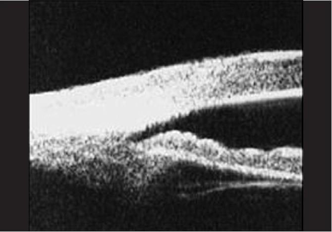
This diagnostic image is an Ultrasound Biomicroscopy (UBM) scan of the anterior segment of a human eye, specifically demonstrating a plateau iris configuration. The cross-sectional view shows the anatomical relationship between the peripheral iris, ciliary body, and cornea. Key findings include a relatively flat iris plane that turns sharply posteriorly at the periphery, and an anteriorly displaced ciliary body that supports the peripheral iris, effectively pushing it against the trabecular meshwork. This results in a significantly narrowed or closed anterior chamber angle, characterized by the close proximity or apposition of the iris root to the posterior corneal surface. The image highlights the absence of significant pupillary block, as the central iris remains relatively flat rather than bowed forward. This UBM finding is clinically significant for diagnosing plateau iris syndrome, a mechanism of angle-closure glaucoma that often persists despite a patent peripheral iridotomy.
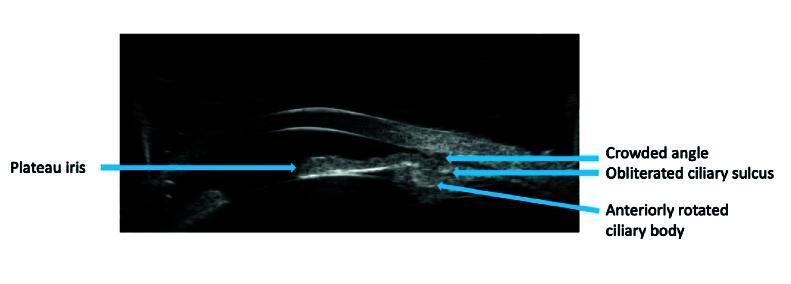
This diagnostic image is an Ultrasound Biomicroscopy (UBM) scan of the anterior segment of the human eye, demonstrating the classic features of plateau iris configuration. The scan provides a cross-sectional view of the iridocorneal angle and associated structures. Key landmarks are identified with blue arrows and text labels. The iris is shown with a flat 'plateau' profile, where the peripheral iris is pushed forward, leading to a crowded iridocorneal angle. The ciliary body is visibly rotated anteriorly, providing structural support to the peripheral iris. A significant diagnostic finding is the obliterated ciliary sulcus, where the space between the posterior iris surface and the ciliary body is lost. This anatomical arrangement is clinically significant in ophthalmology as it represents a primary mechanism for angle-closure glaucoma, where the physical crowding of the angle can impede the outflow of aqueous humor through the trabecular meshwork.
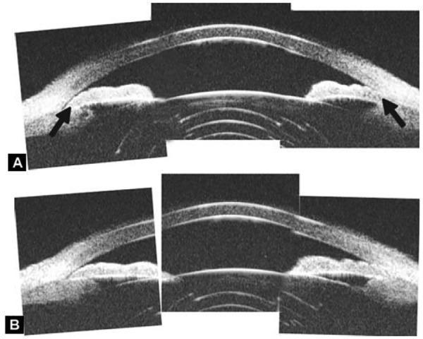
This diagnostic image consists of two Ultrasound Biomicroscopy (UBM) scans, labeled A and B, illustrating the anterior segment of a human eye. The scans clearly depict the cornea, anterior chamber, iris, and ciliary body. Figure A shows a large, anteriorly-rotated ciliary body that provides direct structural support to the peripheral iris, effectively pushing it forward; black arrows highlight the close association between these structures and the anterior insertion of the iris. Figure B shows the same eye following a laser peripheral iridotomy. The comparison demonstrates that despite the iridotomy, the anterior chamber angle remains narrow or closed because the anteriorly displaced ciliary body continues to support and hold the peripheral iris in a forward position. This visual evidence illustrates a non-pupillary block mechanism of angle closure, such as plateau iris syndrome, where structural anomalies of the ciliary body prevent the deepening of the angle even after a patent iridotomy is performed.
Ultrasound Biomicroscopy (UBM)
Definition
- Cornea
- Anterior chamber
- Iridocorneal angle
- Iris
- Ciliary body and ciliary processes
- Zonules
- Lens equator
- Posterior chamber
- Anterior vitreous
- Sclera
- Anterior segment tumors and trauma
Key viva line: UBM is the best routine imaging modality for structures behind the iris, especially the ciliary body and ciliary sulcus.
1. Principle
Frequency, resolution, and penetration
| Characteristic | UBM |
|---|---|
| Frequency | Usually 35-100 MHz |
| Typical frequency | About 50 MHz |
| Axial resolution | About 20-25 µm |
| Lateral resolution | About 40-50 µm |
| Tissue penetration | Approximately 4-5 mm |
| Main region assessed | Anterior segment |
| Medium required | Water bath or coupling fluid |
| Contact with eye | Usually yes, through an eyecup |
Basic physical rule
Higher ultrasound frequency gives better resolution but less penetration.
2. Why UBM Is Needed
- Ciliary body anatomy
- Ciliary body tumors and cysts
- Ciliary sulcus
- Zonules
- Lens equator
- Posterior iris surface
- Retroiridal masses
- Peripheral choroidal detachment
- Anterior cyclodialysis cleft
3. Components of UBM System
- High-frequency ultrasound transducer
- Scanning probe
- Computer/image-processing unit
- Display monitor
- Eyecup or immersion shell
- Sterile saline or water coupling medium
- Topical anaesthetic
- Radial scans
- Transverse scans
- Axial scans
- Longitudinal scans
4. Technique of UBM
Preparation
- Explain the procedure and obtain consent.
- Instill topical anaesthetic.
- Position the patient, commonly supine.
- Insert an appropriately sized eyecup between the lids without exerting pressure on the globe.
- Fill the cup with sterile normal saline or distilled water.
- Place the probe in the water bath, avoiding direct contact with the cornea.
- Ask the patient to fixate in the direction opposite the area being scanned.
- Obtain images in all four quadrants, particularly for angle or ciliary-body assessment.
Important precautions
- Avoid pressure on the globe, especially in:
- Open-globe injury
- Impending corneal perforation
- Markedly thin cornea
- Recent ocular surgery
- Maintain strict asepsis.
- Ensure adequate coupling fluid with no air bubbles.
- Avoid probe contact with cornea or conjunctiva.
5. Normal UBM Anatomy
- Cornea
- Anterior chamber
- Iris
- Anterior chamber angle
- Scleral spur
- Ciliary body
- Ciliary processes
- Ciliary sulcus
- Zonules
- Lens equator
- Sclera
- Anterior vitreous
Appearance of tissues
| Structure | Typical UBM appearance |
|---|---|
| Cornea | Bright anterior and posterior interfaces with a relatively uniform stromal layer |
| Aqueous | Echo-free or dark |
| Iris | Moderately echogenic band |
| Ciliary body | Echogenic structure posterior to iris root |
| Ciliary processes | Folded echogenic projections |
| Lens | Relatively echo-free central area with reflective capsule |
| Sclera | Highly reflective outer coat |
| Ciliary sulcus | Dark potential space between posterior iris and ciliary body |
6. Indications of UBM
A. Glaucoma and angle disorders
- Primary angle closure
- Plateau iris configuration/syndrome
- Pupillary block
- Lens-induced angle closure
- Phacomorphic glaucoma
- Aqueous misdirection syndrome
- Malignant glaucoma
- Secondary angle closure
- Angle recession after trauma
- Peripheral anterior synechiae
- Pigment dispersion syndrome
- Pseudoexfoliation-associated angle assessment
- Ciliary body cyst causing angle closure
- Post-laser peripheral iridotomy persistent narrow angle
- Assessment before and after angle surgery
B. Iris and ciliary-body lesions
- Iris cyst
- Ciliary body cyst
- Iris melanoma
- Ciliary body melanoma
- Iris nevus
- Medulloepithelioma
- Ciliary body metastasis
- Ring melanoma
- Iris bombe
- Retroiridal mass
- Tumor extension into ciliary body, sclera, or angle
C. Trauma
- Angle recession
- Cyclodialysis cleft
- Iridodialysis
- Zonular dialysis
- Lens subluxation
- Traumatic cataract
- Intraocular foreign body in anterior segment
- Occult scleral rupture
- Hypotony after trauma
D. Cornea, sclera, and ocular surface
- Corneal thickness and deep corneal lesion assessment
- Corneal opacity preventing optical imaging
- Scleral thinning
- Scleritis and posterior extension of anterior scleritis
- Peripheral ulcerative keratitis with structural assessment
- Ocular-surface tumors with suspected deep extension
E. Lens, IOL, and postoperative problems
- Phacodonesis and zonular weakness
- Lens subluxation
- Position of posterior chamber IOL
- Sulcus IOL location
- Haptic position
- IOL tilt or decentration
- Retained lens fragment in sulcus
- Suspected uveitis-glaucoma-hyphema syndrome
- Assessment before secondary IOL implantation
- Ciliary sulcus anatomy for IOL planning
F. Uveitis and hypotony
- Ciliary body detachment
- Supraciliary effusion
- Cyclodialysis cleft
- Pars plana exudate
- Ciliary-body membrane
- Hypotony due to occult wound leak or ciliary-body shutdown
7. UBM in Angle-Closure Glaucoma
Why it matters
- Pupillary block
- Plateau iris
- Lens-induced crowding
- Ciliary body rotation
- Ciliary body cyst
- Aqueous misdirection
- Supraciliary effusion
- Tumor-related angle closure
Exam point: Gonioscopy remains the clinical reference standard for angle examination. UBM supplements, rather than replaces, gonioscopy.
A. Pupillary block
Mechanism
UBM findings
- Convex anterior bowing of mid-peripheral iris
- Shallow peripheral anterior chamber
- Narrow or closed angle
- Often relative apposition of iris to anterior lens surface
- No primary anterior rotation of ciliary body
Treatment implication
- Laser peripheral iridotomy is effective if pupillary block is the major mechanism.
B. Plateau iris configuration and syndrome
Definitions
UBM findings
- Anteriorly rotated or anteriorly positioned ciliary processes
- Absent or very narrow ciliary sulcus
- Peripheral iris pushed forward by ciliary body
- Narrow or closed angle
- Relatively flat central iris plane
- Iris root may be short, thick, and inserted anteriorly
Clinical features
- Often relatively young patient with narrow angles
- Central anterior chamber may appear reasonably deep
- Persistent narrow angle despite patent laser peripheral iridotomy
- Double-hump sign on indentation gonioscopy
Management implication
- Peripheral iridotomy is done first to eliminate a coexistent pupillary-block component.
- Persistent appositional angle closure may require argon laser peripheral iridoplasty, lens extraction in selected cases, or individualized management.
C. Lens-induced angle closure
UBM findings
- Thick or anteriorly positioned lens
- Shallow anterior chamber
- Anterior lens vault
- Crowded angle
- Iris convexity may coexist
Clinical relevance
- Phacomorphic glaucoma
- Intumescent cataract
- Short hyperopic eye
- Nanophthalmos
Management
D. Aqueous misdirection or malignant glaucoma
Clinical setting
UBM findings
- Uniformly shallow or flat anterior chamber
- Anterior rotation of ciliary body
- Forward displacement of iris-lens diaphragm
- Supraciliary fluid may or may not be seen
- Patent peripheral iridotomy may be present
Importance
E. Ciliary-body cyst and secondary angle closure
UBM finding
- Round, smooth, echo-free lesion in ciliary body or iridociliary region
- Peripheral iris displaced forward
- Focal angle narrowing or closure
Importance
8. UBM in Ocular Trauma
A. Angle recession
Definition
UBM findings
- Widened ciliary-body band
- Altered angle anatomy
- Irregular iris root
- Associated cyclodialysis or zonular damage may be visible
Clinical relevance
B. Cyclodialysis cleft
Definition
Consequences
- Marked hypotony
- Shallow anterior chamber
- Choroidal effusion
- Hypotony maculopathy
UBM finding
- Separation between ciliary body and scleral spur
- Communication toward supraciliary/suprachoroidal space
- May identify a cleft when gonioscopy is obscured by hyphema, corneal edema, or a very shallow chamber
C. Lens subluxation and zonular dialysis
- Unequal ciliary sulcus configuration
- Lens equator displacement
- Zonular absence or stretching
- Iridodonesis-associated zonular weakness
- Anteriorly displaced lens causing angle closure
9. UBM in Iris and Ciliary-Body Tumors
Uses
- Determines lesion size and thickness
- Identifies cystic versus solid lesion
- Assesses angle involvement
- Detects ciliary-body extension
- Detects scleral extension
- Monitors growth over time
- Guides biopsy and surgical planning
Cyst versus solid tumor
| Feature | Iris/ciliary body cyst | Solid tumor |
|---|---|---|
| Internal reflectivity | Echo-free or very low | Variable internal echoes |
| Shape | Smooth, round, thin-walled | Irregular or lobulated |
| Posterior wall | Usually visible | May be irregular |
| Effect on adjacent structures | Displacement | Infiltration or distortion may occur |
| Follow-up | Often stable | Requires oncologic assessment if suspicious |
Important limitation
- Clinical examination
- Gonioscopy
- Slit-lamp photography
- B-scan ultrasound for larger posterior extension
- MRI in selected tumor cases
10. UBM in Uveitis and Hypotony
Uses in hypotony
- Cyclodialysis cleft
- Ciliary body detachment
- Supraciliary effusion
- Choroidal detachment anteriorly
- Ciliary-body atrophy
- Membranes over pars plicata
- Occult post-surgical wound abnormality
Uses in uveitis
- Ciliary-body edema or thickening
- Pars plana exudates
- Ciliary-body membrane
- Cyclitic membrane
- Anterior vitreous inflammation
- Complicated cataract with ciliary-body involvement
11. UBM versus Anterior-Segment OCT
| Feature | UBM | AS-OCT |
|---|---|---|
| Energy used | Ultrasound | Low-coherence light |
| Typical frequency/wavelength | 35-100 MHz ultrasound | Near-infrared light |
| Contact required | Usually immersion contact | Non-contact |
| Need for coupling medium | Yes | No |
| Resolution | High | Usually higher for superficial anterior structures |
| Penetration through opaque cornea | Good | Poor |
| Imaging behind iris | Excellent | Limited |
| Ciliary body and ciliary sulcus | Excellent | Poor or not visualized |
| Lens equator/zonules | Better | Limited |
| Corneal epithelium and tear film | Less detailed | Better |
| Patient comfort | Less comfortable | More comfortable |
| Dynamic examination | Possible with changes in gaze, indentation, accommodation | Usually static |
| Infection/epithelial defect concern | Contact-related limitation | Safer because non-contact |
| Main value | Hidden anterior anatomy and ciliary body | Cornea, angle configuration, incision, flap, anterior chamber morphology |
One-line answer
AS-OCT is non-contact and better for superficial corneal/anterior chamber imaging, whereas UBM is superior for opaque media and structures posterior to the iris, especially ciliary body, sulcus, and zonules.
12. UBM versus Gonioscopy
| Feature | UBM | Gonioscopy |
|---|---|---|
| Shows angle structures directly | Cross-sectional anatomical image | Direct clinical view through a goniolens |
| Can visualize ciliary body | Yes | Limited |
| Can show structures behind iris | Yes | No |
| Dynamic indentation assessment | Possible but less routine | Excellent |
| Detects pigmentation, neovascularization, PAS | Less detailed | Better |
| Contact | Immersion cup | Contact lens |
| Primary role | Mechanism/anatomical explanation | Clinical angle classification |
Gonioscopy is essential for clinical angle assessment. UBM is especially valuable when the cause of angle closure is uncertain.
13. UBM versus Conventional B-Scan Ultrasound
| Feature | UBM | Conventional B-scan |
|---|---|---|
| Frequency | 35-100 MHz | Approximately 10 MHz |
| Resolution | Microscopic, about 20-50 µm | Lower |
| Penetration | 4-5 mm | Full posterior segment |
| Main use | Anterior segment | Vitreous, retina, choroid, orbit |
| Ciliary body assessment | Excellent | Limited |
| Retinal detachment assessment | Limited | Excellent |
14. Quantitative UBM Parameters
| Parameter | Meaning |
|---|---|
| Anterior chamber depth | Distance from posterior cornea to anterior lens surface |
| Angle opening distance, AOD | Distance between iris and corneal endothelium at a defined point from scleral spur |
| Trabecular-iris angle, TIA | Angular width between trabecular meshwork region and iris |
| Angle recess area, ARA | Area of angle recess in cross-section |
| Trabecular-ciliary process distance | Assesses ciliary-body position |
| Iris thickness | Useful in angle-closure research and selected clinical situations |
| Ciliary body thickness | May assist in inflammation, tumors, and hypotony evaluation |
| Lens vault | Anterior displacement of lens relative to scleral spur plane |
15. Advantages
- High-resolution cross-sectional image of anterior segment
- Visualizes anatomy behind iris
- Visualizes ciliary body, ciliary sulcus, zonules, and lens equator
- Works despite corneal opacity, hyphema, edema, or dense pigmentation
- Helps determine mechanism of angle closure
- Useful in tumors, trauma, hypotony, and postoperative complications
- Allows qualitative and quantitative assessment
- Helps with surgical planning and follow-up
16. Limitations
- Requires topical anaesthesia and immersion technique
- Contact procedure, with some infection and epithelial-trauma risk
- Time-consuming compared with AS-OCT
- Operator dependent
- May be difficult in children, photophobic patients, or uncooperative patients
- Risk of pressure artifact if eyecup or probe is not handled carefully
- Limited penetration, so posterior segment cannot be evaluated adequately
- Does not replace gonioscopy for assessment of pigmentation, neovascularization, or peripheral anterior synechiae
- Interpretation requires familiarity with anterior-segment anatomy and scan orientation
17. Contraindications and Precautions
Relative contraindications
- Suspected open-globe injury
- Corneal perforation or descemetocele
- Recent penetrating ocular surgery
- Active severe infectious keratitis
- Severe ocular-surface disease
- Extreme patient non-cooperation
18. Common Artifacts and Errors
| Problem | Effect | Prevention |
|---|---|---|
| Probe pressure | Artificially narrows angle or deforms anterior chamber | Avoid pressure and ensure correct eyecup position |
| Poor fixation | Wrong quadrant or oblique scan | Give clear fixation instructions |
| Air bubbles in coupling fluid | Acoustic shadow/artifact | Fill cup carefully |
| Inadequate fluid coupling | Poor image quality | Ensure full immersion |
| Off-axis scan | Misleading anatomy and measurements | Obtain radial scans through the area of interest |
| Insufficient dark adaptation in angle study | Underestimates physiologic angle narrowing | Perform standardized light and dark imaging where relevant |
| Failure to identify scleral spur | Incorrect angle measurements | Review anatomy and compare quadrants |
| Failure to compare fellow eye | Missed asymmetry | Image both eyes when relevant |
19. Model Long Answer
Ultrasound biomicroscopy is a high-frequency ultrasound imaging modality used for detailed cross-sectional evaluation of the anterior segment. It commonly employs a 50 MHz transducer, giving an axial resolution of approximately 20-25 µm, a lateral resolution of approximately 40-50 µm, and tissue penetration of about 4-5 mm.UBM works by recording echoes reflected from interfaces of differing acoustic impedance. It requires topical anaesthesia and immersion coupling, usually with an eyecup filled with sterile saline. It can image the cornea, anterior chamber, angle, iris, ciliary body, ciliary sulcus, zonules, lens equator, anterior vitreous, sclera, and anterior segment tumors.Its principal indications include evaluation of angle-closure glaucoma, plateau iris, ciliary-body cysts and tumors, iris lesions, ocular trauma, cyclodialysis cleft, hypotony, lens subluxation, IOL malposition, and occult anterior-segment pathology behind an opaque cornea or iris.In plateau iris, UBM demonstrates anteriorly positioned ciliary processes, absent ciliary sulcus, a relatively flat central iris, and forward displacement of peripheral iris with narrowing of the angle. UBM is superior to AS-OCT for imaging structures posterior to iris, particularly the ciliary body and lens equator. However, it is a contact procedure, has limited penetration, and does not replace gonioscopy for clinical angle assessment.
20. Rapid Viva Questions
-
What is UBM?
High-frequency ultrasound imaging of the anterior segment. -
What frequency is commonly used?
About 50 MHz, though systems may use roughly 35-100 MHz. -
What is the approximate penetration of UBM?
About 4-5 mm. -
What is the major advantage of UBM over AS-OCT?
It images structures behind the iris, especially ciliary body, ciliary sulcus, and lens equator. -
What is the major advantage of AS-OCT over UBM?
It is non-contact, rapid, and gives excellent imaging of superficial corneal and anterior-chamber structures. -
Does UBM replace gonioscopy?
No. Gonioscopy remains essential for clinical angle assessment; UBM helps identify the anatomical mechanism. -
What is the classical UBM finding in plateau iris?
Anteriorly rotated ciliary processes pushing peripheral iris forward, with a narrow or absent ciliary sulcus. -
What is plateau iris syndrome?
Persistent angle closure after a patent peripheral iridotomy due to non-pupillary-block anatomy. -
What is the UBM role in hypotony?
Detection of cyclodialysis cleft, ciliary body detachment, supraciliary effusion, and cyclitic membranes. -
What is the UBM role after blunt trauma?
It detects angle recession, cyclodialysis cleft, zonular dialysis, lens subluxation, and occult anterior-segment damage. -
What is the UBM role in iris/ciliary-body tumors?
It assesses lesion thickness, posterior margin, ciliary-body involvement, angle involvement, and scleral extension. -
Why is UBM useful in corneal opacity?
Ultrasound penetrates opacity better than light-based imaging. -
What is the main limitation of UBM in posterior-segment disease?
Limited penetration depth. -
Why should excessive probe pressure be avoided?
It can artificially alter anterior-chamber depth and angle configuration. -
What is the key difference between a ciliary-body cyst and plateau iris on UBM?
A ciliary-body cyst causes focal displacement, while plateau iris usually shows diffuse anterior ciliary-body rotation and absent/narrow ciliary sulcus.
Teach ophthalmoscopy for MS Ophthalmology exams
AAO direct indirect ophthalmoscopy technique optics field magnification examination
ophthalmoscopy retinal examination systematic review
indirect ophthalmoscopy retinal examination diagram inverted image scleral depression
Ophthalmoscopy for MS Ophthalmology Examinations
Definition
- Retinal disease
- Optic neuropathy
- Glaucoma
- Diabetic and hypertensive retinopathy
- Uveitis
- Retinal detachment and retinal tears
- Neuro-ophthalmology
- Ocular trauma
- Pediatric ophthalmology and ROP
- Systemic vascular and neurologic disease
1. Classification
| Method | Main instrument | Image | Main use |
|---|---|---|---|
| Direct ophthalmoscopy | Hand-held direct ophthalmoscope | Virtual, erect, unreversed | Optic disc, macula, bedside examination, red reflex |
| Monocular indirect ophthalmoscopy | Light source + condensing lens | Real, inverted, laterally reversed | General fundus screening, less commonly used now |
| Binocular indirect ophthalmoscopy, BIO | Head-mounted indirect ophthalmoscope + condensing lens | Real, inverted, laterally reversed | Peripheral retinal examination, retinal tears, detachment, ROP, trauma |
| Slit-lamp indirect biomicroscopy | Slit lamp + 60 D/78 D/90 D lens | Real, inverted, laterally reversed | Detailed posterior-pole examination |
| Contact-lens fundus biomicroscopy | Slit lamp + Goldmann 3-mirror/4-mirror/contact lens | Depends on lens/mirror | Peripheral retina, macula, angle, scleral indentation |
| Digital fundus imaging | Fundus camera, ultra-widefield imaging | Digital image | Documentation, screening, teleophthalmology |
2. Basic Optical Principle
Important optical concepts
A. Emmetropic eye
B. Direct ophthalmoscopy
C. Indirect ophthalmoscopy
3. Direct Ophthalmoscopy
Definition
Components of a direct ophthalmoscope
- Light source
- Mirror or prism directing light into the eye
- Viewing aperture
- Aperture selector
- Lens disc
- Power indicator
- Handle and battery
Apertures and filters
| Aperture/filter | Use |
|---|---|
| Large circular aperture | Dilated pupil, general examination |
| Small circular aperture | Small pupil |
| Microspot aperture | Very small pupil or focal retinal lesion |
| Slit aperture | Estimation of lesion elevation/depression; optic disc cupping |
| Fixation star | Macular fixation testing, especially in children |
| Cobalt-blue filter | Fluorescein examination of cornea, not routine fundus examination |
| Red-free/green filter | Enhances retinal vessels, hemorrhages, nerve fiber layer, and macular lesions |
Image characteristics
| Feature | Direct ophthalmoscopy |
|---|---|
| Image | Virtual |
| Orientation | Erect and unreversed |
| Magnification | Approximately 15x |
| Field of view | Small, approximately 5-10 degrees |
| Stereopsis | Absent |
| Pupil dilation | Helpful but not always essential |
| Peripheral retina | Poorly visualized |
| Media opacity | View easily impaired |
| Main advantage | Portable, simple, bedside use, high magnification |
| Main limitation | Small field and no stereopsis |
Optics and refractive error correction
- Plus lenses, usually shown in green: hypermetropia, aphakia, or examiner needs plus correction
- Minus lenses, usually shown in red: myopia or examiner needs minus correction
Viva pearl
In direct ophthalmoscopy, use plus lenses for hyperopia and minus lenses for myopia.
Technique of direct ophthalmoscopy
Preparation
- Explain the procedure.
- Darken the room.
- Ask the patient to look at a distant target.
- Examine the red reflex from about 30-45 cm first.
- Dilate pupils when clinically appropriate and safe.
- Use the right hand and right eye for the patient's right eye.
- Use the left hand and left eye for the patient's left eye.
Stepwise technique
-
Inspect the red reflex
- Stand 30-45 cm away.
- Use a large circular aperture.
- Compare both eyes.
- Look for brightness, symmetry, and media opacity.
-
Approach the eye
- Start about 15 degrees temporal to the patient's line of sight.
- Ask the patient to look at a distant point.
- Move closer while maintaining the red reflex.
-
Find a retinal vessel
- Follow the vessel centrally toward the optic disc.
-
Examine optic disc
- Shape, size, color, margins, cup-disc ratio, vessels, hemorrhages.
-
Examine retinal vessels
- Artery-vein ratio, caliber, crossings, tortuosity, sheathing, emboli.
-
Examine macula
- Ask the patient to look directly at the light briefly.
- Look for foveal reflex, pigmentation, edema, hemorrhage, drusen, and exudates.
- Warn the patient that this part is uncomfortable.
-
Examine remaining posterior pole
- Sweep systematically through superior, inferior, nasal, and temporal retina.
Red reflex
Definition
Significance
- Bright
- Symmetrical
- Similar in both eyes
- Free of dark shadows or white opacity
Causes of abnormal red reflex
| Finding | Possible cause |
|---|---|
| Diminished reflex | Cataract, corneal opacity, vitreous hemorrhage, small pupil |
| Asymmetrical reflex | Anisometropia, strabismus, media opacity |
| Leukocoria | Retinoblastoma, congenital cataract, Coats disease, ROP, persistent fetal vasculature, retinal detachment |
| Dark shadow | Lens opacity, vitreous opacity, retinal lesion |
| Unequal brightness | Refractive error, ocular misalignment, asymmetric media opacity |
4. Indirect Ophthalmoscopy
Definition
- Real
- Inverted
- Laterally reversed
- Wide field
- Lower magnification than direct ophthalmoscopy
5. Types of Indirect Ophthalmoscopy
A. Monocular indirect ophthalmoscopy
B. Binocular indirect ophthalmoscopy, BIO
- Head-mounted binocular indirect ophthalmoscope
- Light source on head set
- Hand-held condensing lens, commonly +20 D or +28 D
- Dilated pupil, when possible
C. Slit-lamp indirect ophthalmoscopy
- 60 D
- 78 D
- 90 D
- 100 D
- Optic nerve head
- Macula
- Posterior pole
- Posterior vitreous
- Diabetic retinopathy
- Macular edema
- Choroidal neovascularization
- Glaucoma optic neuropathy
6. Indirect Ophthalmoscopy: Optics
Image formation
- Upside down
- Right-left reversed
Orientation rule
In indirect ophthalmoscopy, the image moves in the opposite direction to the examiner's lens movement.
- To view the patient's superior retina, ask the patient to look down.
- To view the patient's temporal retina, ask the patient to look nasally.
- To view the patient's inferior retina, ask the patient to look up.
Magnification
| Condensing lens | Approximate magnification |
|---|---|
| +14 D | About 4.3x |
| +20 D | About 3x |
| +28 D | About 2.1x |
Lens power, field, and magnification
| Lens | Field of view | Magnification | Typical use |
|---|---|---|---|
| +14 D to +15 D | Smaller | Higher | Detailed view in cooperative adult with good dilation |
| +20 D | Good, about 60 degrees dynamic field | Moderate, about 3x | Standard adult BIO lens |
| +28 D | Wider, about 69 degrees dynamic field | Lower, about 2.3x | Small pupil, children, peripheral retina, ROP |
| +30 D | Very wide | Lower | Pediatric and difficult views |
- Field of view increases
- Magnification decreases
- Working distance decreases
7. Technique of Binocular Indirect Ophthalmoscopy
Preparation
- Explain the procedure.
- Check visual acuity and pupils first.
- Dilate the pupil unless contraindicated.
- Position the patient reclining or supine.
- Adjust interpupillary distance and focus of the indirect ophthalmoscope.
- Use appropriate lens:
- +20 D for standard adult examination
- +28 D for children, small pupils, or broad peripheral screening
- Reduce room illumination.
Examination steps
- Align the ophthalmoscope light with the patient's pupil.
- Obtain a red reflex.
- Hold the condensing lens close to the patient's eye.
- Align the reflexes on the condensing lens.
- Move the lens slightly away from the eye until the aerial retinal image comes into focus.
- Examine the optic disc and posterior pole.
- Examine every retinal quadrant systematically.
- Use scleral depression when indicated to inspect the ora serrata and identify tears.
Positioning pearl
8. Scleral Depression
Definition
Principle
- Extreme peripheral retina
- Ora serrata
- Vitreous base
- Retinal breaks
- Lattice degeneration
- Dialysis
- Peripheral retinal tears
Indications
- Flashes and floaters
- Acute posterior vitreous detachment
- Suspected retinal tear
- Ocular trauma
- Retinal detachment assessment
- Lattice degeneration
- High myopia
- Retinal break surveillance
- Preoperative retinal examination before some intraocular surgery
Technique
- Fully dilate the pupil where appropriate.
- Perform indirect ophthalmoscopy first.
- Ask the patient to look in the direction that exposes the quadrant of interest.
- Apply the depressor externally over the sclera opposite the retinal area to be examined.
- Indent gently while viewing the elevated retinal area.
- Examine each clock hour systematically.
Orientation rule
The scleral depressor is placed externally over the sclera corresponding to the retinal area to be brought into view.
Contraindications / precautions
- Suspected or confirmed open-globe injury
- Recent ocular surgery, unless specifically advised
- Severe pain or marked inflammation
- Extremely fragile globe
- Severe scleritis
- Significant hyphema in selected trauma cases
9. Slit-Lamp Fundus Biomicroscopy
Principle
Types of lenses
| Lens | Approximate field | Magnification | Characteristics |
|---|---|---|---|
| 60 D | Smaller field | Higher magnification | Good for disc and macula |
| 78 D | Intermediate | Intermediate | Good balance for routine posterior-pole examination |
| 90 D | Wider field | Lower magnification | Common outpatient lens; can work through relatively smaller pupil |
| 20 D with slit lamp | Wide field | Lower magnification | Often used with indirect viewing techniques |
Technique
- Dilate pupil where possible.
- Ask patient to fixate straight ahead.
- Use low-to-medium slit-lamp magnification initially.
- Hold the lens just in front of the cornea.
- Align illumination, lens, and viewing axis.
- Focus first on the lens surface, then move posteriorly to the aerial image.
- Examine disc, macula, vascular arcades, and posterior pole.
- Use different gaze positions for more peripheral views.
10. Contact Lens Fundus Examination
A. Goldmann three-mirror lens
Components
- Central lens for posterior pole
- Equatorial mirror
- Peripheral retinal mirror
- Gonioscopic mirror
Uses
- Optic disc and macula
- Equatorial retina
- Peripheral retina
- Ora serrata in selected quadrants
- Anterior chamber angle
- Retinal breaks and peripheral lesions
Limitation
- Requires topical anaesthesia and coupling fluid
- Contact procedure
- More time-consuming than non-contact lenses
B. Four-mirror lens
Uses
- Rapid peripheral retinal examination
- Dynamic indentation gonioscopy
- Evaluation of peripheral retinal tears
- Assessment without coupling fluid in many designs
11. Direct versus Indirect Ophthalmoscopy
| Feature | Direct ophthalmoscopy | Binocular indirect ophthalmoscopy |
|---|---|---|
| Examiner distance | Very close to patient | About arm's length |
| Image | Virtual | Real aerial image |
| Orientation | Erect, unreversed | Inverted and laterally reversed |
| Magnification | High, about 15x | Lower, about 2-5x |
| Field of view | Small, about 5-10 degrees | Wide, approximately 45-60 degrees or more |
| Stereopsis | No | Yes |
| Peripheral retina | Poor | Excellent |
| Through media opacity | Poor | Better |
| Pupil dilation | Helpful | Usually necessary for thorough peripheral examination |
| Scleral depression | No | Yes |
| Main use | Optic disc, macula, bedside, red reflex | Retinal tears, detachment, trauma, pediatric retina, ROP |
| Main limitation | Small field, no stereopsis | Lower magnification, technique more difficult |
Best exam line
Direct ophthalmoscopy gives a highly magnified erect image of a small posterior-pole area, whereas binocular indirect ophthalmoscopy gives a lower-magnification, inverted, laterally reversed, stereoscopic wide-field view of the retina.
12. Direct Ophthalmoscopy versus Slit-Lamp Biomicroscopy
| Feature | Direct ophthalmoscopy | Slit-lamp indirect biomicroscopy |
|---|---|---|
| Instrument | Hand-held direct ophthalmoscope | Slit lamp + condensing lens |
| Image | Erect, unreversed | Inverted, laterally reversed |
| Magnification | About 15x | Variable, generally good |
| Stereopsis | No | Yes |
| Portability | Excellent | Poor |
| Peripheral view | Limited | Better but less than BIO |
| Main strength | Bedside disc and red-reflex examination | Detailed disc and macular assessment |
13. Normal Fundus Examination
- Media clarity
- Optic disc
- Retinal vessels
- Posterior pole
- Macula and fovea
- Peripheral retina
- Vitreous
A. Optic disc
Normal features
| Feature | Normal description |
|---|---|
| Color | Pink to orange-pink |
| Shape | Round or slightly vertically oval |
| Margins | Sharp and well-defined |
| Cup | Central pale depression |
| Cup-disc ratio | Usually less than 0.5, but disc size must be considered |
| Neuroretinal rim | Broadest inferiorly, then superiorly, nasally, temporally: ISNT rule |
| Vessels | Central retinal artery and vein emerge from disc |
| Spontaneous venous pulsation | May be present; absence alone is not diagnostic |
Assess in every optic disc
- Disc size
- Color
- Margin
- Cup-disc ratio
- Neuroretinal rim
- Vessel entry and exit
- Hemorrhages
- Pallor
- Edema
- Collaterals
- Drusen
- Peripapillary atrophy
B. Cup-disc ratio
Important points
- Evaluate vertical cup-disc ratio.
- Assess both eyes for asymmetry.
- Inter-eye asymmetry greater than about 0.2 is suspicious, but must be interpreted in relation to disc size.
- A large disc may have a large physiological cup.
- A small crowded disc may have a small cup even in raised intracranial pressure.
Glaucoma clues
- Progressive increase in cup-disc ratio
- Vertical cupping
- Focal rim notching
- Violation of ISNT rule
- Disc hemorrhage
- Retinal nerve fiber layer defect
- Vessel bayoneting
- Nasalization of vessels
- Inter-eye asymmetry
C. Retinal vessels
Normal artery-vein ratio
Assess
- Arteriolar caliber
- Venous caliber
- A:V ratio
- Tortuosity
- Arteriovenous crossing changes
- Sheathing
- Emboli
- Neovascularization
- Venous beading
- Vascular attenuation
Important retinal vascular signs
| Sign | Possible significance |
|---|---|
| Generalized arteriolar narrowing | Chronic hypertension |
| Focal narrowing | Hypertensive retinopathy |
| AV nicking | Arteriolosclerosis |
| Copper wiring | Moderate arteriolosclerosis |
| Silver wiring | Severe arteriolosclerosis |
| Flame hemorrhage | Nerve fiber layer hemorrhage, hypertension, disc edema |
| Dot-blot hemorrhage | Deep retinal hemorrhage, diabetes, vein occlusion |
| Roth spot | White-centered hemorrhage, nonspecific finding |
| Cotton-wool spot | Nerve fiber layer infarct |
| Venous beading | Severe nonproliferative diabetic retinopathy |
| Neovascularization | Proliferative diabetic retinopathy, vein occlusion, ischemic retinopathy |
| Hollenhorst plaque | Cholesterol embolus |
D. Macula and fovea
Normal appearance
- Macula is darker than surrounding retina because of xanthophyll pigment.
- Fovea lies approximately 2 disc diameters temporal and slightly inferior to the optic disc.
- A foveal light reflex may be present in young healthy eyes.
- The foveal avascular zone is central.
Assess
- Pigmentary changes
- Drusen
- Hemorrhage
- Exudates
- Edema
- Subretinal fluid
- Macular hole
- Epiretinal membrane
- Cotton-wool spots
- Choroidal neovascular membrane signs
E. Peripheral retina
- Lattice degeneration
- Retinal holes
- Horseshoe tears
- Operculated tears
- Retinal dialysis
- White without pressure
- Retinal detachment
- Chorioretinal scars
- Peripheral neovascularization
- Tumors
- Snowballs or snowbanking in uveitis
14. Important Pathological Fundus Findings
A. Optic disc edema versus optic atrophy
| Feature | Disc edema | Optic atrophy |
|---|---|---|
| Color | Hyperemic initially | Pale |
| Margins | Blurred | Usually sharp |
| Cup | Obliterated in significant edema | May be normal or enlarged |
| Vessels | Obscured at margins, venous congestion | Attenuated vessels may occur |
| Hemorrhages | May be present | Usually absent |
| Cause | Raised ICP, neuritis, ischemia, infiltration, malignant hypertension | Previous optic nerve injury |
Important clarification
B. Diabetic retinopathy
| Finding | Level / significance |
|---|---|
| Microaneurysms | Earliest clinically visible lesion |
| Dot-blot hemorrhages | Deep retina |
| Hard exudates | Lipid deposition |
| Cotton-wool spots | Nerve fiber layer ischemia |
| Venous beading | Severe ischemic NPDR |
| IRMA | Severe NPDR |
| Neovascularization | Proliferative diabetic retinopathy |
| Preretinal/vitreous hemorrhage | Proliferative disease |
| Macular edema | Major cause of visual loss |
C. Hypertensive retinopathy
- Generalized and focal arteriolar narrowing
- AV crossing changes
- Copper or silver wiring
- Flame-shaped hemorrhages
- Cotton-wool spots
- Hard exudates
- Macular star
- Disc edema in malignant hypertension
D. Retinal detachment
Rhegmatogenous retinal detachment
- Elevated, corrugated, mobile retina
- Retinal tear may be identified
- Subretinal fluid
- Demarcation line in chronic cases
Tractional retinal detachment
- Concave, immobile, taut retina
- Usually associated with proliferative diabetic retinopathy
Exudative retinal detachment
- Smooth, convex, shifting subretinal fluid
- No retinal break
15. Indications for Pupillary Dilatation
Indications
- Diabetes screening and retinopathy assessment
- Flashes, floaters, or suspected retinal break
- Suspected retinal detachment
- Trauma after open-globe injury is excluded
- Uveitis
- Unexplained reduced vision
- Detailed optic disc and macular examination
- Preoperative retinal assessment
- Pediatric retinal examination
Important precautions
- Known occludable/narrow angles
- Symptoms of intermittent angle closure
- Acute red painful eye
- Suspected acute angle closure
16. Limitations of Ophthalmoscopy
- Media opacity: corneal edema, cataract, vitreous hemorrhage
- Small pupil
- Poor patient cooperation
- High refractive error
- Nystagmus
- Inability to document without photography
- Direct ophthalmoscopy has a very limited field
- Subtle macular edema or optic nerve disease may require OCT
- Vascular leakage cannot be assessed by ophthalmoscopy alone and may require FFA
- Peripheral lesions may be missed without indirect ophthalmoscopy and scleral depression
17. Common Errors and How to Avoid Them
| Error | Consequence | Prevention |
|---|---|---|
| Not checking red reflex first | Missed media opacity or leukocoria | Begin from a distance |
| Using wrong eye/hand | Awkward technique and poor view | Right eye/right hand for patient's right eye; left eye/left hand for left eye |
| Trying to examine through a small pupil without dilation | Missed peripheral pathology | Dilate when safe and indicated |
| Examining disc only | Missed macular and peripheral disease | Follow a fixed sequence |
| Failure to document disc size | Incorrect interpretation of cup-disc ratio | Assess cup in relation to disc size |
| Calling all disc edema papilledema | Diagnostic error | Papilledema requires raised intracranial pressure |
| Failure to examine peripheral retina in flashes/floaters | Missed retinal tear | BIO with scleral depression when indicated |
| Excessively bright direct ophthalmoscope light | Patient discomfort and poor fixation | Begin dim and increase only as necessary |
| Not correcting refractive error | Blurred view | Use lens wheel appropriately |
| Not comparing both eyes | Missed asymmetry | Document both eyes systematically |
18. Model Long Answer
Ophthalmoscopy is the examination of the fundus, including the vitreous, retina, retinal vessels, optic nerve head, macula, and peripheral retina. It may be direct, indirect, slit-lamp based, or contact lens based.Direct ophthalmoscopy is performed with a hand-held self-illuminated instrument. It produces a virtual, erect, unreversed image with approximately 15x magnification but a small field of view and no stereopsis. It is useful for bedside examination, red-reflex assessment, optic disc evaluation, and macular examination.Binocular indirect ophthalmoscopy uses a head-mounted light source and a high-plus condensing lens, commonly +20 D. It produces a real, inverted, laterally reversed image with lower magnification, wide field, and stereopsis. It is the preferred method for detailed peripheral retinal examination, retinal tears, retinal detachment, trauma, and pediatric retinal examination. Scleral depression extends the view to the extreme peripheral retina and ora serrata.The fundus examination should be systematic and include media clarity, optic disc, cup-disc ratio, retinal vessels, macula, posterior pole, peripheral retina, and vitreous. Direct and indirect ophthalmoscopy are complementary: direct ophthalmoscopy gives high magnification of a small posterior-pole area, while indirect ophthalmoscopy gives wide-field stereoscopic visualization of the retina.
19. Viva Questions
-
What is ophthalmoscopy?
Examination of the fundus, including retina, optic disc, vessels, and vitreous. -
What is the image in direct ophthalmoscopy?
Virtual, erect, unreversed, and magnified. -
What is the image in indirect ophthalmoscopy?
Real, inverted, laterally reversed, and stereoscopic. -
What is the magnification of direct ophthalmoscopy?
Approximately 15x. -
What is the magnification of a 20 D lens in BIO?
Approximately 3x. -
What lens is commonly used for adult binocular indirect ophthalmoscopy?
A +20 D condensing lens. -
What is the advantage of a 28 D lens?
Wider field and easier examination through a smaller pupil, especially in children. -
Which method is preferred for detecting peripheral retinal tears?
Binocular indirect ophthalmoscopy with scleral depression. -
What is the main disadvantage of direct ophthalmoscopy?
Small field of view and absence of stereopsis. -
How do you examine the right eye by direct ophthalmoscopy?
Use the examiner's right eye and right hand. -
What does a plus lens on the direct ophthalmoscope compensate for?
Hypermetropia, aphakia, or a hyperopic examiner. -
What is the normal artery-vein ratio?
Approximately 2:3. -
What is Shafer sign?
Pigment cells in anterior vitreous, suggesting a retinal break. -
What is papilledema?
Optic disc edema due to raised intracranial pressure. -
What does a high cup-disc ratio alone mean?
It is not diagnostic of glaucoma. It must be interpreted with disc size, neuroretinal rim, inter-eye asymmetry, IOP, OCT, and visual fields. -
What is the significance of a white pupillary reflex in a child?
It is leukocoria and warrants urgent assessment for retinoblastoma and other serious causes. -
What is the role of a 90 D lens?
Non-contact slit-lamp examination of the optic disc, macula, and posterior pole. -
What is the role of Goldmann three-mirror examination?
Contact examination of posterior pole, equator, peripheral retina, and angle. -
Does a normal direct ophthalmoscopy exclude peripheral retinal tear?
No. A complete peripheral retinal assessment generally requires indirect ophthalmoscopy, often with scleral depression. -
Which investigation complements ophthalmoscopy for retinal microstructure?
OCT.