Generate easy microscopic slide diagram of following questions according to textbook of pathology by Harsh Mohan 1.Septic thrombus in lung 2.apostematous nephritis, 3.cryptococcal meningitis (HIV) 4.cytomegaloviral pneumonia (HIV)
Simple easy pathology microscopic slide diagram of Septic Thrombus in Lung (H&E stain). Educational labeled medical illustration. Show a blood vessel cross-section in the lung with: a central thrombus made of fibrin mesh (pink wavy threads) mixed with neutrophils (dark purple multilobed nuclei) and bacterial colonies (tiny dark purple clusters/dots) within the fibrin. The vessel wall shows inflammatory cell infiltration. Surrounding lung tissue shows alveoli with some neutrophils spilling out (septic embolic pneumonia). Label clearly: "Fibrin thrombus", "Neutrophils", "Bacterial colonies", "Vessel wall", "Alveoli with inflammation". Clean white background, simple educational style, H&E color scheme (pink and purple), bold black labels with arrows.
Simple easy pathology microscopic slide diagram of Apostematous Nephritis (acute suppurative pyelonephritis) H&E stain. Educational labeled medical illustration. Show kidney cortex with multiple small abscesses (apostemes): clusters of neutrophils (dark purple multilobed nuclei) packed inside renal tubules and spilling into interstitium. Tubular epithelium appears necrotic and disrupted. Interstitium shows edema and neutrophilic infiltrate. Glomeruli appear relatively spared. Label clearly: "Neutrophil abscess in tubule", "Destroyed tubular epithelium", "Interstitial neutrophilic infiltrate", "Relatively spared glomerulus", "Tubular necrosis". Clean white background, simple educational style, H&E color scheme (pink cortex, purple nuclei), bold black labels with arrows.
Simple easy pathology microscopic slide diagram of Cryptococcal Meningitis in HIV/AIDS patient (Mucicarmine or H&E stain). Educational labeled medical illustration. Show brain tissue cross-section with: meninges expanded by numerous Cryptococcus neoformans yeast organisms - round 5-10 micron yeasts with thick gelatinous capsule (clear halo around each yeast). Virchow-Robin perivascular spaces expanded by yeast clusters creating classic "soap-bubble" lesion. Minimal inflammatory reaction (immunosuppressed patient - no neutrophils, few lymphocytes). Brain parenchyma pushed aside. Label clearly: "Cryptococcus yeast (5-10 um)", "Thick gelatinous capsule (clear halo)", "Soap-bubble lesion in Virchow-Robin space", "Minimal inflammatory reaction", "Brain parenchyma". Clean white background, simple educational style, yeast staining red/pink with clear capsule halos, bold black labels with arrows.
Simple easy pathology microscopic slide diagram of Cytomegalovirus (CMV) Pneumonia in HIV/AIDS patient (H&E stain). Educational labeled medical illustration. Show lung tissue with alveoli. Key feature: enlarged cytomegalic cells (25-35 microns, much larger than surrounding cells) with the classic "owl's eye" appearance - a large central dark basophilic intranuclear inclusion surrounded by a clear halo, then a dark nuclear membrane. Also show smaller cytoplasmic inclusions around the nucleus. Alveolar walls show interstitial thickening and mononuclear infiltrate. Label clearly: "Cytomegalic enlarged cell (25-35um)", "Large central intranuclear inclusion", "Clear halo (owl's eye appearance)", "Cytoplasmic inclusions", "Thickened alveolar wall", "Mononuclear infiltrate". Clean white background, simple educational style, H&E color scheme (pink cytoplasm, dark purple inclusions), bold black labels with arrows.
1. Septic Thrombus in Lung (H&E)
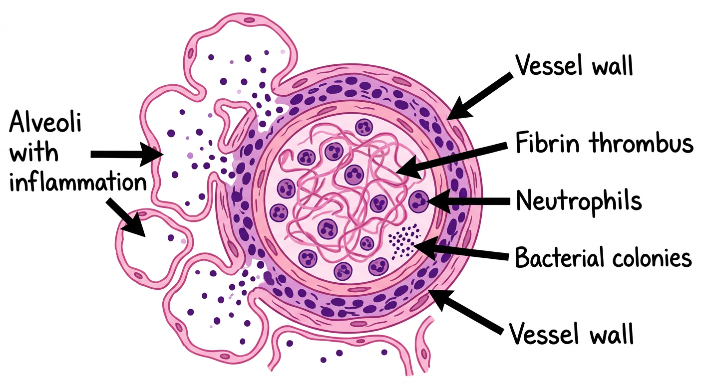
- Fibrin thrombus inside pulmonary vessel - pink wavy mesh
- Thrombus contains neutrophils (polymorphs) and bacterial colonies (dark purple clusters)
- Vessel wall shows inflammatory infiltration
- Surrounding lung parenchyma: alveoli with neutrophilic exudate (embolic pneumonia/abscess formation)
2. Apostematous Nephritis (H&E)
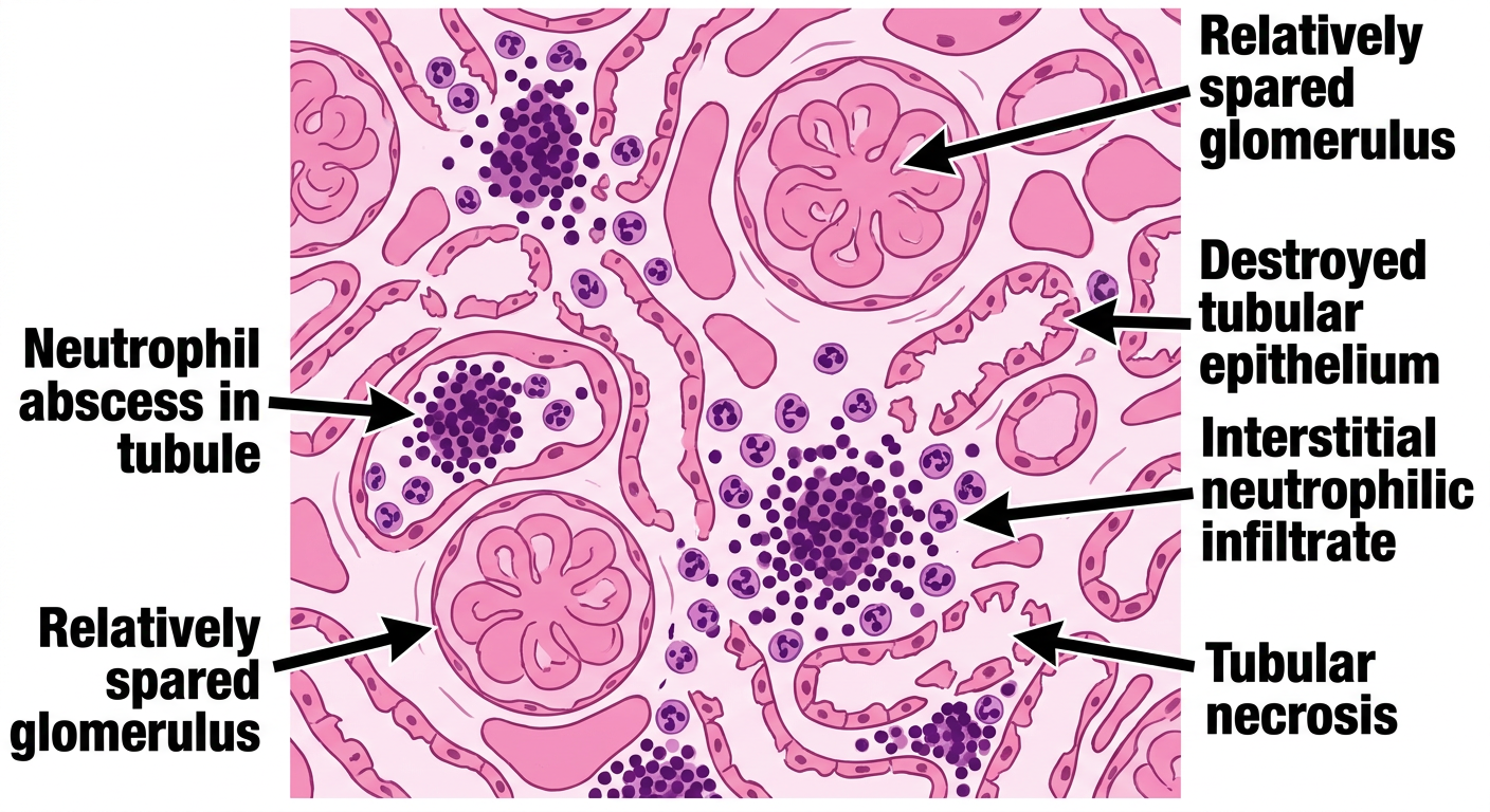
- Multiple small cortical abscesses (apostemes = tiny abscesses) - characteristic feature
- Neutrophil aggregates packed inside renal tubules and interstitium
- Tubular epithelium is necrotic and disrupted
- Interstitial edema with diffuse PMN infiltrate
- Glomeruli relatively spared (hematogenous spread pattern)
3. Cryptococcal Meningitis - HIV (Mucicarmine/H&E)
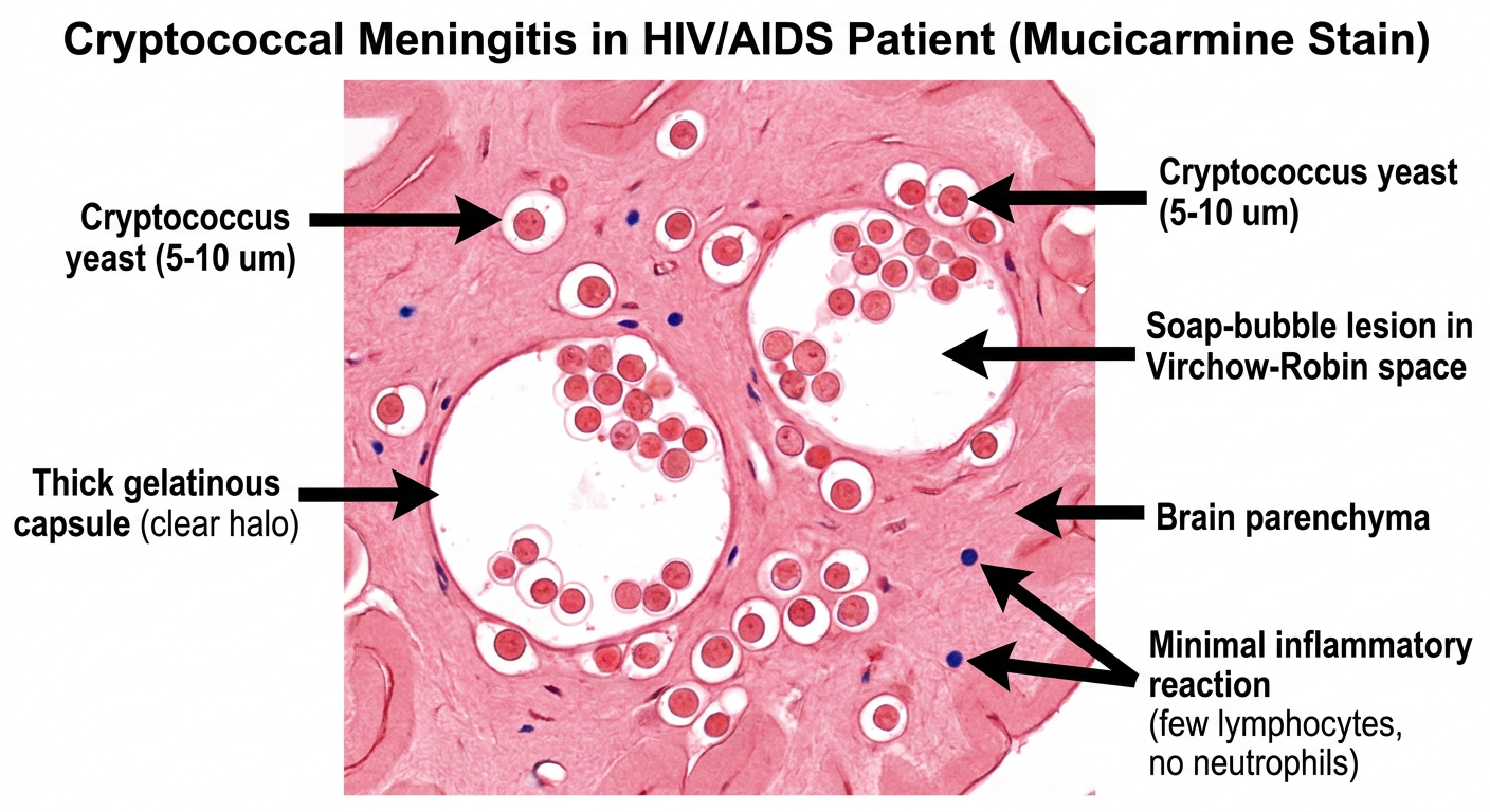
- Cryptococcus neoformans yeast: round, 5-10 µm, with thick gelatinous capsule (stains red with mucicarmine)
- Classic soap-bubble lesion - yeast expand Virchow-Robin perivascular spaces
- Minimal/absent inflammatory reaction - hallmark of immunosuppression in HIV
- Gelatinous masses of fungi in meninges without neutrophils
- India ink prep shows capsule as clear halo around yeast
4. CMV Pneumonia - HIV (H&E)

- Cytomegalic cells: enlarged 25-35 µm cells (3-4x normal size)
- Classic "owl's eye" inclusion: large central basophilic intranuclear inclusion surrounded by a clear halo, bordered by thickened nuclear membrane
- Smaller granular cytoplasmic inclusions also present
- Alveolar wall: interstitial thickening, mononuclear cell infiltrate
- Diffuse alveolar damage pattern in background
Sources: Murray & Nadel's Respiratory Medicine (septic embolism); Robbins & Cotran Pathologic Basis of Disease (cryptococcosis morphology, CMV); Goldman-Cecil Medicine (CMV owl's eye inclusion). Apostematous nephritis features are per Harsh Mohan's Textbook of Pathology standard description.
Give me CMV pneumonia picture again
Simple easy pathology microscopic slide diagram of Cytomegalovirus (CMV) Pneumonia in HIV/AIDS patient (H&E stain). Educational labeled medical illustration. Show lung tissue with alveoli. Key feature: enlarged cytomegalic cells (25-35 microns, much larger than surrounding cells) with the classic "owl's eye" appearance - a large central dark basophilic intranuclear inclusion surrounded by a clear halo, then a dark nuclear membrane. Also show smaller cytoplasmic inclusions around the nucleus. Alveolar walls show interstitial thickening and mononuclear infiltrate. Label clearly: "Cytomegalic enlarged cell (25-35um)", "Large central intranuclear inclusion", "Clear halo (owl's eye appearance)", "Cytoplasmic inclusions", "Thickened alveolar wall", "Mononuclear infiltrate". Clean white background, simple educational style, H&E color scheme (pink cytoplasm, dark purple inclusions), bold black labels with arrows.
CMV Pneumonia - HIV/AIDS (H&E)
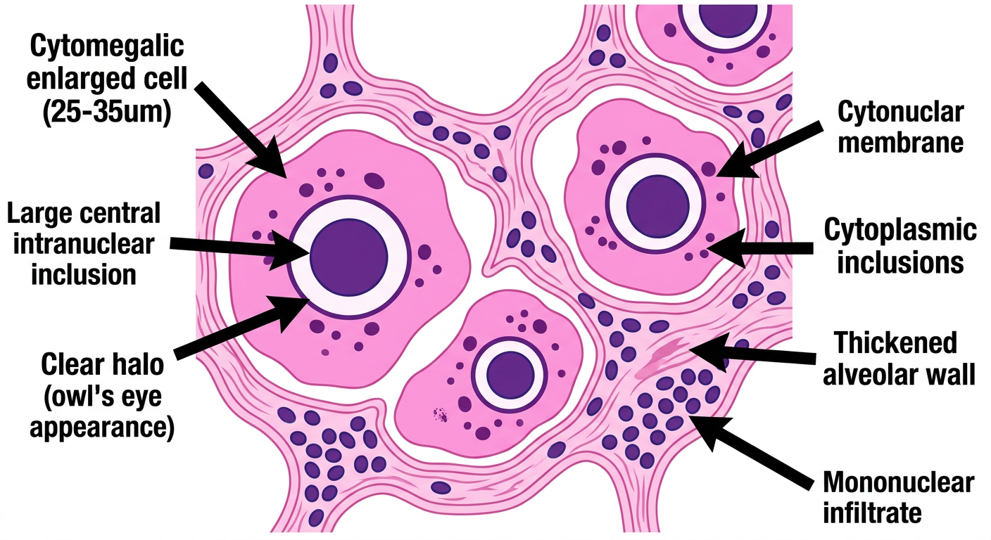
| Feature | Description |
|---|---|
| Cytomegalic cell | Enlarged 25-35 µm cell (3-4x normal size) |
| Owl's eye inclusion | Large central dark basophilic intranuclear inclusion with clear halo |
| Cytoplasmic inclusions | Smaller granular inclusions in the cytoplasm |
| Alveolar wall | Thickened with mononuclear infiltrate |
| Background | Diffuse alveolar damage pattern |