

MBBS Proff and with proper diagrams and flow charts
wound healing phases flowchart diagram
septic shock pathophysiology flowchart
thyroid carcinoma classification histopathology
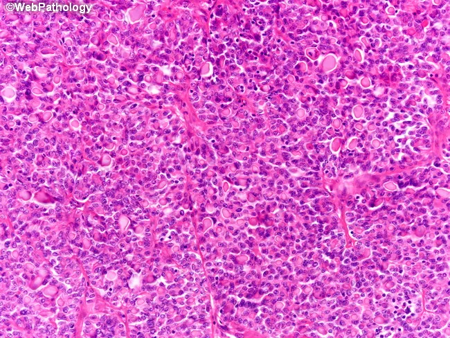
This histopathology image shows a renal neoplasm with thyroid-like follicular differentiation. The section is formalin-fixed and stained with Hematoxylin and Eosin. At low-to-intermediate magnification, the tumor forms numerous small follicles and microfollicles arranged in a predominantly hypercellular pattern; some larger follicles contain colloid-like material resembling thyroid colloid. The epithelial cells are cuboidal to low-columnar with round to oval nuclei, inconspicuous nucleoli, and scant to moderate cytoplasm. Nuclear features show mild to moderate pleomorphism; mitotic activity is infrequent in this field. The architecture mimics thyroid tissue, with follicles ranging from microfollicular to near-solid arrangements and occasional colloid-like intraluminal material giving a heterogeneous appearance. There is no obvious invasion into adjacent stroma within the image; deeper sections may reveal capsular or vascular invasion in other examples. The differential diagnostic considerations include thyroid-like follicular carcinoma of the kidney, other renal cell carcinoma subtypes such as papillary or chromophobe variants, and metastatic thyroid carcinoma. Immunohistochemical profiling (not shown) typically shows PAX8 positivity with TTF-1 and thyroglobulin negativity, supporting a renal origin despite thyroid-like morphology. Clinically, recognition of this pattern is important to distinguish from metastatic thyroid disease and other RCC subtypes for accurate diagnosis and management. Accurate classification guides prognosis, treatment, and surveillance strategies for clinicians.
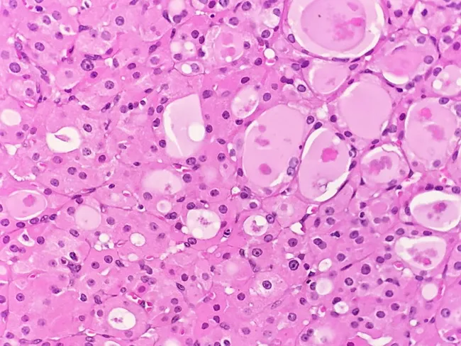
Histology image of Hurthle cell adenoma of the thyroid. This light microscopy slide is stained with Hematoxylin and Eosin (H&E) and shows a predominantly oncocytic (Hurthle) cell population arranged in sheets and microfollicular clusters. The cells are large and polygonal, with abundant granular eosinophilic cytoplasm rich in mitochondria, and round to oval nuclei with often prominent nucleoli. Nuclei may display mild atypia, but mitotic activity is scarce in this field. The lesion appears typically well circumscribed and encapsulated within thyroid parenchyma, with a benign architectural pattern; no definite invasive cords are evident in this view. Colloid material may be scant or variably present within trapping follicles. The surrounding stroma is modest, and vessels are not overtly infiltrated by tumor cells in this image. Clinically, Hurthle cell adenoma represents a benign neoplasm of uncertain function that can simulate carcinoma on cytology; therefore capsule integrity and absence of capsular or vascular invasion are essential for definitive classification. This image is instrumental for histopathology education, differential diagnosis with Hurthle cell carcinoma, and surgical planning discussions, and it supports terminology including oncocytic adenoma, Hurthle cell lesion, and thyroid follicular neoplasm. Further education and clinical correlation.
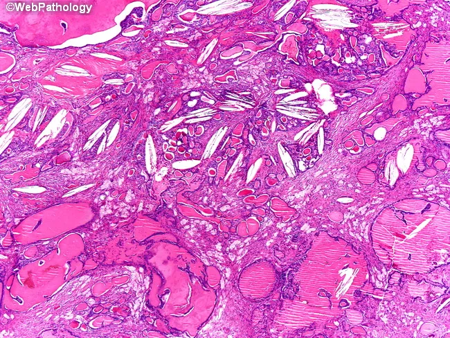
This is a histopathology image from renal tissue demonstrating a thyroid-like follicular variant of renal cell carcinoma. Acquired as a surgical specimen from the kidney, the slide was prepared with standard hematoxylin and eosin staining and examined under bright-field light microscopy at low- to high-power magnification. The tumor exhibits thyroid follicle-like architecture characterized by scattered microfollicles and larger macrofollicles filled with colloid-like eosinophilic material, mimicking benign and malignant thyroid lesions. The follicles are embedded in a fibromyxoid stroma containing chronic inflammatory cells and occasional cholesterol clefts, features that contribute to a distinctive pattern and may complicate cytologic interpretation. Cytologic atypia is minimal, consistent with a low-grade neoplasm; the tumor cells are arranged in a cohesive epithelial lining around colloid-filled lumina, with well-preserved nuclear polarity and subtle mitotic activity if present. Clinically, this entity is rare, often discovered incidental during imaging or nephrectomy for unrelated reasons, and generally portends a favorable prognosis compared with higher-grade RCCs. Differential diagnosis includes metastatic thyroid carcinoma and other follicular-pattern RCCs; immunohistochemistry (e.g., PAX8 positive, TTF-1 negative) and molecular profiling aid accurate classification. Recognize this pattern in diagnostic pathology to prevent misinterpretation and guide appropriate clinical management. This image supports teaching cases of TLCK in pathology education.
burn injury management Parkland formula
shock types classification distributive hypovolemic
papillary thyroid carcinoma Orphan Annie nuclei histology
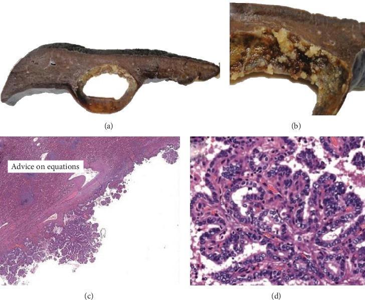
This composite image illustrates the macroscopic and microscopic features of a metastatic papillary thyroid carcinoma within the spleen. Panels (a) and (b) are clinical photographs showing a cross-section of an ovoid, cystic splenic lesion approximately 5.5 cm in diameter. The gross specimen reveals thin cyst walls with internal friable, yellowish papillary projections protruding into the lumen. Panels (c) and (d) are H&E-stained photomicrographs showing the corresponding histology. At low magnification (4x, Panel c), the cyst wall is lined by complex papillary structures with fibrovascular cores extending into the cystic space. At high magnification (20x, Panel d), the neoplastic cells exhibit classic nuclear features of papillary thyroid carcinoma, including cuboidal morphology, clear 'Orphan Annie eye' nuclei, and prominent longitudinal nuclear grooves (nuclear bars). The images demonstrate a rare case of splenic metastasis from a primary thyroid malignancy, emphasizing the importance of recognizing characteristic cytological features in unconventional anatomical sites.
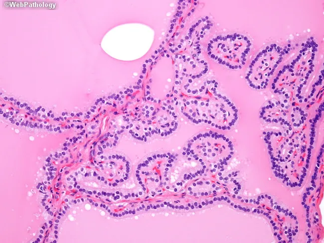
This is a brightfield, hematoxylin and eosin stained histology slide of thyroid tissue examined by light microscopy at high magnification. The specimen is thyroid gland parenchyma showing diffuse follicular hyperplasia characteristic of Graves disease. Follicles appear crowded with epithelial cells lining irregular, papillary-like folds protruding into the lumen, creating pseudo-papillary architectures that could mimic papillary thyroid carcinoma if evaluated in isolation. The nuclei within the thyrocytes are hyperplastic and dense but lack the distinctive nuclear features of papillary carcinoma, such as crowding with nuclear grooves, inclusions, Orphan Annie eye clearing, and powdery chromatin. Colloid is variably present or scant; stroma is vascular, and the overall architecture is polyfollicular with ingrowths into luminal spaces. This pattern reflects autoimmune-driven stimulation by TSH receptor autoantibodies (e.g., thyroid-stimulating immunoglobulin), resulting in diffuse goiter with hyperthyroid state. Clinically, Graves disease presents with weight loss, tachycardia, heat intolerance, and sometimes exophthalmos; histology aids diagnosis when correlated with serology and uptake studies. Key diagnostic significance: avoid mislabeling as papillary thyroid carcinoma; rely on clinical context and absence of malignant nuclear features. Potential educational uses include differential diagnosis training for thyroid pathology, immune-mediated thyroid hyperplasia vs neoplasia, and radiology-pathology correlation. This image supports teaching clinics and pathology review conferences.
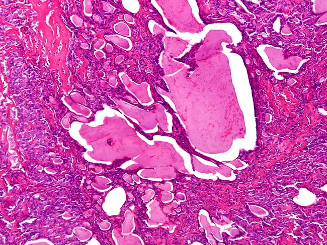
This histopathology image represents renal tissue involved by metastatic thyroid carcinoma, prepared as a hematoxylin and eosin stained slide and examined under light microscopy. The renal cortical/parenchymal field contains variably sized follicles filled with colloid‑like material, features that mimic thyroid follicles. The left side shows microfollicles with abortive lumens and solid appearance, consistent with a thyroid‑type neoplasm invading renal parenchyma. The architectural pattern is compatible with papillary or follicular thyroid carcinoma morphology, including follicle‑forming tumor cells arranged in microacini and acinar clusters amidst desmoplastic stroma. Distinguishing between a primary renal neoplasm and metastatic thyroid carcinoma hinges on immunohistochemical profile and clinical context. If a thyroid primary is confirmed and nuclear features typical of papillary thyroid carcinoma (Orphan Annie eye nuclei, nuclear grooves, grooves/inclusions) are present in metastatic deposits, then immunoreactivity for thyroid transcription factor‑1 (TTF‑1) and thyroglobulin would favor thyroid origin and metastasis. Conversely, a primary renal neoplasm would be unlikely to express TTF‑1 or thyroglobulin. Clinically, recognizing this pattern has implications for staging, prognosis, and treatment, including thyroid cancer‑focused systemic therapy and surveillance for additional metastases. This image underscores the necessity of integrating histology with patient history and immunohistochemistry in diagnostic decision making.
TEERTHANKER MAHAVEER MEDICAL COLLEGE
General Surgery - Final MBBS (3rd Prof Part II) - Complete Answers
Q1. Wound Healing Phases, Classification of Surgical Wounds, and Factors Affecting Wound Healing (3+3+4 = 10 marks)
Part A: Phases of Wound Healing (3 marks)
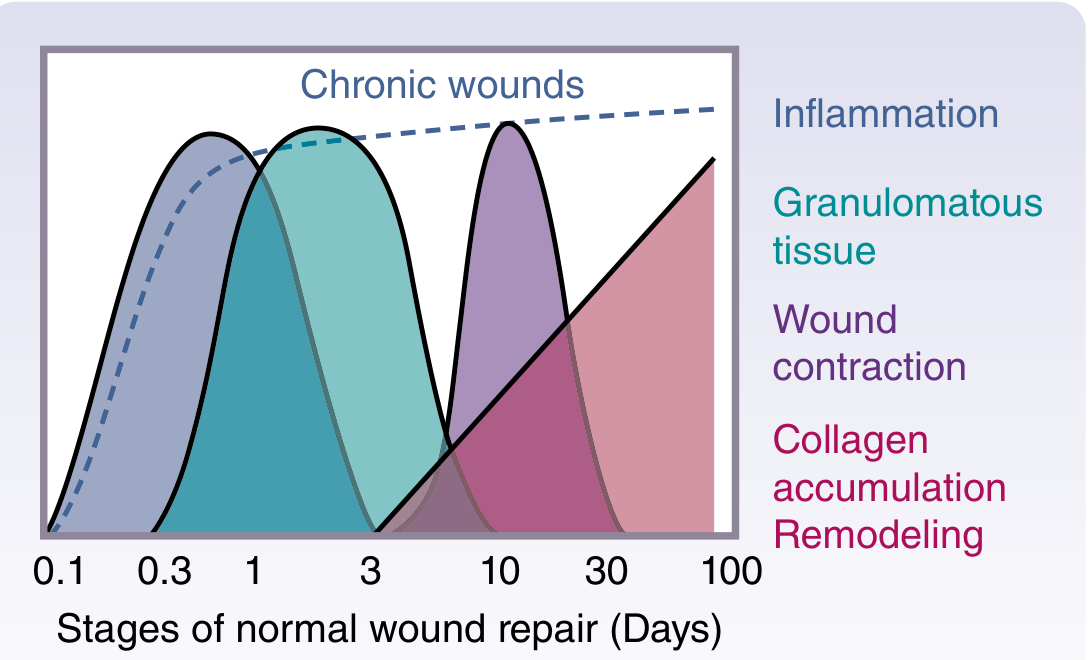
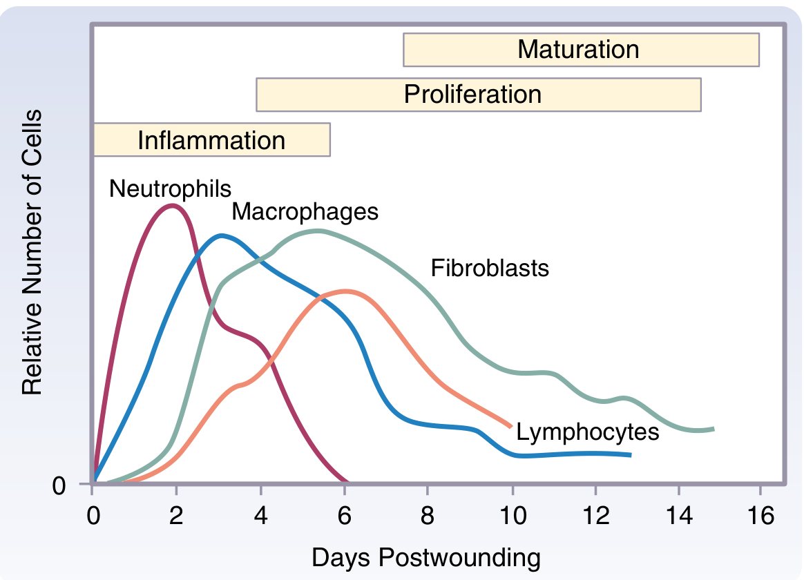
Flowchart: Phases of Wound Healing
TISSUE INJURY
│
▼
━━━━━━━━━━━━━━━━━━━━━━━━━━━━━━━━━━━━━━━━━━━━
PHASE 1: HEMOSTASIS (Immediate - 0 to few hours)
━━━━━━━━━━━━━━━━━━━━━━━━━━━━━━━━━━━━━━━━━━━━
• Vasoconstriction (immediate)
• Platelet adhesion to exposed collagen (via vWF, GPIIb-IIIa)
• Platelet activation → release of alpha granules
(PDGF, TGF-β, IGF-1, fibronectin)
• Dense bodies → serotonin → vasodilation + permeability
• Coagulation cascade → fibrin clot formation
• Provisional wound matrix (fibrin scaffold)
│
▼
━━━━━━━━━━━━━━━━━━━━━━━━━━━━━━━━━━━━━━━━━━━━
PHASE 2: INFLAMMATION (Day 1–5)
━━━━━━━━━━━━━━━━━━━━━━━━━━━━━━━━━━━━━━━━━━━━
Early (Day 1-2): NEUTROPHILS dominate
• PMNs recruited by chemotaxis (IL-8, C5a, LTB4)
• Phagocytosis of bacteria and debris
• Release of proteases, ROS
• Non-essential for wound healing (sterile wounds heal without PMNs)
Late (Day 2-5): MACROPHAGES dominate (MASTER CELLS of wound healing)
• Derived from blood monocytes
• Essential for wound healing
• Release PDGF, TGF-α, TGF-β, IL-1, TNF-α
• Recruit fibroblasts
• M1 phenotype: host defense (TNF-α, NO, IL-6)
• M2 phenotype: wound healing, angiogenesis
│
▼
━━━━━━━━━━━━━━━━━━━━━━━━━━━━━━━━━━━━━━━━━━━━
PHASE 3: PROLIFERATION (Day 4–21)
━━━━━━━━━━━━━━━━━━━━━━━━━━━━━━━━━━━━━━━━━━━━
3 key events:
1. FIBROPLASIA
• Fibroblasts migrated from wound edges
• Collagen synthesis (Type III initially → later Type I)
• Stimulated by PDGF, TGF-β, EGF, FGF
2. ANGIOGENESIS
• New capillary formation (VEGF-driven)
• Granulation tissue = fibroblasts + new capillaries + macrophages
3. EPITHELIALIZATION
• Keratinocytes migrate over moist wound bed
• Contact inhibition stops migration when wound closed
• Growth factors: EGF, TGF-α, KGF (FGF-7)
│
▼
━━━━━━━━━━━━━━━━━━━━━━━━━━━━━━━━━━━━━━━━━━━━
PHASE 4: MATURATION/REMODELING (Day 21 → 2 years)
━━━━━━━━━━━━━━━━━━━━━━━━━━━━━━━━━━━━━━━━━━━━
• Type III collagen replaced by Type I collagen
• Cross-linking by lysyl oxidase
• Collagenase (MMP-1) degrades excess collagen
• Max tensile strength = 80% of original (never 100%)
• Wound contraction by myofibroblasts (α-SMA positive)
• Vascularity decreases → scar becomes avascular, pale, flat
Part B: Classification of Surgical Wounds (3 marks)
CDC Classification of Surgical Wounds
| Class | Type | Definition | Infection Risk |
|---|---|---|---|
| I | Clean | Elective, no inflammation, no breach of GI/GU/respiratory tract, no break in aseptic technique. E.g.: hernia repair, thyroidectomy | 1–2% |
| II | Clean-Contaminated | GI/respiratory/GU tract entered under controlled conditions, minor technique break. E.g.: appendicectomy without perforation, cholecystectomy | 5–10% |
| III | Contaminated | Open traumatic wound, major break in technique, gross GI spillage, acute non-purulent inflammation. E.g.: colostomy closure, penetrating trauma | 15–20% |
| IV | Dirty/Infected | Old traumatic wound with devitalized tissue, existing infection, perforated viscus. E.g.: perforated appendix, faecal peritonitis | >30% |
Classification by Depth
- Superficial incisional SSI: Skin + subcutaneous tissue only
- Deep incisional SSI: Fascia and muscle layers
- Organ/space SSI: Any organ/space opened during operation
Part C: Factors Affecting Wound Healing (4 marks)
FACTORS AFFECTING WOUND HEALING
│
┌────┴────┐
▼ ▼
LOCAL SYSTEMIC
FACTORS FACTORS
LOCAL:
┌─────────────────────────────────────┐
│ Promoting Healing Impairs Healing │
│ • Moist environment • Infection │
│ • Good blood supply • Ischemia │
│ • Debridement done • Foreign body │
│ • Wound closure • Desiccation │
│ • Haematoma │
│ • Radiation │
└─────────────────────────────────────┘
SYSTEMIC FACTORS:
┌──────────────────────────────────────────────────┐
│ Factor Effect │
│────────────────────────────────────────────────── │
│ Diabetes mellitus ↓ PMN function, microangiopathy│
│ Malnutrition ↓ Collagen synthesis (↓ Vit C) │
│ Corticosteroids ↓ Inflammation, ↓ fibroplasia │
│ Chemotherapy ↓ Cell proliferation │
│ Jaundice Impairs collagen synthesis │
│ Uraemia Impairs PMN function │
│ Anaemia/Hypoxia ↓ Collagen hydroxylation │
│ Old age ↓ All healing phases │
│ Obesity Poor vascularity of fat │
│ Zinc deficiency ↓ Epithelialization │
│ Vit C deficiency ↓ Collagen cross-linking │
│ Vit A deficiency ↓ Epithelialization │
└──────────────────────────────────────────────────┘
- Vitamin C: Hydroxylation of proline/lysine for collagen synthesis
- Zinc: DNA synthesis, cell proliferation
- Vitamin A: Epithelialization, counteracts steroid effects
- Protein: Substrate for collagen; negative nitrogen balance impairs healing
Q2. Donor Criteria, Massive Blood Transfusion, and Complications (2.5+2.5+5 = 10 marks)
Part A: Donor Criteria for Blood Transfusion (2.5 marks)
Inclusion Criteria (NACO/WHO Guidelines)
| Parameter | Criteria |
|---|---|
| Age | 18–65 years |
| Weight | ≥45 kg |
| Haemoglobin | ≥12.5 g/dL (female), ≥13 g/dL (male) |
| Blood pressure | Systolic 100–180 mmHg, Diastolic 60–100 mmHg |
| Pulse | 60–100 bpm, regular |
| Temperature | Normal (<37.5°C) |
| Interval between donations | ≥12 weeks (≥3 months) |
| Max donations/year | 4 times |
Exclusion Criteria
- HIV positive / high-risk sexual behaviour
- Hepatitis B or C carrier
- Syphilis / gonorrhoea
- Malaria (within past 3 years or living in endemic area)
- Recent vaccination (defer 2 weeks for live vaccines)
- Pregnancy / recent delivery (defer 6 months)
- Major surgery in last 6 months
- Diabetes on insulin
- Cancer (absolute)
- Epilepsy
- Drug addiction
- Unexplained weight loss >5 kg in 3 months
Part B: Massive Blood Transfusion (MBT) (2.5 marks)
Indications
- Major trauma with haemorrhagic shock
- Ruptured aortic aneurysm
- Obstetric catastrophe (PPH, placenta praevia)
- Major hepatic/vascular surgery
Massive Transfusion Protocol (MTP)
- 1:1:1 ratio: pRBC : Fresh Frozen Plasma (FFP) : Platelets
- Mimics whole blood
- Prevents dilutional coagulopathy
- Tranexamic acid: Give ASAP (within 3 hours of trauma) - CRASH-2 trial
- Avoid crystalloids/colloids as primary resuscitation fluids
- Calcium supplementation: Each unit of blood contains citrate (chelates Ca²⁺)
- Warm all blood products (prevent hypothermia)
- Monitor with TEG (Thromboelastography) or ROTEM
Part C: Complications of Blood Transfusion (5 marks)
Flowchart: Complications
COMPLICATIONS OF BLOOD TRANSFUSION
│
┌─────────┴──────────┐
▼ ▼
IMMUNOLOGICAL NON-IMMUNOLOGICAL
IMMUNOLOGICAL:
├── HAEMOLYTIC REACTIONS
│ ├── Acute (<24 hrs): ABO incompatibility
│ │ • Fever, chills, back/loin pain
│ │ • Haemoglobinuria (red/brown urine)
│ │ • Shock, DIC, acute renal failure
│ │ • Action: STOP transfusion immediately
│ │ → Saline flush, IV fluids, O2, furosemide
│ └── Delayed (1-14 days): Anamnestic reaction
│ • Mild haemolysis, anaemia, jaundice
│
├── FEBRILE NON-HAEMOLYTIC TRANSFUSION REACTION
│ • Fever >1°C rise, rigors
│ • Due to cytokines from donor WBCs
│ • Tx: Paracetamol, slow/stop transfusion
│
├── ALLERGIC / ANAPHYLACTIC
│ • Urticaria, bronchospasm, hypotension
│ • Tx: Chlorpheniramine, adrenaline, steroids
│
└── TRANSFUSION-RELATED ACUTE LUNG INJURY (TRALI)
• Commonest cause of transfusion mortality
• Non-cardiogenic pulmonary oedema
• Within 6 hrs of transfusion (usually FFP)
• Tx: O2, ventilatory support
NON-IMMUNOLOGICAL:
├── INFECTIONS
│ ├── Bacterial (faulty storage)
│ ├── Viral: HIV, Hep B, Hep C, CMV, EBV
│ ├── Parasitic: Malaria, Chagas disease
│ └── Prion: vCJD
│
├── MASSIVE TRANSFUSION COMPLICATIONS
│ ├── Coagulopathy (dilutional)
│ ├── Hypocalcaemia (citrate toxicity)
│ ├── Hyperkalaemia (from old stored blood)
│ ├── Hypokalaemia (later phase)
│ ├── Hypothermia
│ ├── Metabolic alkalosis (citrate → bicarbonate)
│ └── TACO (Transfusion Associated Circulatory Overload)
│ • Pulmonary oedema in elderly/cardiac patients
│ • Tx: Slow rate, furosemide
│
├── IRON OVERLOAD (repeated transfusions, e.g. thalassaemia)
│ • ~250 mg elemental iron/unit pRBCs
│ • Tx: Desferrioxamine
│
├── AIR EMBOLISM
└── THROMBOPHLEBITIS
Q3. SSI: Definition, Organisms, Classification, Prevention and Management (1+1+3+3+2 = 10 marks)
Part A: What is SSI? (1 mark)
Part B: Organisms Causing SSI (1 mark)
| Organism | Wound Type |
|---|---|
| Staphylococcus aureus (most common) | Clean, clean-contaminated |
| Streptococcus pyogenes | Soft tissue |
| E. coli, Klebsiella, Proteus | Abdominal/colorectal |
| Pseudomonas aeruginosa | Burns, ICU wounds |
| Bacteroides fragilis | Colorectal surgery |
| Clostridium spp. | Anaerobic, gas gangrene |
| MRSA | Nosocomial |
Part C: Classification of SSI (3 marks)
CLASSIFICATION OF SSI (CDC Definition)
│
┌─────────┼──────────┐
▼ ▼ ▼
SUPERFICIAL DEEP ORGAN/SPACE
INCISIONAL INCISIONAL SSI
SUPERFICIAL INCISIONAL SSI:
• Within 30 days of surgery
• Involves SKIN + SUBCUTANEOUS tissue only
• At least ONE of:
- Purulent discharge from wound
- Positive culture from wound fluid
- Pain, tenderness, swelling, redness + wound opened by surgeon
- Diagnosis by surgeon/physician
DEEP INCISIONAL SSI:
• Within 30 days (or 1 year if implant)
• Involves FASCIA + MUSCLE layers
• At least ONE of:
- Purulent discharge from deep incision
- Spontaneous dehiscence or opened by surgeon
(when patient has fever >38°C, pain/tenderness)
- Abscess on direct examination, re-operation, imaging
ORGAN/SPACE SSI:
• Any part of body OPENED or MANIPULATED during surgery
• E.g.: intraabdominal abscess after bowel surgery
• Pus from drain placed into organ/space
• Positive culture from organ/space specimen
- Minor SSI: Discharges pus/infected serous fluid; no systemic signs; no delay in discharge
- Major SSI: Significant pus; systemic signs (tachycardia, pyrexia, raised WBC); delayed discharge; needs secondary procedure
Part D: Prevention of SSI (3 marks)
PREVENTION OF SSI
│
┌────┴────┐
▼ ▼
PREOPERATIVE INTRAOPERATIVE POSTOPERATIVE
PREOPERATIVE:
• Treat remote infections before elective surgery
• HbA1c control in diabetics (<7%)
• Smoking cessation (at least 30 days before)
• Nutritional optimization
• Reduce preoperative hospital stay
• Skin decolonization: chlorhexidine body wash
• Hair removal: clippers NOT razors (razors ↑ SSI)
• Shower with antiseptic soap on night before surgery
ANTIBIOTIC PROPHYLAXIS:
• Give within 60 minutes before incision
• Cefazolin is first line (or based on flora)
• Single dose usually sufficient (repeat if surgery >4 hrs)
• NO routine postoperative antibiotics
INTRAOPERATIVE:
• Surgical hand scrub (5 min brushing)
• Sterile gloves + gown
• Maintain surgical site haemostasis
• Gentle tissue handling; avoid devitalized tissue
• Minimize dead space
• Appropriate suture selection
• Irrigation with warm saline
• Maintain normothermia (warming blanket)
• Maintain normoglycaemia (target BSL <10 mmol/L)
• High-flow O2 delivery (FiO2 0.8) intraoperatively
• Minimize operative time
POSTOPERATIVE:
• Aseptic wound dressings
• Remove drains early
• Protect wound for 48 hours (epithelialized by then)
• Monitor glucose, temperature
• Educate patient on wound care
Part E: Management of SSI (2 marks)
MANAGEMENT OF SSI
│
├── MINOR SSI
│ ├── Open wound (remove sutures/staples)
│ ├── Drain pus
│ ├── Wound swab for C&S
│ └── Dressings (moist/saline-soaked)
│ Usually NO antibiotics needed
│
└── MAJOR SSI
├── Open + debride wound
├── Wound swab → C&S
├── IV antibiotics based on organism/sensitivity
│ (empiric: Cefuroxime + Metronidazole for abdominal)
├── Source control (drain abscesses)
├── Nutritional support
├── Consider vacuum-assisted closure (VAC) for large wounds
└── Secondary closure once wound is clean
(granulation tissue present, no infection)
Q4. Shock: Types, Pathophysiology and Management of Septic Shock (1+2+3+4 = 10 marks)
Part A: What is Shock? (1 mark)
Part B: Types of Shock (2 marks)
CLASSIFICATION OF SHOCK
│
┌─────┼──────┬────────┐
▼ ▼ ▼ ▼
HYPO- CARDIO- DISTRIBU- OBSTRUCTIVE
VOLAEMIC GENIC TIVE
1. HYPOVOLAEMIC SHOCK
Cause: ↓ Circadian volume
├── Haemorrhagic (trauma, GI bleed, AAA rupture)
└── Non-haemorrhagic (burns, vomiting, diarrhoea, third spacing)
Haemodynamics: ↓CO, ↑SVR, ↑HR, ↓CVP
2. CARDIOGENIC SHOCK
Cause: Pump failure (>40% myocardium infarcted)
├── MI (most common)
├── Arrhythmia
└── Cardiomyopathy
Haemodynamics: ↓CO, ↑SVR, ↑CVP, ↑PCWP
3. DISTRIBUTIVE SHOCK (vasogenic)
Cause: Abnormal vasodilation → maldistribution of flow
├── Septic shock (most common)
├── Anaphylactic shock
├── Neurogenic shock (spinal cord injury)
└── Addisonian crisis
Haemodynamics: ↑CO (early), ↓SVR, ↓CVP
4. OBSTRUCTIVE SHOCK
Cause: Mechanical obstruction to flow
├── Tension pneumothorax
├── Cardiac tamponade
└── Massive pulmonary embolism
Haemodynamics: ↓CO, ↑SVR, ↑CVP
Classification of Haemorrhagic Shock (ATLS)
| Class | Blood Loss | % Volume | HR | BP | Consciousness |
|---|---|---|---|---|---|
| I | <750 mL | <15% | <100 | Normal | Alert |
| II | 750-1500 mL | 15-30% | 100-120 | Normal/↓ | Anxious |
| III | 1500-2000 mL | 30-40% | 120-140 | ↓ | Confused |
| IV | >2000 mL | >40% | >140 | Very ↓ | Lethargy/unconscious |
Part C: Pathophysiology of Septic Shock (3 marks)
PATHOPHYSIOLOGY OF SEPTIC SHOCK
SOURCE OF INFECTION
(Gram-negative: LPS/Endotoxin | Gram-positive: Teichoic acid, Peptidoglycan)
│
▼
PATTERN RECOGNITION RECEPTORS
(Toll-like receptors on macrophages, monocytes)
│
▼
CYTOKINE STORM
TNF-α, IL-1β, IL-6, IL-8, IL-10, PAF
│
┌────┴──────────────┐
▼ ▼
VASODILATION ENDOTHELIAL DAMAGE
(iNOS → ↑↑ NO) │
│ Capillary leak → Third spacing
▼ │
↓SVR (↓ afterload) ▼
↑CO early HYPOVOLAEMIA
│ │
▼ ▼
WARM/HYPERDYNAMIC ←→ COLD/HYPODYNAMIC
PHASE (early) PHASE (late)
• Warm skin • Cold, clammy skin
• Bounding pulse • Thready pulse
• ↑CO • ↓CO
• ↑HR • Oliguria, MODS
┌────────┬──────────┬─────────┐
▼ ▼ ▼ ▼
CARDIAC RENAL HEPATIC PULMONARY
DEPRESSION FAILURE FAILURE FAILURE
(TNF-α, (elevated (ARDS)
IL-1β, LFTs)
iNOS)
│
▼
MULTIPLE ORGAN DYSFUNCTION SYNDROME (MODS)
│
▼
DEATH
(40-60% mortality with MODS)
THREE CORE MECHANISMS in Septic Shock:
1. HYPOVOLAEMIA - relative + absolute (capillary leak, sweating, poor intake)
2. MYOCARDIAL DEPRESSION - TNF-α, IL-1β, NO impair contractility
3. SYSTEMIC INFLAMMATION - microvascular + mitochondrial dysfunction
Part D: Management of Septic Shock (4 marks)
Surviving Sepsis Campaign / Bundle (Hour-1 Bundle)
MANAGEMENT OF SEPTIC SHOCK
│
┌────┴─────┐
▼ ▼
IMMEDIATE DEFINITIVE
(1st hour) (Ongoing)
IMMEDIATE - THE "SEPSIS 6" (complete within 1 hour):
1. MEASURE LACTATE (target <2 mmol/L)
2. BLOOD CULTURES (×2 sets) BEFORE antibiotics
3. IV BROAD-SPECTRUM ANTIBIOTICS (within 1 hour of recognition)
• Empiric: Piperacillin-tazobactam ± Vancomycin
• Or: Meropenem/Imipenem for high-risk/ICU
4. IV FLUID RESUSCITATION: 30 mL/kg crystalloid (Ringer's lactate preferred)
• Target MAP ≥65 mmHg
5. VASOPRESSORS if MAP <65 mmHg despite fluid:
• NOREPINEPHRINE = first-line vasopressor
(0.1–0.3 mcg/kg/min, titrate)
• Add VASOPRESSIN if refractory (0.03 units/min)
• Add EPINEPHRINE as third line
6. SUPPLEMENTAL O2 / INTUBATION if SpO2 <94%
MONITORING:
• Urine output (target >0.5 mL/kg/hr)
• CVP, MAP monitoring
• Serial lactate every 2 hours
• ScvO2 (target >70%)
SUPPORTIVE CARE:
• SOURCE CONTROL (drain abscess, remove infected device)
• STEROIDS: Hydrocortisone 200–300 mg/day IV
(only if refractory to fluids + vasopressors)
• TIGHT GLYCAEMIC CONTROL (target 7.8–10 mmol/L)
• Lung-protective ventilation if ARDS (TV 6 mL/kg)
• DVT + stress ulcer prophylaxis
• Early enteral nutrition
• Renal replacement therapy if acute kidney injury
ANTIBIOTICS:
• Reassess and de-escalate after 48–72 hrs based on cultures
• Duration: typically 7–10 days (procalcitonin-guided)
Q5. Burns Case (Medico-legal + Management) (2+2+6 = 10 marks)
Part A: Should you inform the police? And why? (2 marks)
-
Medicolegal Case (MLC): Burns from fire in young married women fall under the category of a Medico-Legal Case (MLC). Section 174 CrPC (now BNSS) mandates reporting of all suspicious injuries.
-
Dowry Death / Domestic Violence: Under Section 498A IPC (now BNS) and Section 304B IPC (Dowry Death), burns in newly married women within 7 years of marriage are presumed to be dowry-related until proven otherwise.
-
Dying Declaration: As the patient is conscious, a dying declaration should be recorded in her own words in the presence of a magistrate (if available) or doctor, as it is admissible as evidence even if she recovers.
-
Doctor cannot be neutral in suspected domestic violence/dowry harassment cases. The patient's consent or refusal does NOT absolve the doctor of the reporting duty.
-
Section 302/307 IPC: If criminal act suspected, reporting is compulsory under CrPC.
Part B: Will you delay treatment if police cannot come? (2 marks)
- Doctor's primary duty is to the patient (Hippocratic Oath, NMC guidelines).
- Delay in treatment of burns is life-threatening - every minute matters in airway management and fluid resuscitation.
- Indian law does not permit withholding treatment in an emergency for medicolegal formalities.
- MLC paperwork can be completed simultaneously with/after initiating treatment.
- Inform the police telephonically/via hospital record, and the treatment must start immediately without waiting for police arrival.
Part C: Management of 25% Burns (6 marks)
BURNS MANAGEMENT FLOWCHART
INITIAL ASSESSMENT: ABC + STOP THE BURNING PROCESS
│
┌────┴────┐
▼ ▼
PRIMARY SECONDARY
SURVEY SURVEY
PRIMARY SURVEY (ABCDE):
A - AIRWAY
• Assess for inhalation injury signs:
- Singed nasal hairs, eyebrows
- Hoarseness, stridor
- Carbonaceous sputum
- Burns in enclosed space
• If inhalation injury suspected → EARLY INTUBATION
(airways swell rapidly → late intubation is dangerous)
• High-flow O2 via NRB mask (100% O2 counteracts CO poisoning)
B - BREATHING
• Assess chest wall expansion
• Circumferential chest burns → escharotomy
C - CIRCULATION
• 2 large-bore IV lines (in unburned skin if possible)
• IV fluid resuscitation (PARKLAND FORMULA):
┌─────────────────────────────────────────────────────┐
│ PARKLAND FORMULA: │
│ Total 24-hr fluid = 4 mL × Weight (kg) × %BSA burn │
│ (Only TBSA >20% calculated, 2nd and 3rd degree only)│
│ │
│ This patient: 4 × 55 kg × 25% = 5,500 mL │
│ │
│ Distribution: │
│ • FIRST 8 hours: Give 1/2 = 2,750 mL │
│ (from TIME OF BURN, not hospital arrival) │
│ • NEXT 16 hours: Give 1/2 = 2,750 mL │
│ │
│ Fluid: Ringer's Lactate (NOT normal saline) │
└─────────────────────────────────────────────────────┘
• Foley catheter → target UO: 0.5-1 mL/kg/hr (adults)
• Blood pressure monitoring
D - DISABILITY (Neurological status)
• GCS (patient is conscious here)
• Analgesia: IV morphine (titrated doses)
E - EXPOSURE / ENVIRONMENT
• Remove all clothing and jewellery
• Prevent hypothermia (warm blankets, warm IV fluids)
BURNS ASSESSMENT:
┌──────────────────────────────────────────────┐
│ % BSA CALCULATION - "RULE OF NINES" (Wallace) │
│ │
│ Head + Neck = 9% │
│ Each arm = 9% (×2 = 18%) │
│ Front of trunk = 18% │
│ Back of trunk = 18% │
│ Each leg = 18% (×2 = 36%) │
│ Perineum = 1% │
│ TOTAL = 100% │
│ │
│ Palm of patient's hand = 1% BSA │
│ (useful for scattered burns) │
└──────────────────────────────────────────────┘
DEPTH OF BURNS:
┌────────────────┬────────────┬──────────────────────────┐
│ Depth │ Old Name │ Features │
├────────────────┼────────────┼──────────────────────────┤
│ Superficial │ 1st degree │ Erythema, no blister │
│ Partial thick. │ 2nd degree │ Blisters, painful, moist │
│ (superficial) │ │ Heals in 14 days │
│ Partial thick. │ 2nd degree │ Pale/red mottled, less │
│ (deep) │ │ painful, >21 days │
│ Full thickness │ 3rd degree │ Leathery, insensate, │
│ │ │ no healing without graft │
│ 4th degree │ 4th degree │ Bone/muscle involved │
└────────────────┴────────────┴──────────────────────────┘
WOUND CARE:
• Clean burns: Chlorhexidine solution
• Apply Silver sulfadiazine (SSD) cream (Flamazine)
• OR Mafenide acetate (penetrates eschar)
• Non-adherent dressings
• Tetanus prophylaxis (Tetanus toxoid ± TIG)
NUTRITION:
• Burns are hypermetabolic (↑↑ energy needs)
• Calorie = 25 kcal/kg + 40 kcal/% burn
• Protein = 1-3 g/kg/day
• Start enteral nutrition within 6 hrs
• Curling's ulcer prophylaxis: Ranitidine/PPI
SURGICAL MANAGEMENT:
• Escharotomy: For circumferential full-thickness burns
(chest, limbs) → releases compartment pressure
• Early excision and grafting (within 3-5 days)
- Split-thickness skin graft (STSG)
- Donor sites: thigh, back
• For 25% TBSA: needs ICU admission + multidisciplinary care
SPECIAL CONSIDERATIONS (Kerosene burns):
• Kerosene burns are chemical + thermal
• Remove contaminated clothing
• Wash affected areas copiously with water
• Kerosene can cause aspiration pneumonitis if ingested
CRITERIA FOR BURNS UNIT TRANSFER:
• >10% TBSA partial thickness (adults)
• Any full-thickness burns
• Burns of face, hands, feet, genitalia, perineum
• Inhalation injury
• Chemical/electrical burns
• Circumferential burns of extremities
Q6. Short Notes (4 × 5 = 20 marks)
(a) Refeeding Syndrome
Pathophysiology Flowchart
PROLONGED STARVATION
│
▼
Body switches to fat/protein catabolism
Intracellular electrolytes (P, K, Mg) depleted
(but serum levels remain normal due to shifts)
│
REFEEDING BEGINS (↑ Carbohydrates)
│
▼
↑ Insulin secretion
│
┌────┴───────────────┐
▼ ▼
PHOSPHATE POTASSIUM & MAGNESIUM
shifts intracellular shift intracellular
│
▼
HYPOPHOSPHATAEMIA (KEY ABNORMALITY)
(also: Hypokalaemia, Hypomagnesaemia)
│
┌────┴──────────────────────┐
▼ ▼
↓ ATP production ↓ Cardiac contractility
↓ 2,3-DPG in RBCs (arrhythmias)
(impaired O2 delivery)
│
▼
CARDIAC FAILURE, RESPIRATORY FAILURE,
HAEMOLYTIC ANAEMIA, SEIZURES, RHABDOMYOLYSIS
At-risk Groups
- Anorexia nervosa
- Alcoholism
- Prolonged NPO
- Cancer cachexia
- Post-operative malnourished patients
Prevention and Management
| Step | Action |
|---|---|
| Identify risk | BMI <18.5, >10% weight loss, minimal intake >5 days |
| Start slowly | Begin at 10 kcal/kg/day, increase over 7-10 days |
| Supplement | Thiamine 200–300 mg IV BEFORE feeding (prevents Wernicke's) |
| Monitor | Electrolytes twice daily for first week |
| Replace | Phosphate, K⁺, Mg²⁺ aggressively if low |
| Restrict fluid | Avoid fluid overload |
(b) TPN (Total Parenteral Nutrition)
Composition of TPN
| Component | Requirement | Formulation |
|---|---|---|
| Carbohydrates | 50-60% of calories | Dextrose 25-70% |
| Proteins | 1-2 g/kg/day | Amino acids 10-15% |
| Lipids | 20-30% of calories | Intralipid 20% |
| Water | 30-40 mL/kg/day | Normal saline base |
| Electrolytes | Per requirement | Na, K, Ca, Mg, PO4 |
| Vitamins | Standard doses | Water + fat soluble |
| Trace elements | Required daily | Zn, Cu, Mn, Cr, Se |
Indications
INDICATIONS FOR TPN
├── GI TRACT NON-FUNCTIONAL
│ ├── Short bowel syndrome (<100 cm remaining)
│ ├── High-output fistula (>500 mL/day)
│ ├── Prolonged ileus
│ ├── Severe Crohn's disease / IBD
│ └── Necrotising enterocolitis
├── GI TRACT INACCESSIBLE
│ ├── Oesophageal obstruction
│ └── Post-oesophagectomy
└── GI TRACT MUST REST
├── Acute pancreatitis (severe)
└── Post radiation enteritis
Complications of TPN
COMPLICATIONS OF TPN
│
├── CATHETER-RELATED (Central line)
│ ├── Pneumothorax, haemothorax (at insertion)
│ ├── Air embolism
│ ├── Catheter-related bloodstream infection (CRBSI)
│ │ (most common serious complication)
│ └── Subclavian/superior vena cava thrombosis
│
├── METABOLIC
│ ├── Hyperglycaemia (most common)
│ ├── Hypoglycaemia (abrupt stopping)
│ ├── Refeeding syndrome
│ ├── Electrolyte imbalances
│ ├── Hyperlipidaemia
│ └── Essential fatty acid deficiency
│
└── ORGAN-RELATED
├── TPN-associated liver disease
│ (steatosis → fibrosis → cirrhosis)
├── Cholestasis (no enteral stimulation of bile)
├── Acalculous cholecystitis
└── Metabolic bone disease
(c) Abdominal Compartment Syndrome (ACS)
Pathophysiology
CAUSE (Bowel oedema, haemoperitoneum, massive fluid resuscitation,
burns, packing, ileus)
│
▼
↑ INTRA-ABDOMINAL PRESSURE (IAP)
│
┌─────┼──────────┬────────┐
▼ ▼ ▼ ▼
RESPIRATORY RENAL CARDIAC HEPATIC/GUT
SPLANCHNIC
RESPIRATORY: RENAL:
• Diaphragm pushed up • Renal vein compression
• ↑ Airway pressures • ↓ Renal perfusion
• ↓ FRC, atelectasis • Oliguria → anuria
• Hypoxia + hypercarbia • Acute tubular necrosis
CARDIAC: GUT:
• IVC compression • Bowel ischaemia
• ↓ Venous return • Bacterial translocation
• ↓ Cardiac output • Worsens sepsis
IAP Grading (WSACS)
| Grade | IAP | Action |
|---|---|---|
| I | 12–15 mmHg | Monitor IAH |
| II | 16–20 mmHg | Medical management |
| III | 21–25 mmHg | Consider decompression |
| IV | >25 mmHg | Surgical decompression |
Measurement
- Measured via urinary bladder (intravesical pressure)
- Instil 25 mL saline, measure at end expiration, patient supine
- Abdominal Perfusion Pressure (APP) = MAP - IAP (target >60 mmHg)
Management
MEDICAL MANAGEMENT:
• NG decompression
• Enemas + prokinetics (metoclopramide)
• Diuretics + colloids to remove third-space fluid
• Neuromuscular blockade (paralysis reduces abdominal tone)
• Paracentesis (if ascites/free fluid)
SURGICAL MANAGEMENT:
• DECOMPRESSIVE LAPAROTOMY = definitive treatment
• Midline incision, leave abdomen OPEN
• Temporary abdominal closure:
- Bogota bag (IV bag sutured to fascia)
- Vacuum-assisted closure (VAC) / ABThera
• Re-exploration every 48 hrs; close abdomen when swelling resolved
(d) Buerger's Disease (Thromboangiitis Obliterans)
Key Facts
| Feature | Detail |
|---|---|
| Eponym | Leo Buerger (1908) |
| Pathology | Segmental inflammation + thrombosis of arteries AND veins AND nerves |
| Trigger | Tobacco use (virtually ALL patients) |
| Demographics | Young males 20–40 years; incidence in women rising |
| Incidence | 20/100,000 in USA |
Pathology
- Segmental acute + chronic inflammation in smaller arteries (both upper and lower limbs)
- Initial arterial inflammation → extends to adjacent veins and nerves (all three together = hallmark)
- Fibrous encasement of the neurovascular bundle
- Phlebitis migrans: painful, tender nodules over peripheral artery with reduced/absent pulse
Diagnostic Criteria (Olin's Criteria)
- Smoking history
- Onset before age 50
- Infrapopliteal arterial occlusion
- Either upper limb involvement OR phlebitis migrans
- Absence of atherosclerotic risk factors (other than smoking)
Clinical Features
BUERGER'S DISEASE - CLINICAL FEATURES
LOWER LIMBS (more common):
• Foot/instep claudication (infrapopliteal occlusion)
• Absent foot pulses (normal femoral + popliteal pulses)
• Intense rubor with dependency
• Ischaemic ulcers on toes/fingers
UPPER LIMBS:
• Hand claudication (bilateral, symmetrical)
• Fingertip ulcers
• Digital artery involvement > radial/ulnar
RAYNAUD'S PHENOMENON:
• ~50% patients
• Triphasic colour response (white → blue → red)
PHLEBITIS MIGRANS:
• Early in disease
• Superficial thrombophlebitis
• Tender, red, nodular streaks
Management
MANAGEMENT OF BUERGER'S DISEASE
MOST IMPORTANT: COMPLETE TOBACCO ABSTINENCE
(Only intervention that changes disease course)
│
┌────┴────┐
▼ ▼
STOPS CONTINUES SMOKING
DISEASE → Relentless progression
ACTIVITY → Rest pain → Tissue loss → AMPUTATION
MEDICAL:
• Aspirin (antiplatelet)
• Prostanoids: IV Iloprost/Prostaglandin E1
(most effective pharmacological treatment)
• Calcium channel blockers (for Raynaud's)
• Epidural analgesia for intractable pain
SURGICAL:
• Sympathectomy (lumbar/cervical)
→ Relieves vasospasm, promotes healing of ulcers
• Bypass surgery: limited (distal vessels affected)
• Omental transposition (for neovascularization)
• Amputation: as last resort (good healing capacity means limb salvage often possible)
PROGNOSIS: If smoke-free → benign course
If continues → 43% amputation rate
(e) Phylloides Tumour
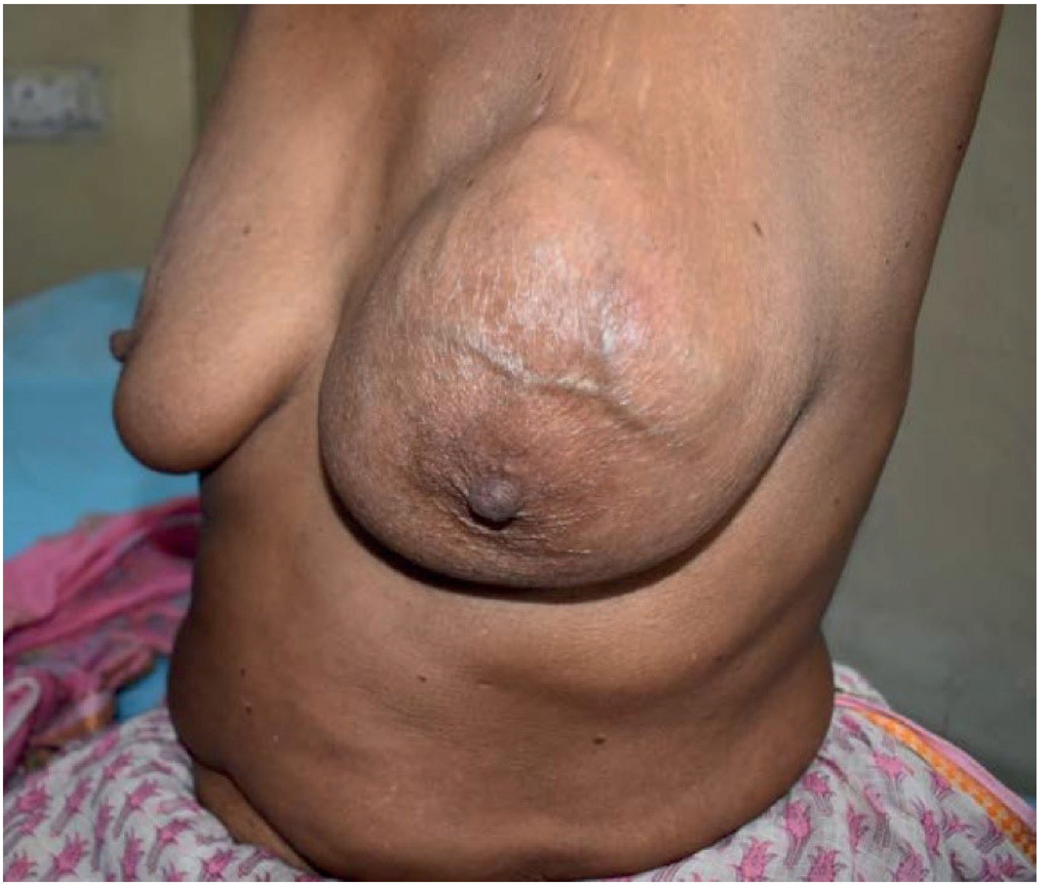
Clinical Features
| Feature | Detail |
|---|---|
| Age | Women >30 years (occasionally younger) |
| Presentation | Large, rapidly growing, bosselated breast mass |
| Consistency | Firm, rubbery |
| Overlying skin | Usually normal; may ulcerate in very large tumours (pressure necrosis) |
| Mobility | Mobile on chest wall (unlike malignant breast carcinoma) |
| Bilaterality | Rare |
Classification (Histological)
PHYLLOIDES TUMOUR CLASSIFICATION
Based on MITOTIC RATE per 10 High-Power Fields (HPF):
BENIGN BORDERLINE MALIGNANT
Mitotic rate Mitotic rate Mitotic rate
< 4/10 HPF 4–9/10 HPF ≥ 10/10 HPF
│ │ │
▼ ▼ ▼
Good prognosis Monitor closely Can metastasise
Local recurrence 10-15% recur Via BLOODSTREAM
possible May progress (NOT lymphatics)
Wide local Wide local MASTECTOMY
excision excision ± Radiotherapy
Histopathology
- Leaf-like intracanalicular projections into cleft-like spaces (Greek: "phyllodes" = leaf-like)
- Cellular stroma with varying atypia
- Both epithelial AND stromal components
- NOT a pure sarcoma (contains epithelium)
Management
TREATMENT OF PHYLLOIDES TUMOUR
1. BENIGN/BORDERLINE:
Wide Local Excision (WLE) with 2-cm clear margins
(including overlying skin + underlying pectoralis fascia)
- High local recurrence if margins inadequate
2. MALIGNANT / LARGE / RECURRENT:
Simple Mastectomy (no axillary clearance needed - lymph node spread rare)
± Postoperative radiotherapy (for malignant/recurrent)
± Systemic chemotherapy (for malignant phyllodes)
AXILLARY SURGERY: NOT routine (lymph node metastases rare)
FOLLOW-UP: Regular (10-year follow-up; late recurrence possible)
Q7. Thyroid Carcinoma: Classification, Papillary Thyroid Carcinoma (2+1+2+5 = 10 marks)
Part A: Classification of Thyroid Carcinoma (2 marks)
CLASSIFICATION OF THYROID CARCINOMA
│
┌─────────┼──────────┬──────────┐
▼ ▼ ▼ ▼
WELL- MEDULLARY UNDIFFEREN- LYMPHOMA
DIFFERENTIATED TIATED (rare)
│ │ (Anaplastic)
├── PTC │ │
│ (80%) │ Worst prognosis
│ │ 6% of thyroid Ca
└── FTC │
(10%) │
│
C-cell origin
(parafollicular)
Calcitonin ↑
RET proto-oncogene
MEN 2A & 2B
ORIGIN SUMMARY:
┌────────────────┬─────────────────────────────────┬────────────┐
│ Type │ Cell of Origin │ % of cases │
├────────────────┼─────────────────────────────────┼────────────┤
│ Papillary (PTC)│ Follicular cell │ 80% │
│ Follicular (FTC│ Follicular cell │ 10% │
│ Hurthle cell │ Follicular cell (oncocytic) │ 3-5% │
│ Medullary (MTC)│ Parafollicular C-cell │ 5-8% │
│ Anaplastic │ Follicular cell (dedifferentiated│ 1-2% │
│ Lymphoma │ Lymphocytes │ <1% │
└────────────────┴─────────────────────────────────┴────────────┘
SPECIAL ASSOCIATIONS:
• PTC: BRAF V600E mutation (most common), RET/PTC rearrangement
• FTC: RAS mutations, PAX8-PPARγ rearrangement
• MTC: RET proto-oncogene mutation (hereditary: MEN 2A, 2B)
• Anaplastic: TP53 mutation, often from dedifferentiated PTC/FTC
• Lymphoma: Associated with Hashimoto's thyroiditis
Part B: Clinical Features of Papillary Thyroid Carcinoma (1 mark)
Clinical Features
- Solitary thyroid nodule: Most common presentation - painless, firm, mobile lump in thyroid
- Cervical lymphadenopathy: Very common (30–80%) - especially young patients; nodes may present before the primary
- Slow-growing, often asymptomatic for years
- Rarely: hoarseness (RLN involvement), dysphagia, dyspnoea
- Rare distant metastases (lung, bone)
- Associated with: Prior radiation exposure (Chernobyl), childhood radiation to neck
Part C: Histopathology of PTC (2 marks)
Hallmark Nuclear Features of PTC (PATHOGNOMONIC)
HISTOPATHOLOGY OF PAPILLARY THYROID CARCINOMA
ARCHITECTURE:
• Papillary fronds with fibrovascular cores
(true papillae = fibrovascular stalk covered by neoplastic epithelium)
• May show follicular areas
NUCLEAR FEATURES (DIAGNOSTIC - any 2 of these is diagnostic):
┌─────────────────────────────────────────────────────────────┐
│ 1. "ORPHAN ANNIE EYE" NUCLEI (Ground-glass/clear nuclei) │
│ → Empty-looking nuclei with marginated chromatin │
│ → Most characteristic feature │
│ │
│ 2. NUCLEAR GROOVES │
│ → Longitudinal grooves (nuclear bars) │
│ │
│ 3. NUCLEAR PSEUDO-INCLUSIONS │
│ → Cytoplasmic invaginations into nucleus │
│ → Round, eosinophilic intranuclear inclusions │
│ │
│ 4. Nuclear crowding and overlapping │
│ 5. Large irregular nuclei │
└─────────────────────────────────────────────────────────────┘
PSAMMOMA BODIES:
• Laminated calcified concentric rings
• Pathognomonic of PTC (present in ~50%)
• Represent calcified foci of infarcted papillary tips
IMMUNOHISTOCHEMISTRY:
• TTF-1 positive (thyroid transcription factor)
• Thyroglobulin positive
• CK19 positive (cytokeratin)
• HBME-1 positive
• BRAF V600E staining
VARIANTS OF PTC:
• Classic PTC (most common)
• Follicular variant (lymph node mets less common)
• Tall cell variant (aggressive)
• Diffuse sclerosing variant (young patients, bilateral)
• Columnar cell variant
• Micropapillary (<10 mm, usually indolent)
Part D: Management of Papillary Thyroid Carcinoma (5 marks)
Risk Stratification (ATA Guidelines)
| Risk | Features |
|---|---|
| Low risk | Intrathyroidal PTC, <4 cm, no extrathyroidal extension, no vascular invasion, no mets |
| Intermediate | Aggressive histology, minimal ETE, vascular invasion, >5 nodes 0.2-3 cm |
| High risk | Gross extrathyroidal extension, incomplete resection, distant metastases, pT4 |
Management Flowchart
DIAGNOSIS OF PTC
(FNAC/Core biopsy + USS + CT/MRI neck + TFTs + Thyroglobulin)
│
▼
STAGING (TNM system):
T - Primary tumour size + extrathyroidal extension
N - Regional lymph node status
M - Distant metastases
│
┌────┴────┐
▼ ▼
MICROCARCINOMA CLINICALLY SIGNIFICANT PTC
(<10 mm) (≥10 mm or symptomatic)
│ │
▼ ▼
CONTROVERSIAL: TOTAL THYROIDECTOMY
- Active surveillance │
(Japan: 2/3 do not ┌───┴────────────────────┐
progress) ▼ ▼
- OR hemithyroidectomy LOW RISK HIGH RISK
│ │
Hemithyroidectomy Total thyroidectomy +
(lobectomy) if Central neck dissection
unifocal, <4 cm, (Level VI)
no ETE, no mets ±
Lateral neck dissection
(if N1b - level II-V)
SURGICAL DETAILS:
• Total thyroidectomy = removal of both lobes + isthmus
• AVOID injury to:
- Recurrent laryngeal nerve (RLN) - hoarseness
- External laryngeal nerve (ELN) - voice pitch loss
- Parathyroid glands (4) - hypocalcaemia
• Intraoperative neuromonitoring of RLN
POST-SURGICAL MANAGEMENT:
RADIOACTIVE IODINE (RAI / I-131):
• Remnant ablation: destroys remaining thyroid tissue
• Allows use of thyroglobulin as tumour marker
• Required for: High-risk, intermediate-risk PTC
• NOT required for: Low-risk PTC
• Preparation: Stop T4 for 4-6 weeks (or give recombinant TSH)
• Dose: 30–150 mCi depending on risk
TSH SUPPRESSION THERAPY:
• Levothyroxine (T4) given at doses to suppress TSH
• Rationale: TSH is a growth factor for thyroid cancer cells
• Target TSH:
- High risk: <0.1 mU/L (suppressed)
- Low risk: 0.5–2.0 mU/L (normal)
SURVEILLANCE:
• Serum thyroglobulin (Tg) levels - tumour marker
(should be undetectable post total thyroidectomy + RAI)
• Anti-Tg antibodies
• Neck USS (6-12 monthly)
• Whole body scan if Tg rises
PROGNOSIS:
• Excellent: 10-year survival >95% for low-risk PTC
• Worse in: Elderly, large tumour, ETE, distant mets
• Lymph node mets in young patients do NOT worsen prognosis
(unique feature of PTC - unlike other cancers)
TARGETED THERAPY (for advanced/refractory disease):
• Sorafenib (BRAF/VEGFR inhibitor)
• Lenvatinib (multi-kinase inhibitor)
• Dabrafenib + Trametinib (for BRAF V600E-positive)
• External beam radiotherapy (for unresectable/palliative)
Summary Table - AMES / MACIS Prognostic Scoring for PTC
| Factor | Low Risk | High Risk |
|---|---|---|
| Age | Female <50, Male <40 | Female >50, Male >40 |
| Metastases | None | Present |
| Extent | Intrathyroidal | ETE/incomplete resection |
| Size | <5 cm | >5 cm |
Sources: Sabiston Textbook of Surgery (wound healing); Bailey & Love's Short Practice of Surgery 28th Edition (SSI, blood transfusion, phylloides, thyroid); Rosen's Emergency Medicine (shock, septic shock, Buerger's disease); Robbins & Kumar Basic Pathology (thyroid carcinoma histopathology)