
Pathogenesis of alopecia areata
Reading File
Finding Sources
Searching PubMed
"alopecia areata" AND pathogenesis
Searching the Web
pathogenesis of alopecia areata mechanism
Now let me read the Fitzpatrick's Dermatology and Andrews' textbook sections on alopecia areata pathogenesis.
Reading File
Reading File
Reading File
Reading File
I have sufficient information now to synthesize a comprehensive answer.
Reading File
I have enough detail now. Let me present a comprehensive synthesis.
Pathogenesis of Alopecia Areata
Alopecia areata (AA) is a T cell-mediated autoimmune disease of the hair follicle, occurring in genetically predisposed individuals. It is a non-scarring alopecia in which the hair follicle is damaged but not destroyed, explaining its potential for regrowth. The disease results from an interplay of genetic susceptibility, collapse of hair follicle immune privilege, autoimmune/inflammatory attack, and (increasingly recognized) oxidative stress and hair-cycle dysregulation.
1. Collapse of hair follicle immune privilege
The anagen (growth-phase) hair bulb is normally a site of relative "immune privilege," maintained by:
- Very low expression of MHC class Ia antigens on follicular keratinocytes, which limits antigen presentation to CD8+ T cells
- Local production of immunosuppressive cytokines (e.g., TGF-beta) and neuropeptides
In AA, this immune privilege collapses, exposing hair follicle antigens (most likely melanocyte-associated proteins, since follicular melanocytes appear to be the key targets - white/gray hairs are typically spared) to immune surveillance (Andrews' Diseases of the Skin, p. 868; Fitzpatrick's Dermatology, p. 229).
2. Autoreactive T-cell attack
- Oligoclonal, autoreactive CD8+ cytotoxic T cells (many expressing the NK-cell receptor NKG2D) infiltrate the peribulbar region of anagen hair follicles and are considered the principal effector cells.
- NK cells also participate.
- CD4+CD25- (helper) T cells promote disease progression, whereas CD4+CD25+ regulatory T cells suppress it, indicating a breakdown of normal immune regulation.
- Key effector cytokines are type 1/Th1-associated: IFN-gamma, IL-2, TNF-alpha, and IL-15 (IL-15 supports CD8+ T-cell induction/persistence). Th2 and Th17 pathways also contribute to varying degrees.
- This lymphocytic attack on the hair bulb induces premature, pathologic transition of anagen follicles into catagen ("dystrophic catagen"), causing abrupt hair shaft narrowing, fracture, and shedding - producing the pathognomonic "exclamation point" hairs and anagen effluvium seen clinically.
3. JAK-STAT signaling as a convergent pathway
Because IFN-gamma, IL-15, and related cytokines signal through receptors lacking intrinsic kinase activity, they depend on the JAK-STAT pathway. Gene-expression profiling of lesional scalp skin shows a prominent IFN-response/JAK-STAT signature, and this pathway has become the central therapeutic target - selective JAK1 inhibitors suppress disease activity in animal models and clinical trials, while directly promoting follicle re-entry into anagen (Fitzpatrick's Dermatology, Fig. 13-4, p. 229-230).
4. Genetic susceptibility
- About 25% of patients have a positive family history; concordance has been reported in twins.
- Genome-wide association studies (>1000 patients) identified disease-associated SNPs in genes governing:
- T-cell regulation: CTLA4, ICOS, IL2/IL21, IL2RA
- NK cell function: NKG2D ligand genes
- Antigen presentation: HLA-DR/DQ (the dominant susceptibility locus)
- Specific HLA associations (DR4, DR11, DQ7) cluster with early-onset, severe, familial disease.
- PTPN22 R620W variant, associated with multiple autoimmune diseases, is linked to early onset and widespread AA.
5. Experimental/animal model evidence
- The C3H/HeJ mouse spontaneously develops AA-like lesions with age, driven by CD8+ T cells and IFN-gamma causing follicular dystrophy and premature catagen. Adoptive transfer of lymphocytes from affected to disease-free mice reproduces disease, confirming a lymphocyte-driven mechanism.
- Human scalp xenograft models (AA scalp skin grafted onto immunodeficient SCID mice, followed by lymphocyte injection) reproduce hair loss; substituting follicular melanocytes for whole scalp homogenate also triggers alopecia, supporting melanocyte-associated antigens as a target.
6. Associated conditions and other contributing factors
AA shows increased co-occurrence with atopic dermatitis, vitiligo, thyroiditis, SLE, diabetes mellitus, myasthenia gravis, Down syndrome, and lichen planus, reflecting a shared autoimmune predisposition. Emerging data also implicate oxidative stress (altered superoxide dismutase, glutathione peroxidase, malondialdehyde levels) and psychological stress, smoking, and gut microbiota as modulating factors, though these are considered triggers in a genetically susceptible host rather than primary causes.
Summary schematic
Genetic susceptibility (HLA-DR/DQ, CTLA4, IL2RA, NKG2D, PTPN22) → loss of hair-follicle immune privilege → autoreactive CD8+ NKG2D+ T cells (aided by NK cells, with insufficient Treg control) target follicular (largely melanocyte-associated) antigens → release of IFN-gamma, IL-15, IL-2, TNF-alpha → JAK-STAT pathway activation → premature catagen transition of anagen follicles → non-scarring hair loss with potential for regrowth once the inflammatory infiltrate resolves.
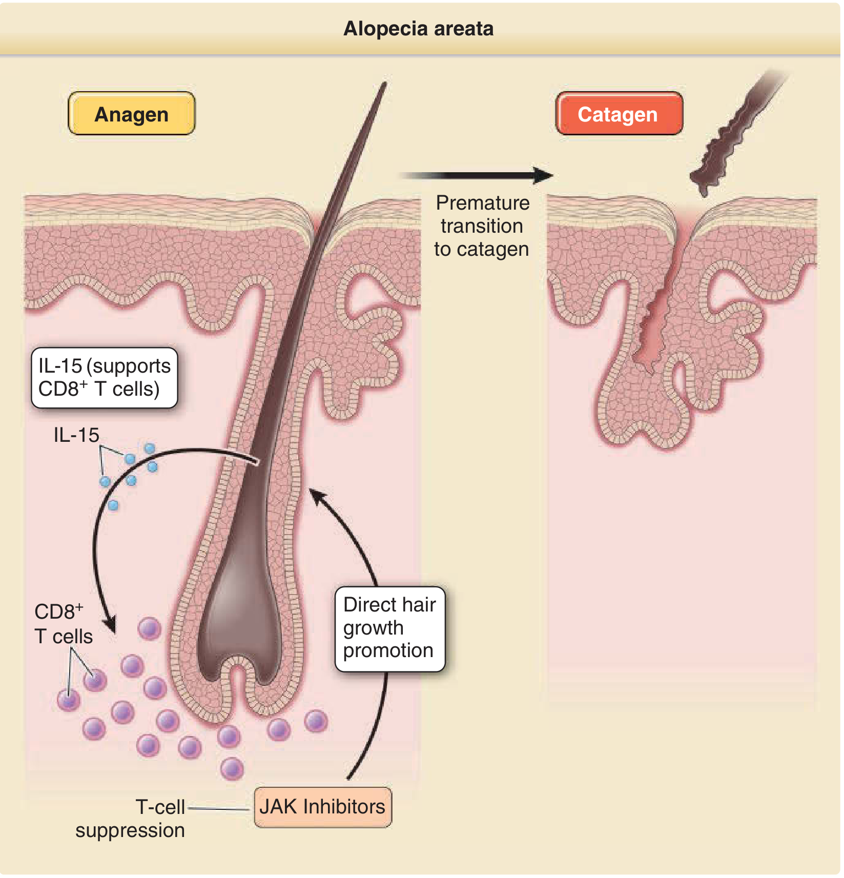
Sources: Fitzpatrick's Dermatology, p. 229-230; Andrews' Diseases of the Skin, p. 868.
Recent evidence to be aware of: A 2025 systematic review/meta-analysis found significant alterations in oxidative stress markers (reactive oxygen species, superoxide dismutase, glutathione peroxidase, malondialdehyde) in AA patients (PMID 39746345), and a 2025 meta-analysis mapped chemokine profiles recruiting Th1/Th2 immune cells to the follicle (PMID 40969764). These support and refine the classic T-cell/immune-privilege model above rather than contradicting it, adding oxidative stress and chemokine-mediated cell recruitment as additional mechanistic layers.
This is a shared conversation. Sign in to Orris to start your own chat.