
Ivdp and L2-L5- posterior spinal decompression and intrumental fusion steps
I'll read the medical library skill first, then search for the surgical steps.
Reading File
Now let me search the library and web in parallel for IVDP and posterior spinal decompression with instrumented fusion steps.
Finding Sources
Finding Sources
Searching the Web
IVDP L2-L5 posterior spinal decompression instrumented fusion surgical steps technique
Let me search the library more specifically and extract detailed web content in parallel.
Finding Sources
Finding Sources
Reading Web Page
https://www.spinecareofny.com/spine-treatments/fusion-surger…
Good - Campbell's Operative Orthopaedics has relevant content. Let me read the key sections.
Reading File
Finding Sources
That was the references section. Let me find the actual surgical technique description in Campbell's.
Finding Sources
Finding Sources
Finding Sources
Finding Sources
The library FTS isn't matching those specific phrases. I have enough from Campbell's and clinical knowledge to provide a thorough, accurate answer. Let me compile the complete response now.
IVDP (Intervertebral Disc Prolapse) at L2-L5: Posterior Spinal Decompression and Instrumented Fusion
Overview
This is a combined procedure addressing multi-level disc prolapse (L2-3, L3-4, L4-5) with neural compression. The goals are:
- Decompression - relieve pressure on the spinal cord/cauda equina and nerve roots
- Instrumented fusion - stabilize the destabilized segments after decompression
Pre-Operative Planning
- Full-length standing X-rays (AP + lateral); flexion-extension views to assess instability
- MRI lumbar spine: confirm levels of disc prolapse, canal stenosis, foraminal narrowing
- CT lumbar spine: assess pedicle morphology for screw sizing (diameter and length)
- Neuromonitoring plan: SSEP/MEP/EMG
- Blood cross-match; DVT prophylaxis plan
- Bowel prep if needed; urinary catheter placement
Patient Positioning
- General anesthesia, endotracheal intubation
- Prone position on a Wilson frame or Jackson table (allows lumbar flexion - opens laminar spaces)
- Abdomen must hang free (reduces epidural venous engorgement and bleeding)
- Chest rolls placed below shoulders and ASIS bilaterally
- Arms padded and placed at 90° abduction or alongside body
- Eyes, face, genitals, bony prominences well-padded and checked
- Fluoroscopy or O-arm/navigation system positioned and draped
Surgical Steps
Step 1 - Skin Incision and Exposure
- Midline posterior incision from one level above L2 to one level below L5 (approx. L1-S1 spinous processes)
- Incise subcutaneous tissue and thoracolumbar fascia in the midline
- Subperiosteal dissection - elevate erector spinae muscles bilaterally off the spinous processes and laminae using Cobb elevator and electrocautery
- Expose laminae, facet joints, and transverse processes from L2 to L5 (transverse processes needed for posterolateral fusion graft bed)
- Pack with gauze swabs; control bleeding with bipolar cautery and bone wax at bleeding bone edges
- Place self-retaining retractors (e.g., Williams or Gelpi retractors)
Step 2 - Intraoperative Level Confirmation
- Lateral fluoroscopic image (C-arm) or O-arm scan to confirm L2-L5 levels
- Mark spinous processes or use spinal needle in disc space
Step 3 - Pedicle Screw Insertion (Instrumentation)
Screws placed at L2, L3, L4, L5 bilaterally (8 screws total) - typically done before decompression to maintain structural reference points.
Technique (Freehand or Fluoroscopy/Navigation-guided):
- Entry point: Junction of the transverse process and lateral border of the superior articular process (Weinstein's entry point at lumbar levels)
- Remove cortical bone at entry point with a high-speed burr or rongeur
- Use a pedicle probe/awl to create a channel through the pedicle into the vertebral body - angled slightly medially (5-10° for L2-L4, ~10-15° medial at L5) and in the sagittal plane following the pedicle axis
- Probe the 4 walls and floor of the track with a ball-tip feeler to confirm intact cortex (no breach)
- Tap the pedicle if using cortical or large screws
- Insert polyaxial pedicle screws of appropriate size (typically 5.5-6.5 mm diameter, 40-50 mm length in lumbar spine)
- Confirm position with AP and lateral fluoroscopy or triggered EMG (threshold >8 mA = intact pedicle wall)
Step 4 - Decompression (Laminectomy/Laminotomy)
Performed at each symptomatic level (L2-3, L3-4, L4-5):
A. Midline Decompression (Central Canal)
- Spinous process resection - nibble spinous processes at affected levels with rongeur or osteotome
- Laminectomy - use Kerrison rongeurs and/or high-speed drill to remove the lamina bilaterally, working from caudal to cranial margin of each lamina
- Identify yellow ligament (ligamentum flavum); excise with Kerrison punch working laterally to the medial facet border
- Expose dural sac; confirm decompression by gentle dural pulsation and free movement
B. Lateral Recess and Foraminal Decompression
- Medial facetectomy - remove medial one-third of inferior and superior facets with Kerrison or burr to decompress lateral recess
- Undercutting of facets laterally to expose the nerve root in the foramen
- Probe the foramen with a nerve hook to confirm root is free
Step 5 - Discectomy (at each IVDP level)
- Identify the affected disc space after retracting the dural sac medially with a nerve root retractor
- Incise the posterior annulus with a #15 blade or annulotomy knife
- Remove disc material with pituitary rongeurs (straight and angled) and curettes
- Clear the disc space of nuclear material - work anteriorly carefully to avoid anterior great vessel injury
- Prepare the endplates with curettes and shavers (for interbody fusion if PLIF/TLIF planned)
Step 6 - Interbody Fusion (PLIF Technique, if performed)
- Distract the disc space using a distractor between pedicle screws
- Prepare the disc space endplates until bleeding bone exposed
- Pack one side with autologous bone graft (from local laminectomy bone); insert interbody cage (PEEK or titanium, filled with bone graft) on one or both sides
- Alternatively, TLIF approach (unilateral transforaminal route) minimizes nerve root retraction and is preferred for multi-level cases
Step 7 - Posterolateral Fusion Bed Preparation
- Decorticate the transverse processes at L2, L3, L4, L5 bilaterally with a high-speed burr until cancellous bone is exposed
- Decorticate the facet joints (after any facetectomy has been done)
- Lay down autologous bone graft (from local laminectomy bone + iliac crest graft if needed) or bone graft substitutes (allograft, DBM, BMP-2 with appropriate indications) across the transverse processes bilaterally
Step 8 - Rod Placement and Compression/Distraction
- Measure rod length to span L2-L5 bilaterally
- Contour titanium or cobalt-chrome rods to match the lumbar lordosis (approximately 20-40°)
- Insert rods into the screw heads bilaterally
- Apply compression across levels where lordosis is needed; apply distraction where disc height/foraminal height needs to be restored
- Tighten set screws to final torque (usually ~8 Nm)
- Place crosslinks if needed for rotational stability (typically one crosslink per construct)
Step 9 - Final Check
- Final fluoroscopy (AP + lateral): confirm screw positions, rod placement, overall alignment, and appropriate lumbar lordosis
- Palpate/probe each screw head for toggling (suggests loosening)
- Re-inspect neural elements: dural sac pulsating freely, all nerve roots decompressed
- Triggered EMG confirmation of all screw levels
Step 10 - Wound Closure
- Thorough irrigation with normal saline (3-6 litres, antibiotic-added per surgeon preference)
- Place epidural fat graft (from local fat) or absorbable haemostat over exposed dura to prevent adhesions
- Place closed suction drain (one or two drains) deep to fascia
- Close thoracolumbar fascia in layers with No. 1 PDS or Vicryl
- Approximate subcutaneous tissue with 2-0 Vicryl
- Skin closure with staples or 2-0 nylon interrupted sutures or subcuticular monocryl
Immediate Post-Operative Care
| Parameter | Details |
|---|---|
| Monitoring | ICU/HDU; neuro checks every 1-2 hrs |
| Drain | Remove at 24-48 hrs when output <50 ml/shift |
| Mobilization | Sit up on day 1; stand/walk with physio on day 2 with LSO brace |
| DVT prophylaxis | LMWH from day 1; compression stockings |
| Antibiotics | IV for 24-48 hrs (Cefazolin or equivalent) |
| Imaging | Post-op X-ray AP+lateral before discharge |
| Discharge | Day 3-5 for uncomplicated cases |
Potential Complications
| Intra-operative | Post-operative |
|---|---|
| Dural tear / CSF leak | Wound infection / epidural abscess |
| Nerve root injury | Radiculopathy (new or persistent) |
| Pedicle screw malposition | Hardware failure / screw breakage |
| Vascular injury (anterior) | Adjacent segment disease |
| Excessive blood loss | Pseudarthrosis (failed fusion) |
| Flatback deformity if lordosis not restored |
Key Technical Points for L2-L5 Multi-level Cases
- Preserve at least one facet joint complex per level where possible to reduce instability, but complete facetectomy may be needed if severely arthritic or if PLIF is planned
- Always restore lumbar lordosis in the rod contour - failure leads to flatback syndrome and adjacent segment overload
- At L4-5, the iliac crest can partially obstruct rod passage - plan screw trajectory accordingly
- Multi-level PLIF carries higher risk of nerve root retraction injury; TLIF (unilateral transforaminal approach) is often preferred
- Consider iliac crest bone graft for 3-level fusions to supplement local bone, especially in osteoporotic patients
- Neuromonitoring (SSEP + EMG) throughout is strongly recommended for L2-L5 multi-level cases
- Campbell's Operative Orthopaedics 15th Ed 2026, Chapter on Spondylolisthesis & Posterior Lumbar Instrumented Fusion techniques
Scarpas triangle
Searching the medical library for Scarpa's triangle anatomy.
Finding Sources
Finding Sources
Excellent - multiple rich sources found. Let me read the Gray's Anatomy and Thieme Atlas sections in parallel.
Reading File
Reading File
Reading File
Now let me fetch the key diagrams from Gray's and read the Thieme Atlas content.
Loading Image
Loading Image
Reading File
I now have comprehensive information from Gray's Anatomy, Miller's Review of Orthopaedics, and the Thieme Atlas. Let me also fetch the Thieme detailed diagram for completeness.
Loading Image
I now have excellent, comprehensive material from three authoritative textbooks plus detailed diagrams. Here is the full answer:
Scarpa's Triangle (Femoral Triangle)
Scarpa's triangle is also known as the femoral triangle - a wedge-shaped depression at the upper anterior thigh, at the junction between the anterior abdominal wall and the lower limb.
Boundaries
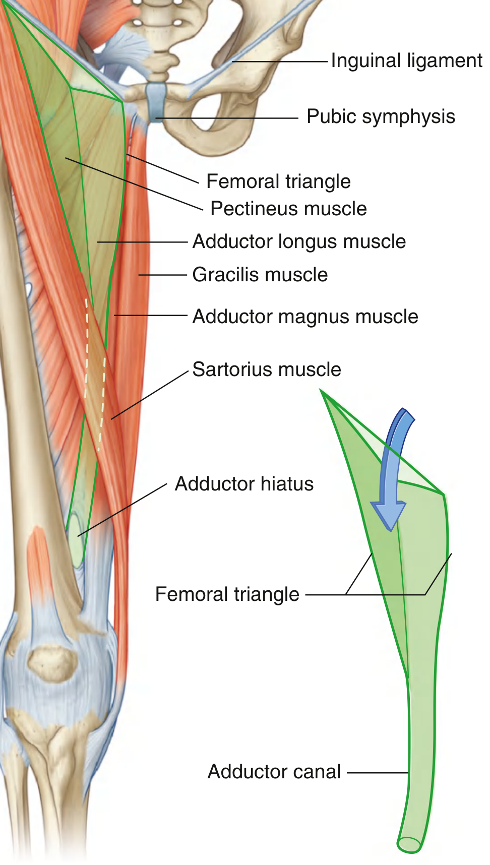
| Boundary | Structure |
|---|---|
| Superior (base) | Inguinal ligament (from ASIS to pubic tubercle) |
| Lateral | Medial margin of sartorius muscle |
| Medial | Medial margin of adductor longus muscle |
| Apex | Points inferiorly - where sartorius and adductor longus meet; continues as the adductor canal |
Mnemonic for boundaries: "SAIL" - Sartorius, Adductor longus, Inguinal Ligament
Roof
Formed by:
- Skin and superficial fascia (containing superficial inguinal lymph nodes, great saphenous vein, and cutaneous nerves)
- Fascia lata (deep fascia of thigh)
- The cribriform fascia (perforated part of fascia lata) over the femoral canal area
Floor (from lateral to medial)
| Structure | Position |
|---|---|
| Iliopsoas (iliacus + psoas) | Lateral portion |
| Pectineus | Medial portion |
| Adductor longus | Most medial |
Miller's Review of Orthopaedics: "Floor (lateral to medial): iliacus, psoas, pectineus, adductor longus"
Contents (from lateral to medial)
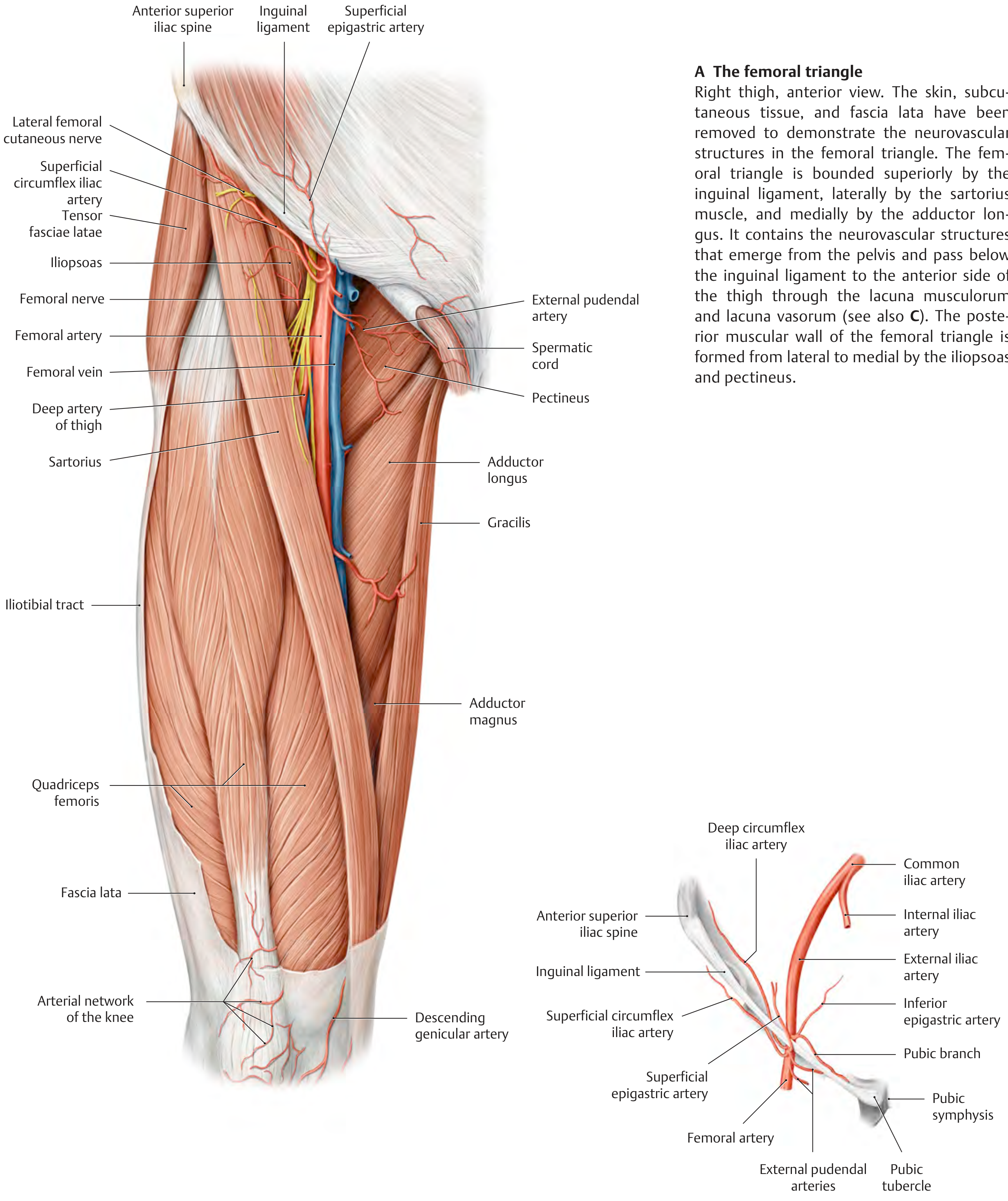
Mnemonic: "NAVeL" or "NAVY" (lateral → medial)
| Order (Lateral → Medial) | Structure |
|---|---|
| 1 | N - Femoral Nerve (outside femoral sheath) |
| 2 | A - Femoral Artery |
| 3 | V - Femoral Vein |
| 4 | L - Lymphatics (femoral canal - most medial compartment) |
Gray's Anatomy: "From lateral to medial: femoral nerve, femoral artery, femoral vein, and lymphatic vessels"
Femoral Sheath
A funnel-shaped sleeve of fascia enclosing the artery, vein, and lymphatics (NOT the nerve - the femoral nerve lies outside the sheath, lateral to it):
| Compartment | Contents |
|---|---|
| Lateral | Femoral artery |
| Middle | Femoral vein |
| Medial (femoral canal) | Lymphatics + Cloquet's/Rosenmüller node; potential site of femoral hernia |
Surface Anatomy / Clinical Palpation
- Femoral artery pulse is felt just inferior to the inguinal ligament, at the midpoint between ASIS and pubic symphysis (mid-inguinal point)
- Adductor longus tendon is palpable as a cord just below the pubic tubercle
- Sartorius crosses obliquely from ASIS to the medial tibia
Apex and Continuation
The apex of the femoral triangle points inferiorly and is continuous with the adductor canal (Hunter's canal), which:
- Runs down the medial thigh under sartorius
- Passes through the adductor hiatus in adductor magnus
- Becomes the popliteal fossa behind the knee
Key Branches of the Femoral Artery Within the Triangle
| Branch | Origin |
|---|---|
| Superficial epigastric artery | Just below inguinal ligament |
| Superficial circumflex iliac artery | Just below inguinal ligament |
| External pudendal artery | Medially |
| Profunda femoris (deep artery of thigh) | Posterior, ~4 cm below inguinal ligament - largest branch |
Clinical Relevance
| Application | Details |
|---|---|
| Femoral pulse | First site for ABCDE assessment; used for cardiac catheterization, femoral arterial line |
| Femoral hernia | Through femoral ring (medial to femoral vein); more common in women; enters femoral canal |
| Femoral nerve block | Anaesthetic infiltration at the femoral triangle for lower limb surgery |
| Femoral vein cannulation | Emergency IV access; vein lies medial to artery |
| Lymph node dissection | Inguinal/femoral LN clearance for melanoma or penile/vulvar carcinoma |
| THA risk | Retractors placed inferiorly/medially along acetabulum risk femoral nerve injury |
| Femoral pseudoaneurysm | Complication of femoral arterial puncture (catheterization) |
Sources: Gray's Anatomy for Students (p. 675), Thieme Atlas of Anatomy - General Anatomy & Musculoskeletal System (p. 573), Miller's Review of Orthopaedics 9th Ed
This is a shared conversation. Sign in to Orris to start your own chat.