
Long answer on virtual autopsy
Virtual autopsy is a medical/forensic topic, so I need to invoke the medical library skill first.
Quick answer: Virtual autopsy (also called "virtopsy") is a non-invasive or minimally invasive post-mortem examination technique that uses advanced imaging modalities - primarily CT and MRI - to document, analyze, and interpret findings in a deceased individual without the need for traditional surgical dissection. It has gained significant traction in forensic pathology, offering advantages in certain cases while complementing conventional autopsy.
Searching sources...
Reading File
Now let me search the medical library and PubMed in parallel for virtual autopsy content.
Finding Sources
Finding Sources
Searching PubMed
"virtual autopsy" OR "virtopsy" forensic imaging
Excellent findings. Now let me read the detailed content from both textbooks in parallel.
Reading File
Reading File
Finding Sources
Good content collected. Now let me read more from Parikh's (Q 9.2) and from the Brogdon radiology book sections, plus the Essentials of Forensic Medicine section.
Reading File
Reading File
Reading File
Excellent - rich content gathered. Let me now read the comparison table from Parikh and the Brogdon MSCT/MRI sections, and also fetch key images.
Reading File
Reading File
Searching Images
virtual autopsy postmortem CT scan forensic body imaging
Let me fetch the CT scan figure from Brogdon and also read more from Brogdon about specific applications.
Loading Image
Reading File
Finding Sources
I now have comprehensive content from multiple textbook sources. Let me do one final read to get the Virtobot/team structure details and specific pediatric findings, plus fetch the full-body scan image.
Reading File
Loading Image
I now have comprehensive information to write a thorough long answer. Let me compose it.
Virtual Autopsy (Virtopsy)
1. Definition and Origin
Virtual autopsy - widely known by the trademark term "Virtopsy" - is a non-invasive or minimally invasive post-mortem examination technique that determines the cause of death and documents forensic findings using advanced imaging modalities, without requiring the traditional surgical dissection of a conventional autopsy.
The term "Virtopsy" was coined by Professor Richard Dirnhofer, former Head of the Institute of Forensic Medicine (IFM), University of Bern, Switzerland, in collaboration with Michael J. Thali and their team. Dirnhofer stated that virtopsy "fully satisfies the requirement that forensic medical findings provide a complete and true picture of the examined object" and helps any reader to visually follow the expert's reasoning. The first landmark feasibility study - "Virtopsy, a new imaging horizon in forensic pathology: Virtual autopsy by postmortem multislice computed tomography (MSCT) and magnetic resonance imaging (MRI)" - was published in the Journal of Forensic Sciences in 2003 (Thali MJ, Yen K, Schweitzer W et al., J Forensic Sci. 48(2):386-403, 2003).
- Parikh's Textbook of Medical Jurisprudence Forensic Medicine and Toxicology, p. 158-159
- Brogdon's Forensic Radiology, p. 415-416, 475
2. Core Concept
Virtual autopsy is not a real autopsy involving dissection and cutting of organs. Instead, it involves imaging of the body using:
- 3D surface scanning
- Multislice computed tomography (MSCT)
- Magnetic resonance imaging (MRI)
- Photogrammetry (3D/CAD)
It results in non-invasive, non-subjective, digitally stored, and web-transmittable findings that can be presented before a court of law. All records can be shared for second opinions across institutions. Coloured 3D pictures in multiple sections allow visualization of the surface, deeper tissues, organs, coronary arteries, pulmonary emboli, soft tissue trauma, and bone injuries.
3. Imaging Modalities Used
3.1 Multislice Computed Tomography (MSCT / pmCT)
MSCT (postmortem CT, pmCT) is the workhorse of virtual autopsy. Technical requirements per the Bern Virtopsy protocol:
- A 16-row multidetector CT with large bore is sufficient; even 4- or 6-row MDCT works with concessions
- Whole-body data acquired with slice thickness <3 mm in axial sections
- Sagittal and coronal reformations calculated
- Selected regions (larynx, small implants, coronaries) additionally scanned at sub-millimeter slice thickness
- Image reconstruction in both soft-tissue and bone-weighted kernels
- 3D reconstructions post-processed for court presentation (software: Leonardo, Siemens Medical Solutions, OsiriX)
Applications of pmCT:
- Morbid anatomical findings
- Firearm injuries: entrance/exit wound determination via inward/outward beveling of bone
- Projectile trajectory tracking through brain and organs
- Explosions, charred bodies, decomposed bodies
- Child abuse - fractures in various stages of healing
- Sex and age estimation
- Emphysema, air embolism, pneumothorax, hyperbaric trauma
- Multi-planar reconstruction (MPR) and volume rendering technique (VRT)
3.2 Magnetic Resonance Imaging (MRI / pmMRI)
MRI provides superior soft-tissue contrast and is best performed using a whole-body imaging MRI unit (TIM - Total Imaging Matrix) with a wide inner bore diameter. Coronal, sagittal, and axial images with different signal weightings are acquired.
Applications of pmMRI:
- Soft tissue injury and organ trauma
- Pathologies of the CNS - focal cortical injuries, shearing injuries, intraparenchymal hemorrhage
- State of blood vessels
- Mastoid fluid, small brain tears that may be confused with artifacts of organ removal
- Superior to CT in gross cranial, pulmonary, and vascular abnormalities
- MR spectroscopy measures metabolites formed during decomposition, enabling post-mortem interval (time since death) estimation
3.3 Postmortem CT Angiography (pmCTA)
Conventional pmCT cannot directly visualize the vascular system, relying only on indirect signs (perilesional hematoma, collapse of great vessels). Postmortem CT angiography (pmCTA) was introduced to close this gap.
In 2005, the first minimally invasive whole-body pmCTA of an adult cadaver was performed (Jackowski et al., J Forensic Sci, 50(5):1175-1186, 2005). pmCTA uses contrast agents injected into the circulatory system post-mortem, providing:
-
3D volumetric vascular imaging (vs. 2D plain projectional images)
-
Visualization of coronary stenoses, traumatic vascular lesions, pulmonary emboli
-
Display of neck, skull base, and pelvic structures not reviewable in classic autopsy without major damage
-
Extension of diagnostic spectrum to identifying natural causes of death
-
Brogdon's Forensic Radiology, p. 475
3.4 3D Surface Scanning / Photogrammetry
The body surface is scanned using robot-guided 3D photogrammetric techniques, producing a digital surface model. This allows:
- Precise documentation of external wounds
- Pattern injury analysis (linking wound to weapon)
- Virtual model simulation: computer software can create a virtual model of an injury using a 3D image simulation of a similar weapon
3.5 Image-Guided Postmortem Biopsy
CT and MRI cannot assess histopathologic changes (image resolution insufficient for cellular-level examination). Therefore, postmortem image-guided needle biopsies are performed to obtain tissue specimens for:
- Classic histopathologic examination (gold standard)
- Toxicologic analysis of body fluids (blood, urine, cerebrospinal fluid)
- Sampling techniques include: fine needle aspiration (FNA), Tru-Cut/core biopsies with automated biopsy guns, coaxial technique, CT-guided step-and-shoot, CT-fluoroscopy, navigated/robot-assisted biopsies
- Applications: cause of death investigations, forensic vitality assessment, pulmonary fat embolism detection, aspiration/inhalation of gas determination
4. The Virtobot
The Virtobot is the robotic system developed at the IFM Bern that integrates all virtopsy modalities into a semi-automated forensic examination pipeline. As described in Brogdon's Forensic Radiology (p. 415), the IFM has had a forensic examination process line capable of subjecting a corpse to:
- Robot-guided 3D photogrammetry-supported surface area scanning
- CT examination
- Postmortem biopsy
- Postmortem angiography
- Whole-body MR scanning (supplementary)
The fourth generation Virtopsy team at Bern - consisting of graduate engineers, radiologists, imaging specialists, and forensic doctors under the direction of Michael J. Thali - reported that implementing steps 1-3 alone resolves approximately 60-80% of forensic case material arriving at the institute.
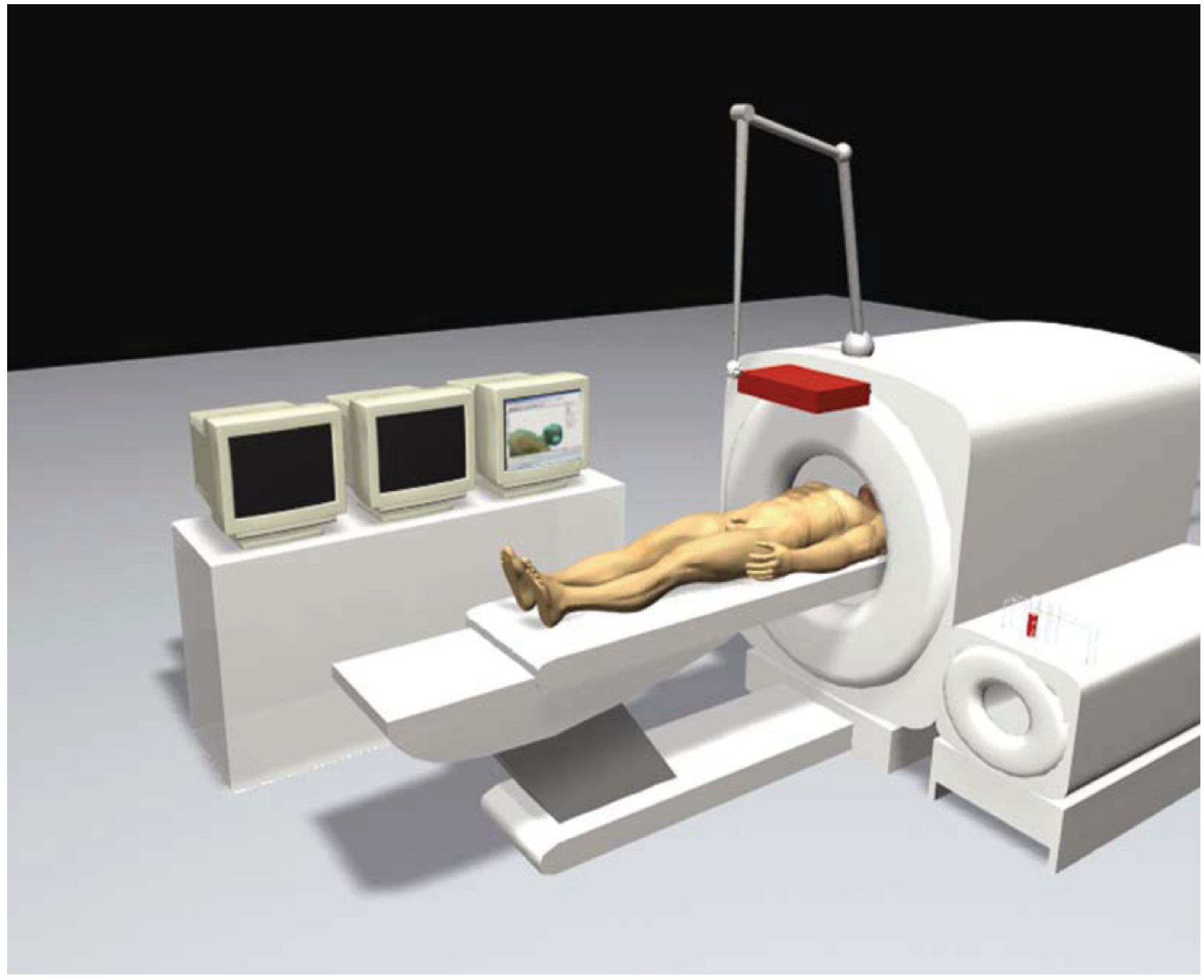
Figure: The Virtobot concept - a person in an MRI machine with three monitors for simultaneous imaging at the NIH presentation (Brogdon's Forensic Radiology)
5. Applications of Virtual Autopsy
| Area | Application |
|---|---|
| Traumatic deaths | Gunshot wounds, stab wounds, blunt force trauma - wound documentation without disturbing body structure |
| Firearm injuries | Entrance/exit wound analysis by beveling pattern; projectile trajectory tracking |
| Explosions | Internal and external injuries mapped in 3D |
| Charred/burned bodies | Skeletal and soft tissue analysis when surface examination is unreliable |
| Decomposed bodies | Imaging through maceration and putrefaction stages |
| Child abuse | Occult fractures, intracranial injuries, shearing tears; subdural hematomas |
| Natural causes of death | Coronary artery disease (via pmCTA), pulmonary embolism, aortic dissection |
| Drowning | Pulmonary findings, diatom presence |
| Air/gas embolism | CT is superior - directly visualizes intravascular gas |
| Identification | Dental anatomy, skeletal age and sex estimation, implant identification |
| Toxicology | MR spectroscopy of metabolites; guided fluid sampling |
| Time of death | MR spectroscopy of decomposition metabolites |
- The Essentials of Forensic Medicine and Toxicology (36th ed., 2026), p. 126
- Brogdon's Forensic Radiology
6. Virtual Autopsy in Infants and Children
Virtual autopsy has particular utility in pediatric deaths, including suspected child abuse cases and perinatal deaths.
Historical milestones (per Brogdon's Forensic Radiology):
- 1985-2003: Tsukuba Medical Center Hospital (Japan) performed >500 postmortem CT examinations
- 1990: Ros and colleagues - first postmortem preautopsy MRI in the pediatric age group, including stillborns of 29-42 weeks gestational age; found MRI superior for gross cranial, pulmonary, vascular abnormalities, and detecting air/fluid in potential body spaces
- 1990-1993: Hart et al. - studied 11 cases of unexplained death or suspected child abuse (≤2 years); MRI advantages included mastoid fluid, focal cortical injuries, shearing injuries, and small tears; preautopsy imaging directed pathologist attention to abnormal areas
- Consensus: Postmortem CT and MRI each have advantages in detecting certain abnormalities, and combining both is optimal - Brogdon's Forensic Radiology, p. 7383-7414
A 2025 review in NeoReviews [PMID: 40744461] addresses virtual autopsy in perinatal pathology and highlights the ongoing need for standardization across centers.
A 2023 systematic review (Wickramasinghe et al., SAGE Open Medicine [PMID: 37197019]) confirmed usefulness of virtual autopsy in diagnosing pathologies in the pediatric population.
7. Comparison: Virtopsy vs. Conventional Autopsy
| Feature | Virtopsy | Conventional Autopsy |
|---|---|---|
| Technique | No scalpel, non-invasive, no dissection | Involves opening and dissection |
| Wound study | Without disturbing body structure | Requires opening |
| Mutilation/artifacts | No mutilation; no dissection artifacts | Mutilation and dissection artifacts occur |
| Time | Less time consuming (for imaging) | More time consuming |
| Religious/cultural acceptance | More willingly accepted | Often objected to on religious/cultural grounds |
| Evidence preservation | Digitally stored; reproducible; transmissible | Tissue preserved but not digitally reproducible |
| Legal use | Full digital record presented in court | Written report + histology samples |
| Air/gas detection | CT superior (direct visualization) | Difficult; gaseous findings may be lost |
| Histopathology | Requires image-guided biopsy | Direct tissue sampling (gold standard) |
| Toxicology | Guided sampling; MR spectroscopy | Direct organ and fluid sampling |
| Cost | High (scanner, software, robots) | Relatively lower |
| Infection hazard to staff | Greatly reduced | Present |
| Small tissue injuries | May be missed | Better detection by direct examination |
| Infection status | Cannot be discerned | Can be determined by culture/histology |
- Parikh's Textbook, p. 158-159
8. Advantages of Virtual Autopsy
- Non-invasive - no scalpel, no dissection
- No mutilation - body returned intact to family
- Culturally and religiously acceptable - particularly important in communities that prohibit conventional autopsy
- Digitally stored and reproducible - findings can be reviewed, re-analyzed, and transmitted globally
- Objective documentation - reduces inter-observer variability
- Evidence preservation - complete digital record admissible in court, allowing expert witnesses to be examined remotely
- No disturbing of body structure - wounds and injuries examined in situ without artifact
- Superior air/gas detection - CT directly visualizes pneumothorax, gas embolism, air in body cavities
- 3D visualization - multi-planar reconstruction and volume rendering allow anatomical perspectives impossible with conventional autopsy
- Vascular imaging - pmCTA reveals coronary stenoses, vascular tears, pulmonary emboli
- Reduced biohazard to forensic staff
- Pediatric advantage - particularly in detecting intracranial injuries and subtle skeletal trauma
9. Disadvantages and Limitations
- High cost of equipment (CT scanner, MRI unit, 3D scanners, software, robots)
- Limited resolution - CT and MRI cannot replace histopathology; cellular-level changes cannot be detected
- Metal foreign objects - cause artifacts and degrade image quality
- Colour of internal organs - cannot be clearly appreciated; important in assessing congestion, icterus, pallor
- Insufficient database - normative postmortem imaging data is still being built
- Infection status - cannot determine microbiological cause of death
- Antemortem vs. postmortem wound differentiation - difficult on imaging alone (vital reaction requires histology)
- Colour changes and postmortem artefacts - may be misinterpreted
- Small tissue injuries - may be missed due to resolution limitations
- Toxicology - MR spectroscopy is promising but not a viable alternative to classic toxicologic analysis yet
- Operator expertise - requires radiologists experienced in forensic imaging; not widely available
- Not a complete autopsy - still requires supplementary biopsy, angiography, and toxicological sampling
- Parikh's Textbook of Medical Jurisprudence, p. 159
- The Essentials of Forensic Medicine and Toxicology, p. 126
10. Technical Workflow - The Bern Protocol
The standard Virtopsy protocol (IFM Bern) includes:
- External inspection of the undisturbed body
- 3D surface scan (photogrammetric surface documentation)
- pmCT - whole body without contrast; slice <3 mm; soft tissue and bone kernels; 3D reconstruction
- pmMRI (if indicated) - whole body, multiple weightings
- pmCTA (if indicated) - for vascular lesions, coronary disease, pulmonary emboli
- Image-guided postmortem biopsy - CT-guided needle biopsy for histology and toxicology
- Conventional autopsy - still performed alongside imaging at Bern as complementary, not replacement
The complete data set is analyzed preferably by a radiologist experienced in forensic imaging, using PACS workstations for intra- and inter-individual comparison.
11. Current Status and Future Directions
A 2025 scoping review (Cergan R et al., Journal of Clinical Medicine [PMID: 39941453]) assessed the current status of virtual autopsy using combined imaging modalities and emphasized the complementary nature of CT, MRI, and angiography. A 2025 review in Frontiers in Radiology by Gascho (PMID: 40421097) specifically evaluated CT and MRI applications in modern forensic death investigations.
Key developments include:
- Micro-CT and micro-MRI for "virtual histology" - the first steps toward examining tissue specimens non-invasively (Thali et al., 2004); still under research
- Oral and maxillofacial radiology as a component of virtual autopsy (systematic review, Azizah et al., Forensic Sciences Research, 2023 [PMID: 39633891])
- Standardization efforts - protocols vary between institutions; consensus guidelines are being developed
- AI and machine learning integration for pattern recognition in postmortem imaging
- Global adoption - Japan, Switzerland, UK, Scandinavia have been leaders; developing-world adoption is limited by cost
Summary
Virtual autopsy (Virtopsy) represents a significant evolution in forensic pathology, integrating multislice CT, MRI, postmortem CT angiography, 3D surface scanning, and image-guided biopsy to document and interpret post-mortem findings with minimal invasiveness. It does not replace conventional autopsy entirely - histopathology, microbiology, and toxicology still require tissue and fluid sampling - but its strengths in documentation, objectivity, cultural acceptability, vascular imaging (via pmCTA), and pediatric forensic cases make it an increasingly important adjunct or, in select circumstances, the primary investigative method. The Virtopsy project at Bern, Switzerland, remains the world's most developed operational model, demonstrating that implementing CT scanning, postmortem biopsy, and angiography can resolve 60-80% of forensic questions in routine case material.
Key sources:
- Parikh's Textbook of Medical Jurisprudence, Forensic Medicine and Toxicology, p. 158-165
- Brogdon's Forensic Radiology, p. 415-416, 438, 475
- The Essentials of Forensic Medicine and Toxicology (36th ed., 2026), p. 126
- Thali MJ et al. (2003) - the original Virtopsy feasibility paper, J Forensic Sci 48(2):386-403
Ai in FMT
Artificial Intelligence in Forensic Medicine and Toxicology (FMT) is a growing field. Let me search the medical library and recent literature simultaneously.
Quick answer: AI applications in forensic medicine and toxicology include age and sex estimation from skeletal/radiological data, wound pattern analysis, identification, toxicological screening, cause of death determination, and facial reconstruction. Machine learning models are being applied to postmortem imaging data, fingerprint analysis, DNA profiling, and handwriting examination.
Searching sources...
Reading File
Finding Sources
Finding Sources
Searching PubMed
"artificial intelligence" forensic medicine pathology
Good PubMed results. Now let me get the textbook content from the library and fetch key abstracts simultaneously.
Finding Sources
Finding Sources
Reading Article
PMID: 40776925
Reading Article
PMID: 38767078
Good - the textbook library doesn't have specific AI in FMT sections, but I have strong PubMed content. Let me fetch more abstracts and search the library more broadly.
Reading Article
PMID: 37761359
Reading Article
PMID: 37634174
Reading Article
PMID: 39450596
Finding Sources
Excellent - rich PubMed abstracts collected. Let me now search the textbook library for specific FMT topics where AI is applied (age estimation, fingerprints, toxicology, wound analysis), and do a web search for current 2025-2026 developments.
Finding Sources
Searching PubMed
"machine learning" OR "deep learning" forensic toxicology postmortem
Searching the Web
AI artificial intelligence forensic medicine toxicology applications 2025 2026
Excellent - I now have very rich content from multiple sources including a 2026 paper on AI in forensic toxicology. Let me get one more PubMed abstract and the forensic nursing CE page for details.
Searching PubMed
"artificial intelligence" forensic toxicology
Reading Web Page
https://nursingcecentral.com/lessons/utilizing-ai-in-forensi…
I now have comprehensive information from textbooks, PubMed reviews (up to 2026), and web sources. Let me compose the full long answer.
Artificial Intelligence (AI) in Forensic Medicine and Toxicology (FMT)
1. Introduction
Artificial Intelligence (AI) refers to the ability of a computer system to simulate human cognitive capabilities such as reasoning, learning, planning, pattern recognition, and decision-making. In forensic medicine and toxicology - a discipline that produces medical-biological evidence for legal purposes - AI is increasingly being applied to overcome the limitations of traditional methods including conventional autopsy, expert subjective opinion, and manual laboratory analysis.
Forensic medicine is entering the "big data era," where massive datasets of imaging, genomic, biochemical, and trace-evidence data require computational tools far beyond human manual capacity. AI provides exactly this - with algorithms that analyze complex data, detect anomalies, and generate objective, reproducible decisions.
A 2024 systematic review in La Clinica Terapeutica (Volonnino et al., [PMID: 38767078]) identified six macro-domains of AI application in forensic medicine: forensic pathology, toxicology, radiology, personal identification, forensic anthropology, and forensic psychiatry. A 2025 systematic review in Frontiers in Medicine (Orsini et al., [PMID: 40776925]) analyzing 18 studies confirmed significant success across all these domains.
2. AI Technologies Used in FMT
Before examining applications, it is important to understand the AI tools employed:
| Technology | Description | FMT Use |
|---|---|---|
| Artificial Neural Networks (ANN) | Multilayer networks mimicking neural architecture | Age/sex estimation, drug identification |
| Convolutional Neural Networks (CNN) | Deep learning for image pattern recognition | Wound analysis, histopathology, radiology |
| Machine Learning (ML) | Statistical models trained on data to make predictions | Toxicology screening, PMI estimation |
| Deep Learning | Multi-layered neural networks for complex pattern recognition | Neurological forensics, diatom detection |
| Natural Language Processing (NLP) | AI processing of text/reports | Forensic report generation, literature mining |
| Generative AI | AI that creates new content from learned patterns | Facial reconstruction, crime scene simulation |
| Expert Systems | Rule-based AI encoding human expert knowledge | Toxicological decision support |
| Computer Vision | AI-driven image interpretation | Fingerprints, bite marks, wound patterns |
3. Applications in Forensic Pathology
3.1 Wound Analysis and Classification
AI-powered wound analysis is one of the most clinically impactful applications. Deep learning and CNN models have achieved 87.99-98% accuracy in gunshot wound classification (Orsini et al., 2025, Frontiers in Medicine [PMID: 40776925]).
Key applications:
- Gunshot wounds: Distinguishing entrance from exit wounds; range-of-fire estimation; bullet trajectory reconstruction in 3D
- Sharp force injuries: Automated classification of stab wounds by weapon type (knife, blade dimensions)
- Blunt force trauma: Pattern analysis linking injury morphology to weapon characteristics
- Strangulation: Detection of subtle ligature marks and petechial hemorrhage patterns
- Bite mark analysis: CNN-based comparison of bite mark patterns (particularly in sexual assault cases)
3.2 Postmortem Interval (PMI) / Time of Death Estimation
PMI determination is one of the most challenging problems in forensic pathology. AI addresses it through multiple biochemical and biological approaches:
- Biochemical markers: AI analyzes lactate dehydrogenase (LDH), AST, triglycerides, and pH levels from femoral vein blood to correlate with time elapsed since death
- Cadaveric microbiome analysis: Next-generation sequencing (NGS) of the postmortem microbiome, analyzed with ML algorithms, provides precise PMI estimates and ecological data about decomposition stage
- MR spectroscopy + AI: Metabolite profiling of decomposition products combined with ML regression models to predict PMI with precision previously unattainable
- Histological changes: CNN analysis of histological slides (e.g., autolytic changes, leukocyte emigration) to estimate survival interval and PMI
A 2024 review (Szeremeta et al., [PMID: 39450596]) confirmed AI's role in thanatology and postmortem microscopic diagnostics.
3.3 Neurological Forensics and Cause of Death
Deep learning models applied to postmortem brain imaging (CT/MRI) and neuropathological specimens have achieved 70-94% accuracy in neurological forensic assessments (Orsini et al., 2025 [PMID: 40776925]).
Applications include:
- Automated detection of intracranial hemorrhages (subdural, subarachnoid, epidural) from postmortem CT
- Differentiation of hypoxic-ischemic encephalopathy from other patterns
- Detection of shear injuries in shaken baby syndrome / abusive head trauma
- AI-assisted cardiac cause-of-death determination from postmortem angiography and histology
3.4 Drowning Detection - AI-Enhanced Diatom Analysis
Diatom testing (detecting diatoms in internal organs as evidence of ante-mortem aspiration of water) has historically been subjective and tedious. AI-enhanced diatom analysis using deep learning achieved:
- Precision: 0.90
- Recall: 0.95 (Orsini et al., 2025 [PMID: 40776925])
CNN models can scan digital slides of lung, liver, kidney, and bone marrow samples far faster than manual microscopy, automating what was previously a painstaking manual process.
3.5 Virtual Autopsy + AI Integration
AI supercharges virtual autopsy (Virtopsy):
- Automated segmentation of organs and anatomical structures in whole-body postmortem CT/MRI
- 3D weapon-wound simulation: Computer software creates virtual models of injuries with 3D simulation using similar weapons for court presentation
- AI-assisted image interpretation: Reducing inter-observer variability in postmortem imaging interpretation
- Crime scene reconstruction: Integrating wound data, ballistic trajectories, and environmental data using AI-based reconstruction software
4. Applications in Forensic Toxicology
4.1 Drug Identification and Screening
AI connects to massive chemical databases - for example, the Chemical Abstracts Service (CAS) with over 160 million substances - enabling:
- Automated identification of novel psychoactive substances (NPS) from spectrometric data
- Toxin and metabolite identification in biological specimens
- Reduction of human errors in mass spectrometry and chromatography interpretation
- Significant improvements in efficiency and cost-effectiveness of forensic toxicological screening
A dedicated 2026 review (La Clinica Terapeutica, Marinelli et al., [PMID: 41525127]) confirmed that AI technologies including ML, deep learning, generative AI, and expert systems substantially improve operational practices in forensic toxicology, with potential explored over three decades.
4.2 Counterfeit Drug Detection
Spectroscopic imaging combined with ML has become a key tool:
- Hyperspectral and multispectral imaging generates high-dimensional "data cubes" capturing spatial and spectral information simultaneously
- AI models process these data cubes to detect adulterants, counterfeit medications, and seized pharmaceuticals
- These methods provide explainability critical for legal defensibility (Spectroscopy Online, 2025-2026)
4.3 Poison / Substance Classification
- AI classifies poisoning patterns from clinical and autopsy biochemistry
- Differentiates accidental, suicidal, and homicidal poisoning scenarios based on tissue concentration patterns and metabolite ratios
- Drug interaction modeling using AI to predict synergistic or antagonistic toxicity in polypharmacy deaths
4.4 Alcohol and Drug Impairment Assessment
- ML models correlate blood/breath alcohol concentrations with behavioral impairment metrics
- Prediction models for pharmacokinetic back-calculation (estimating blood alcohol level at time of incident vs. time of testing)
- AI-assisted analysis of hair and nail specimens for long-term drug exposure timelines
5. Applications in Forensic Identification
5.1 Facial Recognition and Reconstruction
- CNNs for facial recognition from CCTV footage, even under occlusion, variable lighting, or decomposed remains
- Facial reconstruction from skull: Generative AI (GANs - Generative Adversarial Networks) can produce probabilistic facial reconstructions from skeletal remains
- Age progression modeling - predicting current appearance of missing persons
5.2 Fingerprint Analysis
- Deep learning models for automated fingerprint identification (AFIS enhancement)
- Latent fingerprint enhancement using CNNs even from degraded, partial, or superimposed prints
- 3D fingerprint reconstruction from crime scene surfaces
- AI reduces false positive and false negative rates significantly compared to conventional ridge-counting methods
5.3 DNA Analysis
- STR (Short Tandem Repeat) mixture interpretation: AI resolves complex mixed DNA profiles (e.g., from sexual assault evidence with multiple contributors) that were previously ambiguous or uninterpretable
- Phenotypic DNA profiling: ML models predict physical traits (eye color, hair color, ancestry, age) from DNA - enabling investigative leads even without database matches
- Low-template and degraded DNA: AI improves allele calling accuracy in challenging samples
5.4 Forensic Odontology (Dental Identification)
A 2023 review in the Journal of Forensic Odonto-Stomatology (Vodanović et al., [PMID: 37634174]) highlighted:
- ANN and CNN models for automated dental age and sex estimation
- Bite mark analysis: CNN comparison of bite mark patterns in skin with dental casts
- Dental radiograph comparison for mass disaster victim identification
- Mandible reconstruction using AI-assisted virtual modeling
- Tartar (dental calculus) analysis for socioeconomic and health profiling
5.5 Forensic Age Estimation
Age estimation from biological specimens is critical in cases involving unidentified remains and asylum seekers/undocumented individuals. AI applications:
- Automated analysis of dental panoramic radiographs (Demirjian/Willems method enhanced with deep learning)
- Bone density and growth plate assessment from CT/MRI using CNN models
- Wrist/hand radiograph analysis (Greulich-Pyle atlas enhanced with ML)
- Combining multiple skeletal, dental, and biochemical markers using ensemble ML models for superior accuracy
5.6 Sex Determination from Skeletal Remains
- CNN analysis of pelvic and cranial morphology from CT scans
- Decision tree and random forest models analyzing skeletal metric data
- AI outperforms traditional morphological scoring in accuracy and reproducibility
6. Applications in Forensic Anthropology
- Ancestry determination from craniofacial metrics using ML
- Stature estimation from long bone measurements - AI regression models incorporating population-specific data
- Trauma analysis on skeletal remains - distinguishing perimortem vs. postmortem fractures
- Burned bone analysis - AI-assisted determination of burning temperature and circumstances
- Taphonomy - ML models analyzing decomposition data to reconstruct postmortem circumstances
7. Applications in Forensic Psychiatry
- Violence risk assessment: ML algorithms predicting recidivism and violence risk from structured clinical and behavioral data
- Insanity evaluations: AI-assisted consistency analysis in forensic psychiatric reports
- Malingering detection: Pattern recognition in neuroimaging and behavioral testing to differentiate genuine from feigned mental illness
- Competency assessment: AI tools assisting structured evaluations for court proceedings
8. Applications in Forensic Radiology (Postmortem Imaging + AI)
Postmortem CT and MRI combined with AI represent a powerful fusion:
- Automated organ segmentation and volumetry from whole-body pmCT
- Pneumothorax and gas embolism detection algorithms
- Fracture detection and classification from skeletal CT
- Intracranial injury classification with deep learning
- Ballistic analysis: AI-aided trajectory reconstruction from CT imaging of gunshot wounds and projectile paths
- Pattern matching between wound channels and weapon databases
9. Ballistics and Crime Scene Reconstruction
- AI models for muzzle-to-target distance estimation in gunshot wounds based on stippling patterns, soot deposition, and wound morphology
- Bloodstain pattern analysis (BPA): AI algorithms analyzing blood spatter patterns to reconstruct mechanism and directionality of injury
- Crime scene 3D reconstruction: Combining photogrammetry, LiDAR data, and AI to create accurate virtual crime scenes admissible as court evidence
- Firearm identification: CNN matching of bullet striations and cartridge case marks to specific firearms
10. AI in Forensic Report Writing and Education
A 2026 paper in Frontiers in Medicine (Wu and Du, [DOI: 10.3389/fmed.2026.1732967]) specifically addressed AI-driven transformation in forensic medicine education:
- AI-generated 3D reconstructions for teaching forensic anatomy
- Simulation-based training for autopsy techniques
- ChatGPT-4 and similar LLMs assessed for forensic report generation (Aydogan et al., 2025)
- AI-assisted quality evaluation of medical acts and medicolegal opinions
11. Advantages of AI in FMT
- Objectivity - reduces human subjectivity and cognitive bias in interpretation
- Speed - processes large datasets and digital slides far faster than manual methods
- Reproducibility - same algorithm produces the same result every time
- Accuracy - in specific tasks (wound classification, diatom detection), exceeds human performance
- Scalability - handles mass disaster scenarios (MH370, Uttarakhand disasters) with hundreds of victims
- Pattern recognition - detects subtle patterns invisible to the naked eye
- Cost-effectiveness - reduces laboratory labor costs in screening
- Transmissibility - digital AI-analyzed data can be shared globally for second opinions
- Big data integration - connects to chemical, genomic, and imaging databases of enormous scale
- Documentation - produces objective, quantified, legally defensible records
12. Limitations and Challenges
- Data quality dependence - AI performance is only as good as the training data; biased or small datasets produce unreliable models
- Small sample sizes - most forensic AI studies have small sample sizes with variable performance across populations (Orsini et al., 2025 [PMID: 40776925])
- Interpretability / "Black box" problem - many deep learning models cannot explain why they made a decision, which is problematic in court ("explainability" is legally essential)
- Ethnic and population bias - models trained on predominantly Western populations perform poorly on Asian, African, or mixed-ancestry individuals for age/sex estimation
- Legal admissibility - AI-generated evidence must meet evidentiary standards (Daubert standard in the US, similar in other jurisdictions)
- Ethical concerns - privacy, data security, discrimination, accountability
- Algorithmic correctness - errors may propagate without detection in automated pipelines
- Regulatory framework - no established global standards for validation or certification of AI tools in forensic contexts
- Cannot replace expertise - AI serves best as a supportive tool; final forensic opinions must remain with qualified human experts (Vodanović et al., 2023 [PMID: 37634174]; Szeremeta et al., 2024 [PMID: 39450596])
- Cost of infrastructure - high initial investment in hardware, software, and training
13. Ethical and Medico-Legal Dimensions
The integration of AI into forensic medicine raises profound medico-legal questions:
- Legal liability: If an AI-assisted diagnosis leads to a wrongful conviction, who is responsible - the developer, the deploying institution, or the forensic expert?
- Transparency: Courts require that forensic methods be explainable, reproducible, and peer-reviewed. Opaque "black box" algorithms may fail this standard.
- Consent and privacy: Forensic databases containing biometric, genomic, and imaging data of deceased individuals require governance frameworks.
- AI as auxiliary tool: Medico-legal consensus (2024) is clear - AI in forensic medicine should be treated as an auxiliary diagnostic tool; final diagnostic and therapeutic decisions remain the responsibility of humans (Szeremeta et al., [PMID: 39450596]).
14. Current Status and Future Directions
The field is rapidly evolving. Key 2025-2026 developments include:
- Generative AI (LLMs) for forensic report drafting and courtroom expert communication
- Multimodal AI combining imaging, genomics, microbiome, and biochemistry for comprehensive case analysis
- Federated learning - training AI models across institutions without sharing sensitive data
- Micro-CT + AI for "virtual histology" - non-invasive tissue examination at near-microscopic resolution
- Standardization efforts - international working groups developing validation protocols for forensic AI tools
- AI in mass disaster victim identification (DVI) - interpol-compatible AI platforms for rapid matching of dental, fingerprint, and DNA data
Summary Table
| Domain | AI Application | Accuracy / Performance |
|---|---|---|
| Wound classification (gunshot) | CNN-based pattern analysis | 87.99-98% |
| Diatom detection (drowning) | Deep learning image scan | Precision 0.90, Recall 0.95 |
| Neurological forensics | Deep learning on brain imaging | 70-94% |
| Microbiome-based PMI | NGS + ML | Improved precision over traditional methods |
| Dental age/sex estimation | CNN on panoramic radiographs | Comparable to expert, higher throughput |
| DNA mixture interpretation | ML-based deconvolution | Superior to manual in complex mixtures |
| Toxicological screening | AI-CAS database matching | Millions of compounds screened simultaneously |
| Fingerprint identification | AFIS + deep learning | Reduced false positives |
| Facial reconstruction | Generative AI (GAN) | Probabilistic reconstruction from skeletal data |
Key References:
- Orsini F et al. (2025) - AI in forensic pathology systematic review - Frontiers in Medicine [PMID: 40776925]
- Volonnino G et al. (2024) - AI and future perspectives in Forensic Medicine - La Clinica Terapeutica [PMID: 38767078] (Systematic Review)
- Piraianu AI et al. (2023) - How AI is transforming forensic medicine - Diagnostics [PMID: 37761359]
- Vodanović M et al. (2023) - AI in forensic medicine and forensic dentistry - J Forensic Odonto-Stomatology [PMID: 37634174]
- Szeremeta M et al. (2024) - AI in forensic medicine - selected issues - Arch Med Sadowej Kryminol [PMID: 39450596]
- Marinelli S et al. (2026) - Forensic toxicology and AI - La Clinica Terapeutica [PMID: 41525127]
This is a shared conversation. Sign in to Orris to start your own chat.