

1000538208.jpg
what do you make of thsis prescription
analyse_media
Here is a clear breakdown of this prescription from R.G. Kar Medical College & Hospital, Kolkata.
Patient Details
- Name: Amar Krishna Ghosh
- Age: 60 years, Male
- Date: 22-07-2026
- Department: Neuro Medicine
- Doctor: Dr. Bidhan Chandra Mandal, Assistant Professor, Neuro Medicine
Clinical Notes (What the doctor found)
| Finding | Detail |
|---|---|
| Chief complaint | Left hand clumsiness for ~6 months |
| Newly diagnosed | Hypertension (HTN) |
| Blood pressure | 150/90 mmHg (elevated) |
| Pronator drift test | Positive on the LEFT side |
| Finger tapping coordination | Impaired on the LEFT side |
The combination of left-hand clumsiness + positive pronator drift is a classic sign of a subtle upper motor neuron (UMN) lesion - meaning there may be an issue in the brain or upper spinal cord affecting the right hemisphere (which controls the left side of the body). This is why the neurologist is investigating urgently.
Investigations Ordered
- MRI Brain (Plain) - to look for stroke, TIA, tumour, or demyelinating disease
- MRA (Magnetic Resonance Angiography) of Brain & Neck vessels - to check for narrowing or blockage in the arteries supplying the brain (carotid, vertebral, intracranial vessels)
These are appropriate and important investigations given the presentation.
Medications Prescribed
| Drug | Dose | Timing | Purpose |
|---|---|---|---|
| Amlodipine 5 mg | 1 tablet | Morning (once daily) | Calcium channel blocker to lower blood pressure |
| Atorvastatin 10 mg | 1 tablet | Night (once daily) | Statin to lower cholesterol and reduce stroke/cardiovascular risk |
Summary & What to Know
This is a neurology OPD visit for a 60-year-old man presenting with 6 months of left hand clumsiness, newly found to have hypertension (BP 150/90) and a positive pronator drift on the left - a subtle but important sign of upper motor neuron weakness.
The doctor is rightly investigating for a possible TIA (transient ischemic attack), early stroke, or other brain lesion with MRI and MRA. The medications started are standard for stroke risk reduction - blood pressure control (Amlodipine) and cholesterol/plaque management (Atorvastatin).
Key next steps for the patient:
- Get the MRI Brain and MRA Brain & Neck done as soon as possible (they appear urgent/"P" likely means "priority" or "plain")
- Take both medicines daily as prescribed
- Follow up in the Neuro Medicine OPD with the imaging reports
- Monitor blood pressure regularly
Disclaimer: This is a transcription and educational explanation only. Always follow the treating doctor's instructions directly and do not change any medication without consulting Dr. Mandal or your neurologist.
What do pronator drift and finger tapping tests indicate clinically?
Reading File
Searching sources...
Finding Sources
Finding Sources
Reading File
Reading File
Reading File
Loading Image
Finding Sources
Reading File
Here is a thorough clinical explanation of both tests, drawn directly from neurology textbook sources.
Pronator Drift Test
How It Is Performed
The patient holds both arms fully extended, palms facing upward, parallel to the ground, and closes their eyes. The position is maintained for about 10 seconds.
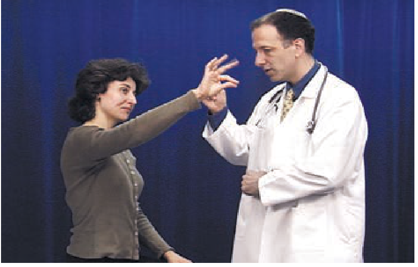
What a Positive Result Looks Like
On the affected side, the forearm involuntarily rotates inward (pronates) and may drift downward. Even subtle curling of the fingertips on one side counts as positive.
Why It Happens - The Mechanism
The corticospinal (upper motor neuron) tract normally maintains a balance between flexor and extensor muscle tone in the upper limb. When this tract is damaged, extensors and supinators are disproportionately weakened relative to flexors. With the eyes closed removing visual correction, the unopposed flexors and pronators pull the arm inward - producing the characteristic pronation drift.
As Frameworks for Internal Medicine explains: "UMN lesions disproportionately affect the upper limb extensors (causing pronator drift) and lower limb flexors."
What It Localizes To
A positive pronator drift is a sign of upper motor neuron (UMN) / corticospinal tract dysfunction anywhere along its path:
- Cerebral cortex
- Subcortical white matter
- Internal capsule
- Brainstem (if associated with cranial nerve signs)
- Cervical spinal cord (if no facial involvement)
In Amar Krishna Ghosh's case, the drift is on the left side, meaning the lesion is in the right hemisphere (the corticospinal tract crosses at the medullary decussation).
Rapid Finger Tapping Test
How It Is Performed
The patient rapidly taps the index finger against the thumb repeatedly, or taps each finger to the thumb in sequence. Each hand is tested separately and compared.
What a Positive Result Looks Like
Slowness, clumsiness, or reduced amplitude on one side - especially if the non-dominant hand is worse than expected compared to the dominant hand.
Why It Happens - The Mechanism
Fine rapid finger movements have a very large cortical representation and are exquisitely sensitive to subtle corticospinal damage. As Neuroanatomy through Clinical Cases (3rd Ed.) states:
"Slow or awkward fine finger movements or toe tapping in the absence of weakness can signify a subtle abnormality of the corticospinal pathways."
This test catches deficits that formal muscle strength testing (grading 0-5) can miss entirely - a patient can have full 5/5 power and still have slowed finger tapping.
What It Localizes To
Like pronator drift, impaired finger tapping points to corticospinal (UMN) pathway dysfunction. However, it is not exclusive - slowness can also occur with:
- Cerebellar lesions (though typically with dysmetria and other cerebellar signs)
- Basal ganglia disease (e.g., Parkinson's - bradykinesia)
The clinical context distinguishes these: in Amar's case, both tests being abnormal on the same (left) side strongly points to a contralateral (right-sided) corticospinal lesion, not cerebellar or basal ganglia disease.
UMN vs LMN - Quick Comparison
| Feature | UMN Lesion (Amar's picture) | LMN Lesion |
|---|---|---|
| Weakness pattern | Proximal > distal, extensors > flexors | Individual muscle / distal |
| Tone | Increased (spasticity) | Decreased (flaccid) |
| Reflexes | Hyperreflexia | Hyporeflexia / absent |
| Atrophy | Mild / disuse only | Prominent |
| Fasciculations | Absent | Present |
| Pronator drift | Positive | Negative |
| Babinski sign | Positive (extensor plantar) | Absent |
Clinical Significance in This Patient
Both tests positive on the left side in a 60-year-old hypertensive man with 6 months of left hand clumsiness is a classic presentation of a subtle right-hemisphere corticospinal lesion. The differential diagnosis includes:
- Lacunar infarct (small vessel stroke from hypertension) - most likely given his BP of 150/90 and age
- TIA with residual deficit
- Cortical/subcortical infarct
- Demyelinating disease (MS - less typical at age 60)
- Mass lesion (tumour - less likely but MRI will exclude)
This is exactly why the neurologist ordered MRI Brain and MRA of the brain & neck vessels - to find where the corticospinal tract is being affected and whether there is arterial disease driving it.
- Harrison's Principles of Internal Medicine, 22E - Motor Examination chapter
- Neuroanatomy through Clinical Cases, 3rd Ed. - Table 3.3 & Section 6.4
https://2.sdl1.info/SURKSA/63c33777f9 Recived the MRI report
Let me fetch the MRI report from that link right away.
Reading Web Page
https://2.sdl1.info/SURKSA/63c33777f9
Unfortunately, that link failed to load - I wasn't able to retrieve any content from it. This could be because:
- The URL requires a login or authentication
- The link has expired or is restricted
- The server is not publicly accessible
Here are a few ways to share the MRI report with me:
- Take a photo of the printed report and attach it to your next message (like you did with the prescription)
- Copy and paste the text of the report directly into the chat
- Share a different link - if it's stored on Google Drive, Dropbox, or another public sharing platform
Once I can see the report, I'll break down all the findings and explain what they mean in the context of Amar Krishna Ghosh's left hand clumsiness and the clinical picture we already discussed.Let me read the PDF skill first, then extract the MRI report.
Reading File
Running Command
python3 -c " import pdfplumber with pdfplumber.open('/tmp/workspace/attachments/febcbe07-7e1c-47da-ab71-3788a387dd7e/PR2534509.pdf') as pdf: print(f'Pages: {len(pdf.pages)}') for i, page in enumerate(pdf.pages): print(f'--- PAGE {i+1} ---') text = page.extract_text() print(text) "
Got the report. Here is the full breakdown.
MRI Brain Report - Amar Krishna Ghosh
Lab No: PHL/23-07-2026/PR2534509 | Date: 23 July 2026
Sequences Performed
T1, T2, T2 FLAIR, GRE, and Diffusion Weighted Imaging (DWI) - a thorough standard brain MRI protocol.
Findings - Point by Point
| Finding | What It Means |
|---|---|
| No restricted diffusion on DWI | No acute stroke or acute ischaemic lesion |
| Normal signal intensity of cerebral hemispheres | No tumour, no haemorrhage, no demyelination seen |
| Normal ventricular system & subarachnoid spaces | No hydrocephalus, no atrophy |
| No midline shift | No space-occupying lesion pushing the brain |
| Normal pituitary, optic chiasma | No sellar/suprasellar pathology |
| Normal brainstem & cerebellum | No posterior fossa lesion |
| Normal CP angle & internal auditory canals | No acoustic neuroma or nerve pathology |
| Mucosal thickening in bilateral maxillary & ethmoid sinuses | Incidental finding - chronic sinusitis, not neurologically relevant |
Impression (official)
MRI of brain is within normal limits.
What This Means for Amar
This is actually a reassuring but not fully conclusive result. Here is why:
The Good News
- No acute stroke - the DWI sequence (most sensitive for acute infarction within hours to days) is completely clear.
- No brain tumour, no haemorrhage, no demyelinating plaques detected.
- No serious structural lesion found.
Why the Symptoms Still Matter - The Critical Point
A normal MRI Brain does NOT fully rule out the cause of his left hand clumsiness and positive pronator drift. This is because:
- Small lacunar infarcts in the internal capsule, corona radiata, or pons can sometimes be missed on a plain MRI, especially if they are old or very small - this is why the MRA (brain & neck vessels) was ordered separately and is still pending.
- TIA (Transient Ischaemic Attack) - by definition leaves no permanent brain lesion on MRI, yet produces real neurological symptoms. A TIA is a medical emergency even with a normal MRI.
- Chronic small vessel disease may not be dramatically visible on a plain non-contrast MRI.
What Must Happen Next
The MRA of brain and neck vessels (also ordered by Dr. Mandal) has not been done yet based on this report. That investigation is still needed to check:
- Carotid artery stenosis in the neck
- Intracranial arterial narrowing (MCA, ACA territory)
- Vertebrobasilar disease
The neurologist needs to see both reports together before concluding the cause. The clinical picture (positive pronator drift + left hand clumsiness + hypertension) still warrants full investigation even with this normal MRI.
In summary: The MRI is reassuring - there is no acute stroke, tumour, or bleed. But the MRA is still pending and the symptoms must be taken seriously. Amar should attend his follow-up with Dr. Mandal as scheduled and get the MRA done promptly.
This is a shared conversation. Sign in to Orris to start your own chat.