
Vasovagal complex
vasovagal syncope neural pathway reflex arc diagram
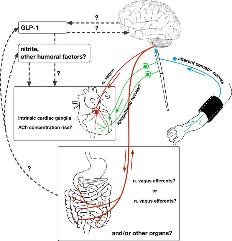
This pathophysiology diagram illustrates the 'remote preconditioning reflex arc,' a complex neural and humoral pathway involved in remote ischemic preconditioning (RIPre). The diagram depicts a stimulus applied to an arm via a blood pressure cuff, which triggers afferent somatic nerves that project to the brain. Efferent signals are shown descending from the central nervous system via the vagus nerve (n. vagus) and sympathetic nerves to the heart and other visceral organs, specifically the intestine. Key cardiovascular features include the involvement of intrinsic cardiac ganglia and a potential rise in acetylcholine (ACh) concentration in the myocardium. The diagram highlights several scientific uncertainties using question marks, specifically regarding the role of sympathetic nerves, whether abdominal vagal fibers are afferent or efferent, and the origin of humoral factors like GLP-1 and nitrite. Humoral pathways are shown as dashed lines connecting systemic factors to the brain and heart, indicating a parallel mechanism of cardioprotection alongside the primary neural reflex arc.
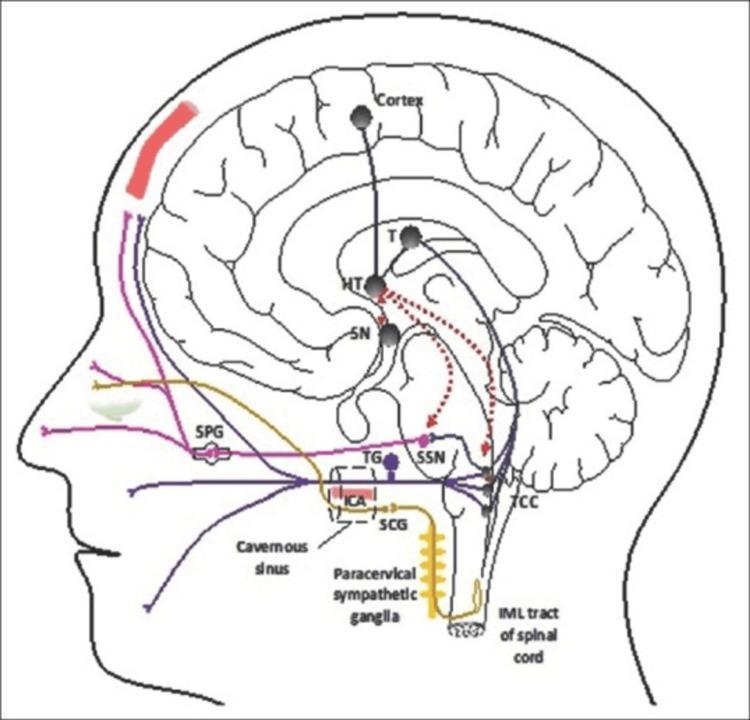
A pathophysiology diagram illustrating the neural pathways involved in cluster headaches within a sagittal view of the human head and brain. The diagram highlights the trigeminal-autonomic reflex arc. Key neural structures depicted include the hypothalamus (HT), which serves as a central hub with connections to the thalamus (T), suprachiasmatic nucleus (SN), superior salivatory nucleus (SSN), and the trigeminocervical complex (TCC). Sensory pathways are shown in purple and blue, originating from the dural structures and trigeminal nerve, synapsing in the TCC and projecting to the cortex. The parasympathetic pathway (pink) involves the SSN and sphenopalatine ganglion (SPG), leading to autonomic symptoms. The sympathetic pathway (yellow) is traced from the intermediolateral (IML) tract of the spinal cord through the superior cervical ganglion (SCG) and the plexus surrounding the internal carotid artery (ICA) in the cavernous sinus. Red dashed lines indicate the hypothalamic modulation of the SSN and TCC, which are critical in the pathogenesis of trigeminal autonomic cephalgias.
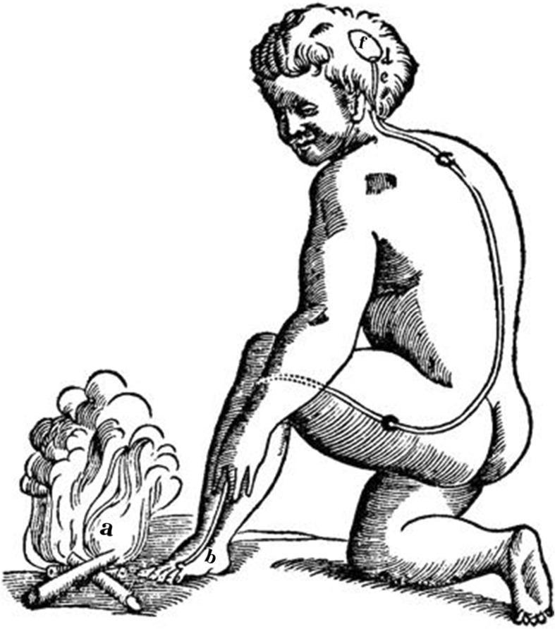
This historical anatomical illustration depicts René Descartes' early 17th-century mechanistic theory of the reflex arc. The diagram shows a human figure kneeling by a fire, illustrating the physiological response to a noxious stimulus. Key elements are labeled to represent a functional pathway: (a) indicates the external stimulus (fire); (b) represents the peripheral sensory site on the skin of the foot; (c) shows a 'fine thread' or nerve fiber extending from the foot, up the leg and back, to the head. The path includes nodes representing the spinal column or ganglia. Within the brain, (d) and (e) represent mechanical valves that govern the release of 'animal spirits' from a central cavity (f). This anatomical diagram serves as a foundational concept in the history of neuroscience, demonstrating an early understanding of the stimulus-response pathway, where sensory input travels to the central nervous system to trigger a motor reflex. It is a critical visual resource for teaching the evolution of neurophysiology and the history of medicine.
The Vasovagal Complex
Definition
- Harrison's Principles of Internal Medicine 22E describes it as "the final pathway of a complex central and peripheral nervous system reflex arc" with "increased parasympathetic outflow, plus sympathoinhibition, resulting in bradycardia, vasodilation, and/or reduced vasoconstrictor tone."
Neural Anatomy of the Reflex Arc
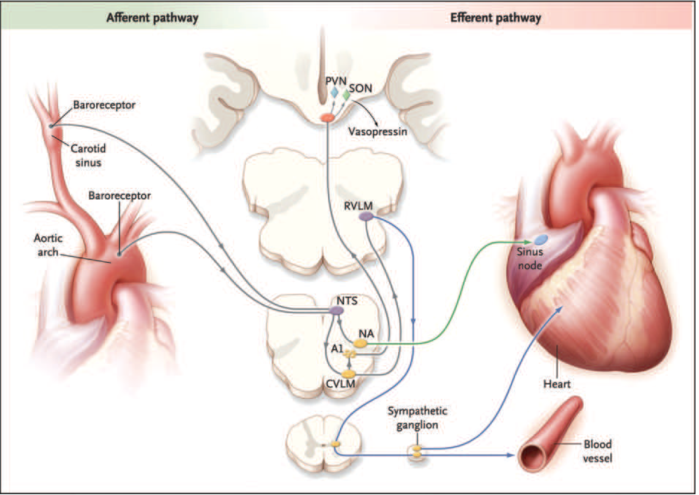
- Baroreceptors in the carotid sinus (CN IX - glossopharyngeal) and aortic arch (CN X - vagus) detect arterial pressure changes
- Afferent impulses travel to the Nucleus Tractus Solitarius (NTS) in the dorsomedial medulla
- NTS projects to the Nucleus Ambiguus (NA) - controls vagal (parasympathetic) output to the sinus node
- NTS projects to the Caudal Ventrolateral Medulla (CVLM) then Rostral Ventrolateral Medulla (RVLM) - controls sympathetic outflow to blood vessels and heart
- Higher cortical and hypothalamic centers (emotional inputs, fear, pain) can also override and trigger the reflex directly - explaining why the sight of blood or extreme emotion alone can precipitate syncope
- Increased vagal tone (via NA) → bradycardia via the sinus node
- Sympathoinhibition (via RVLM withdrawal) → peripheral vasodilation, hypotension
- Both combine to reduce cardiac output and systemic vascular resistance simultaneously
Pathophysiological Mechanism
- Trigger (orthostatic stress, emotional stress, pain, etc.) causes venous pooling
- Reduced venous return → decreased ventricular preload
- Baroreceptors sense the drop in BP → increased sympathetic drive → catecholamine surge
- A volume-depleted ventricle contracts vigorously against low filling
- Cardiac mechanoreceptors (C fibers) in the atria, ventricles, and pulmonary artery are activated by this paradoxical vigorous contraction
- These afferent C fibers project centrally to the dorsal vagal nucleus of the medulla
- This triggers a "paradoxical" withdrawal of sympathetic tone + surge in vagal tone
- Result: vasodilation + bradycardia → BP falls → cerebral blood flow drops below autoregulatory limits → syncope
Triggers and Classification
| Category | Examples |
|---|---|
| Classic vasovagal | Intense emotion, fear, pain, sight of blood, orthostatic stress, venipuncture |
| Pulmonary (situational) | Cough syncope, weightlifter's syncope, sneeze syncope |
| Urogenital | Postmicturition syncope, prostatic massage |
| Gastrointestinal | Swallow syncope, defecation syncope, glossopharyngeal neuralgia |
| Cardiac | Bezold-Jarisch reflex (inferior MI), outflow obstruction |
| Carotid sinus | Carotid sinus hypersensitivity, carotid massage |
| Ocular | Ocular pressure, oculocardiac reflex |
Clinical Features (Symptom Complex)
- Prodrome: lightheadedness, generalized muscle weakness, giddiness, visual blurring, tinnitus, nausea/GI symptoms, diaphoresis, pallor
- Loss of consciousness: brief (seconds to minutes), usually gradual onset
- During the faint: motionless or brief myoclonic jerks (never tonic-clonic), weak and slow pulse, shallow breathing, barely obtainable BP
- Recovery: rapid on assuming the horizontal position; some residual weakness, but no postictal confusion (key differentiator from seizure)
VASIS Classification (Types of Vasovagal Response)
| Type | Response |
|---|---|
| Type 1 (Mixed) | Both HR and BP fall; HR does not drop below 40 bpm |
| Type 2A (Cardioinhibitory) | HR drops to <40 bpm or asystole for <3 sec; BP falls before HR |
| Type 2B (Cardioinhibitory) | Asystole >3 sec; BP falls simultaneously with or after HR |
| Type 3 (Vasodepressor) | BP falls >30 mmHg; HR rises or stays the same |
Diagnosis
- Clinical history is most important - prodrome, triggers, rapid recovery without postictal state
- Tilt-table test (head-up tilt at 60-80 degrees) - gold standard for confirming NMS
- ECG: usually normal; important to exclude arrhythmias (long QT, Brugada, WPW, AV block)
- Carotid sinus massage: for suspected carotid sinus hypersensitivity (>3 sec asystole or >50 mmHg BP drop = diagnostic)
Management
| Approach | Details |
|---|---|
| Education & avoidance | Avoid known triggers, prolonged standing, dehydration, hot environments |
| Physical counter-maneuvers | Leg crossing, handgrip/arm tensing at prodrome - shown to abort episodes |
| Volume expansion | Increased salt/fluid intake; compression stockings |
| Tilt training | Repeated orthostatic stress ("stand training") |
| Pharmacotherapy | Midodrine (alpha-agonist); fludrocortisone (mineralocorticoid); beta-blockers (controversial); SSRIs in selected patients |
| Cardiac pacing | For highly symptomatic cardioinhibitory type (Type 2B) with asystole - closed-loop pacing systems (dual-chamber) preferred |
Key Differentiations
| Feature | Vasovagal Syncope | Seizure | Cardiac Arrhythmia |
|---|---|---|---|
| Prodrome | Yes (prolonged) | Aura (brief/absent) | Often none |
| Trigger | Emotional/orthostatic | None or specific | None |
| Duration | Seconds-minutes | Minutes | Variable |
| Movements | Brief myoclonic jerks | Tonic-clonic | Absent or brief |
| Postictal confusion | No | Yes | No |
| Recovery | Rapid horizontal | Slow | Rapid |
| Incontinence | Uncommon | Common | Uncommon |