
If only bondys technique of mastoidectomy is asked in long answers of 20 marks in ms ent final exam, then how to start it, what to write, what to draw. Give me proper answer on this along with perfect diagrams. Also according to standard ent textbook
Reading File
Now let me search the medical library for Bondy's mastoidectomy content.
Finding Sources
Finding Sources
Excellent! Found the key sections. Now let me read all the relevant content in parallel.
Reading File
Reading File
Reading File
Reading File
Reading File
Reading File
Reading File
Finding Sources
Reading File
Finding Sources
Now let me get the Scott-Brown content and look for diagrams/images related to Bondy mastoidectomy.
Reading File
Finding Sources
Finding Sources
Reading File
I found a Bondy mastoidectomy diagram. Let me fetch this image and also search for more images.
Loading Image
Finding Sources
I now have all the information I need from standard ENT textbooks (Shambaugh Surgery of the Ear, Cummings Otolaryngology, Scott-Brown's) to write a comprehensive MS ENT exam answer on Bondy's technique of mastoidectomy. Let me now compose the full model answer.
Bondy's Modified Radical Mastoidectomy - Model Answer for MS ENT Final (20 Marks)
How to Start, Structure, and What to Draw
Here is a complete model answer structured exactly as expected in an MS ENT Final examination.
BONDY'S MODIFIED RADICAL MASTOIDECTOMY
(Shambaugh Surgery of the Ear; Cummings Otolaryngology; Scott-Brown's Otorhinolaryngology)
I. INTRODUCTION / DEFINITION (2 marks)
Bondy's modified radical mastoidectomy is a canal wall-down mastoidectomy in which disease confined to the epitympanum (pars flaccida cholesteatoma) is exteriorized by removing the superior and posterior canal wall, while the uninvolved mesotympanum and ossicular chain are left intact. It is the most conservative of the open-cavity mastoid operations.
- Described by Bondy in 1910 as the first procedure to actively address hearing preservation in mastoid surgery.
- Classically referred to as the "Modified Radical Mastoidectomy" in the strict historical sense (Cummings Otolaryngology, 3633).
II. HISTORICAL NOTE (1 mark)
- 1873 - Schwartze: cortical (simple) mastoidectomy
- 1890 - Zaufal: radical mastoidectomy (removed TM, ossicles, posterior canal wall)
- 1910 - Bondy: modified the radical procedure - preserved the mesotympanum and ossicular chain when disease was limited to the attic (epitympanum)
- 1938 - Lempert's fenestration operation redirected attention to hearing preservation
- 1950s - Zollner & Wullstein: tympanoplasty techniques
- Bondy's procedure eventually gave way to modified radical mastoidectomy with tympanoplasty due to limitations
III. INDICATIONS (2 marks)
Bondy's procedure is indicated when:
- Attic/epitympanic cholesteatoma limited strictly to the epitympanum (pars flaccida type)
- Ossicular chain is intact - especially when the malleus head and incus body are surrounded by matrix but the chain remains mobile
- Mesotympanum is uninvolved - no disease in mesotympanum or hypotympanum
- Small cholesteatomas where disease has not extended to mastoid air cells
- Cholesteatoma matrix lying lateral to the ossicular heads (not medial) - can be used as a cavity lining
IV. CONTRAINDICATIONS (1 mark)
- Disease extending to mesotympanum
- Disease extending to mastoid air cells (requires full modified radical)
- Acute coalescent mastoiditis
- Active suppurative otitis media with significant mucosal disease
- Labyrinthine fistula with extensive erosion
V. PREOPERATIVE ASSESSMENT (1 mark)
- Pure tone audiogram (to document hearing)
- HRCT temporal bone to assess extent of disease
- Microscopic ear examination: confirm attic disease, status of ossicular chain
- Control active infection preoperatively with 1.5% acetic acid irrigation + antibiotic ear drops for 2-4 weeks
VI. ANESTHESIA & POSITION
- General anesthesia (GA) preferred
- Patient supine, head turned to opposite side
- Operating table: 15-20° reverse Trendelenburg to reduce venous oozing
VII. SURGICAL TECHNIQUE - STEP BY STEP (8 marks)
A. Incision
Endaural (Lempert) incision is the classic approach for Bondy's operation (can also use postauricular).
Two components of the endaural incision:
Step 1: Starting at "12 o'clock" on the superior canal wall, 1 cm from the outer edge of the canal, the first incision extends down the posterior canal wall in the incisura terminalis nearly to "6 o'clock," then at right angles outward 2-3 mm to the edge of the conchal cartilage.
Step 2: From the same starting point at "12 o'clock," the second incision extends directly upward in the incisura terminalis to a point halfway between the meatus and the upper edge of the auricle.
- Both incisions are deepened through skin and periosteum
- A broad periosteal elevator elevates periosteum over the entire mastoid process
- The Shambaugh self-retaining endaural retractor is inserted
DRAW FIGURE 1 HERE - Endaural incision diagram
B. Atticotomy (Bone Removal)
- A surgical cutting burr removes the outer cortex just above and behind the meatus over a semilunar area
- Watch for pink color shining through bone (middle fossa dura approaching)
- The notch of Rivinus is located by passing a narrow periosteal elevator along the superior osseous meatal wall
- The epitympanum is encountered - the white smooth wall of the cholesteatoma sac is identified
- The sac is opened cautiously (distinguish from dura), contents removed by suction
- Bone cortex and overhang removed with cutting burr, curet, or rongeur until entire cholesteatoma sac is exposed
DRAW FIGURE 2 HERE - Atticotomy showing removal of scutum/outer attic wall
C. Key Feature: Preservation of Middle Ear
In Bondy's operation:
- A limited intact canal wall procedure is performed, staying superior in the dissection
- The antrum is identified; the superior and posterior canal wall is removed until only a thin rim of bone remains over the ossicles
- A medium-sized burr is drawn medially to laterally for bone removal
- The final rim of bone is removed with a small curette (to avoid traumatizing the ossicular chain)
- The ossicular chain is preserved - specifically malleus head and incus body (if matrix only lies lateral to them)
- If matrix lies against and lateral to the ossicles, matrix may be left as lining; ossicles left undisturbed
- If matrix envelops the medial surface of the malleus head or incus, these ossicles must be removed
D. Taking Down the Bridge and the Facial Ridge
This is the most critical and most commonly poorly performed step:
- The remaining superior osseous meatal wall (the "bridge") bridging the notch of Rivinus is removed in small bites with a narrow rongeur
- Anterior and posterior spines of the notch of Rivinus (the "buttresses") are taken down with a small curet, always working outward away from the fallopian canal
- The tympanic segment of the facial canal is identified and kept in view at all times
- The facial ridge (posterior osseous meatal wall housing the vertical facial nerve) is taken down slowly with a drill or curet, under the operating microscope, always parallel to and never across the direction of the facial nerve
- Landmarks to locate the facial nerve:
- Bony horizontal semicircular canal (above)
- Tympanomastoid suture (in posterior meatal wall)
- Digastric ridge in mastoid tip (Note: in Bondy's operation, tip cells are rarely removed, so this landmark is less useful)
DRAW FIGURE 3 HERE - Facial ridge lowering, facial nerve landmarks
E. Preparation of the Meatal Skin Flap
- The plastic-pedicled skin flap consists of the skin and periosteum of the entire superior osseous meatal wall and most of the posterior meatal wall
- As atticotomy proceeds and the bridge is taken down, a narrow periosteal elevator separates skin and periosteum from superior and posterior meatal walls
- With a curved meatal knife and iris scissors, an incision along the anterosuperior angle of the meatus frees the flap
- This flap is turned posteriorly to cover the facial ridge and the floor of the completed operative cavity
F. Meatoplasty
- A large meatoplasty is mandatory in Bondy's procedure
- The meatus must be an adequate size relative to the cavity for ventilation and postoperative cleaning
- Conchal cartilage is removed from the posterior meatus to create a properly sized meatal opening
- Without an adequate meatoplasty, postoperative cavity problems (discharge, debris accumulation) are guaranteed
G. Final Cavity
- The bowl of the surgical cavity after removal of disease should be flush with the intact (or perforated) tympanic membrane
- KEY DIFFERENCE from radical mastoidectomy: Tympanic membrane and ossicular chain are preserved; middle ear is not entered; Eustachian tube is not obliterated
- KEY DIFFERENCE from modified radical mastoidectomy (complete mastoidectomy + tympanoplasty): In Bondy's, the posterior canal wall is not completely removed; limited dissection only; no tympanoplasty performed; no mastoid air cell exenteration
VIII. DIAGRAMS TO DRAW (Key for 20-mark answer)
Diagram 1: Endaural (Lempert) Incision
        Helix
         |
    _____↓_____
   /  Incision  \
  |  (2 limbs:   |
  |  superiorly  |    ← Vertical limb going upward
  |  + posteriorly→  ← Horizontal limb in incisura terminalis
  |______________|
       EAC

Label: 12 o'clock start point, incisura terminalis,
       Concha, 2 limbs meeting at 12 o'clock
Diagram 2: Atticotomy - Opening the Epitympanum (Cross-section)
              TEGMEN (middle fossa dura)
         ___________________________
        |  EPITYMPANUM              |
        |  Malleus head  Incus body |
        |      ↑              ↑     |
        |   [PRESERVED OSSICLES]    |
        |___________________________|
              ↑↑↑↑↑↑↑
         SCUTUM REMOVED
         (attic wall / outer wall of
          epitympanum removed)

        EAC lumen below

Labels: Scutum, Epitympanum, Tegmen,
        Malleus head, Incus body,
        Cholesteatoma matrix (lateral to ossicles)
Diagram 3: Bondy's Cavity (Completed) - Coronal Cross-section
              TEGMEN
        ________________________
       |   MASTOID             |
       |   ANTRUM              |
       |_______________________|
              |
       ________|_________
      |   EPITYMPANUM     |
      |  (cavity now open)|← Saucerized
      |   Malleus  Incus  | ← PRESERVED
      |___________________|
       |               |
       |   EAC         |
       |               |
        _______________
          MEATOPLASTY
          (enlarged)

Labels: Tegmen, Antrum, Facial ridge (lowered),
        Malleus head, Incus body (intact),
        Tympanic membrane (intact),
        Meatoplasty, EAC
Here is the actual intraoperative photograph from Shambaugh Surgery of the Ear showing the completed Bondy modified mastoid cavity:
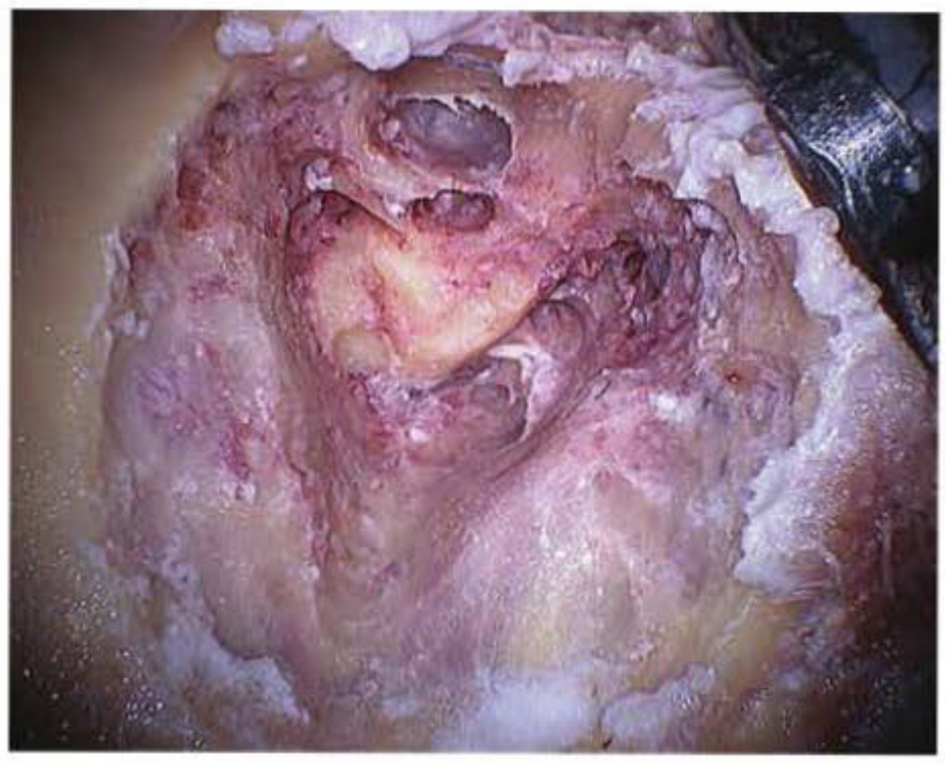
Figure: Bondy modified mastoid cavity demonstrating removal of the external canal wall with preservation of the tympanic membrane and ossicular chain (Shambaugh Surgery of the Ear, Fig. A-21)
IX. COMPARISON TABLE - Bondy vs Radical vs Modified Radical (1 mark)
| Feature | Radical | Bondy's Modified Radical | Modified Radical (Complete) |
|---|---|---|---|
| Canal wall | Removed | Partially removed (attic region only) | Completely removed |
| Tympanic membrane | Sacrificed | Preserved | Reconstructed |
| Ossicular chain | Removed | Preserved (if lateral disease only) | May be preserved/reconstructed |
| Eustachian tube | Obliterated | Patent | Patent |
| Middle ear | Exteriorized with mastoid | Not entered | Sealed with tympanoplasty |
| Mastoid air cells | All exenterated | Limited/not exenterated | All exenterated |
| Meatoplasty | Yes | Yes (mandatory) | Yes |
| Hearing | Lost | Preserved or minimal loss | Reconstructed |
X. POSTOPERATIVE CARE (1 mark)
- Sterile dressing for 5-7 days
- Aural toilet and regular cavity cleaning (lifelong commitment)
- Antibiotic-steroid ear drops if any discharge
- Avoid water entry into ear (use ear plug while bathing)
- Regular outpatient follow-up every 6 months for cavity inspection and debris removal
- Annual pure tone audiogram
XI. COMPLICATIONS (1 mark)
Intraoperative:
- Facial nerve injury (most feared - especially during facial ridge lowering)
- Labyrinthine fistula (if semicircular canal eroded by cholesteatoma)
- Dural exposure/injury
- Sigmoid sinus injury/bleeding
- Ossicular chain damage
Postoperative (specific to open cavities):
- Persistent cavity discharge - due to:
- Inadequate meatoplasty
- High facial ridge
- Dependent tip cell (sump effect)
- Perforation in tympanic membrane
- Small EAC meatus
- Recurrent/residual cholesteatoma
- Granulation tissue in cavity
XII. DRAWBACKS OF BONDY'S PROCEDURE (why it fell out of favor) (1 mark)
- Incomplete removal of infected mastoid air cells → recurrent/persistent aural discharge
- Retained cholesteatoma matrix in attic → continued bone erosion by osteolytic enzymes
- Squamous debris accumulation due to incomplete tip cell removal and high facial ridge
- These drawbacks led to refinement into the modified radical mastoidectomy with tympanoplasty (complete mastoidectomy), which became the preferred open cavity technique (Shambaugh, p. 540-541)
SUMMARY POINTS TO REMEMBER FOR EXAM
- "Disease limited to epitympanum" = hallmark indication
- Ossicular chain preserved = key difference from radical mastoidectomy
- Middle ear NOT entered = key difference from modified radical with tympanoplasty
- Endaural incision is the classic approach
- Meatoplasty is mandatory
- Facial ridge lowering is the most critical (and most commonly poorly done) step
- Landmarks for facial nerve: horizontal SCC, tympanomastoid suture, digastric ridge
- Matrix on lateral surface of ossicles = can be left as lining; matrix on medial surface = ossicles must be removed
- Cavity should be flush with tympanic membrane at completion
- Bondy (1910) was the first to address hearing preservation in mastoid surgery
References for your answer:
- Shambaugh Surgery of the Ear, Chapter on Canal-Wall-Down Mastoidectomy, pp. 540-544
- Cummings Otolaryngology Head and Neck Surgery, Chapter 143 "Mastoidectomy: Surgical Techniques," p. 2742
- Scott-Brown's Otorhinolaryngology Head & Neck Surgery, Vol. 2, Canal Wall-Down Mastoidectomy section
This is a shared conversation. Sign in to Orris to start your own chat.