
Hormonal regulation of human reproductive system, menustral cycle, Gynecology examination according to bereks and nova gynecologic
Reading File
Finding Sources
Finding Sources
Finding Sources
Finding Sources
Finding Sources
Finding Sources
Finding Sources
Finding Sources
Now let me read the key sections in parallel:
Reading File
Reading File
Reading File
Reading File
Reading File
Reading File
Reading File
Reading File
Reading File
Reading File
Reading File
Reading File
I now have comprehensive content from Berek & Novak's Gynecology. Let me update the todos and compile the comprehensive answer.Here is a comprehensive review drawn directly from Berek & Novak's Gynecology:
Hormonal Regulation of the Female Reproductive System, the Menstrual Cycle, and the Gynecologic Examination
PART I: NEUROENDOCRINOLOGY — HORMONAL REGULATION
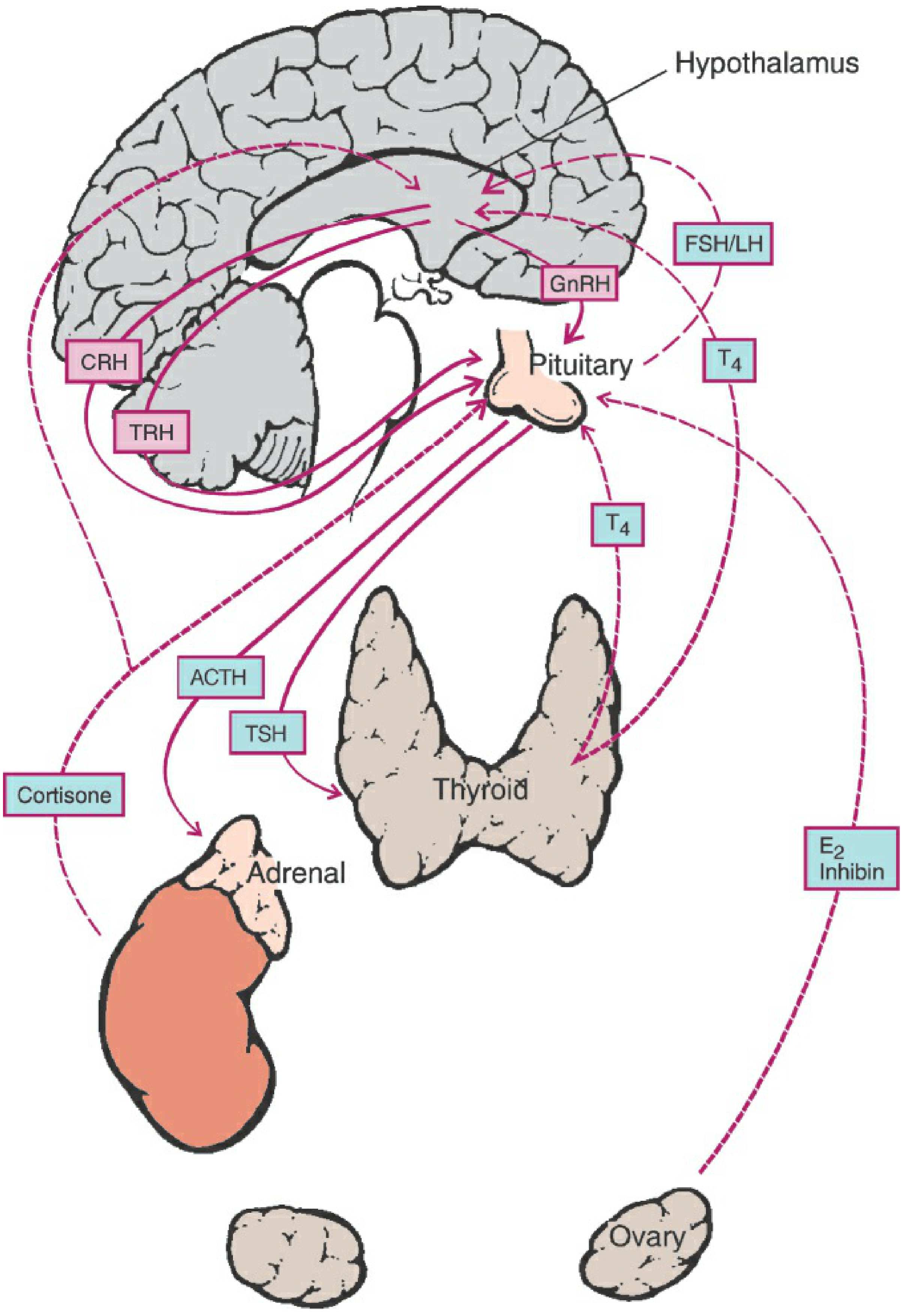
The HPO Axis: The Big Picture
The female reproductive system is governed by a precisely orchestrated interaction among four anatomical levels: the central nervous system (hypothalamus), the anterior pituitary, the ovary, and the uterus (endometrium). All must function in concert for normal reproduction to occur.
1. The Hypothalamus
The hypothalamus is a small neural structure at the base of the brain, above the optic chiasm and below the third ventricle. It is anatomically divided into three zones:
- Periventricular (adjacent to the 3rd ventricle)
- Medial (primarily cell bodies)
- Lateral (primarily axonal)
It has extensive interconnections with the limbic system (amygdala, hippocampus), thalamus, and pons, forming feedback loops.
Major Hypothalamic Secretory Products:
| Releasing Factor | Controls |
|---|---|
| GnRH (Gonadotropin-releasing hormone) | LH and FSH secretion |
| CRH (Corticotropin-releasing hormone) | ACTH secretion |
| GHRH (Growth hormone-releasing hormone) | GH secretion |
| TRH (Thyrotropin-releasing hormone) | TSH secretion |
Feedback Loops:
- Long-loop: Circulating sex steroids (estrogen, androgens) feed back onto hypothalamic steroid receptors
- Short-loop: Pituitary hormones feed back to the hypothalamus
- Ultrashort-loop: Hypothalamic secretions feed back onto the hypothalamus itself
2. GnRH — The Master Regulator
GnRH is a 10-amino-acid decapeptide (Fig. 7-4) produced by neurons with cell bodies primarily in the arcuate nucleus of the hypothalamus. These neurons originate embryologically in the olfactory pit and migrate to their adult positions.

Key properties of GnRH:
- Must be secreted in a pulsatile fashion to be effective — continuous infusion leads to gonadotroph downregulation (decreased surface GnRH receptors) and suppression of LH/FSH
- Intermittent exposure upregulates ("autoprimes") GnRH receptors on pituitary gonadotrophs
- Half-life is only 2–4 minutes (rapid proteolytic cleavage)
- The GnRH gene produces a 92-amino-acid precursor that also encodes GnRH-Associated Peptide (GAP) — a prolactin inhibitor and gonadotropin stimulator
Pulse variation across the cycle:
- Follicular phase: frequent, small-amplitude pulses
- Late follicular phase: increased frequency and amplitude
- Luteal phase: progressive lengthening of pulse intervals; higher amplitude but declining over 2 weeks
- Decreasing GnRH pulse frequency → ↓ LH but ↑ FSH (important for late-luteal FSH rise)
3. Anterior Pituitary — Gonadotropins (LH and FSH)
LH and FSH are glycoprotein hormones secreted by pituitary gonadotrophs in response to GnRH pulses.
FSH functions:
- Recruits a cohort of primordial follicles
- Stimulates granulosa cell proliferation and aromatase activity
- Drives estrogen synthesis in growing follicles
- Suppressed by rising estradiol and inhibin B
LH functions:
- Acts on theca cells to produce androgens (substrate for estrogen synthesis via two-cell theory)
- LH surge (triggered by high estradiol levels) → ovulation 24–36 hours later
- Maintains corpus luteum and drives progesterone production
Other anterior pituitary hormones relevant to reproduction:
- Prolactin: Secreted by lactotrophs; elevated prolactin suppresses GnRH pulsatility and causes anovulation. Regulated by dopamine (inhibitory) from the hypothalamus
- GH: Secreted in pulses during sleep; plays a role in ovarian function and follicular responsiveness
- ACTH: Controls adrenal glucocorticoids; diurnal variation with AM peak
4. Posterior Pituitary
The neurohypophysis is a direct extension of the hypothalamus (derived from neuroectoderm). Its neurons originate in the supraoptic and paraventricular nuclei (the magnocellular system).
Oxytocin:
- 9-amino-acid peptide produced by the paraventricular nucleus
- Functions: (1) stimulates uterine contractions during parturition, (2) triggers milk letdown (lactiferious duct myoepithelial contraction) via nipple stimulation → thoracic nerves → spinal cord → hypothalamus
Arginine Vasopressin (AVP):
- Controls water reabsorption; also linked (in animal models) to sexual activity, learning, temperature regulation, and maternal behavior
- Receptor variants linked to autistic spectrum disorders and altruistic behavior
PART II: THE MENSTRUAL CYCLE
Normal Menstrual Cycle — Overview
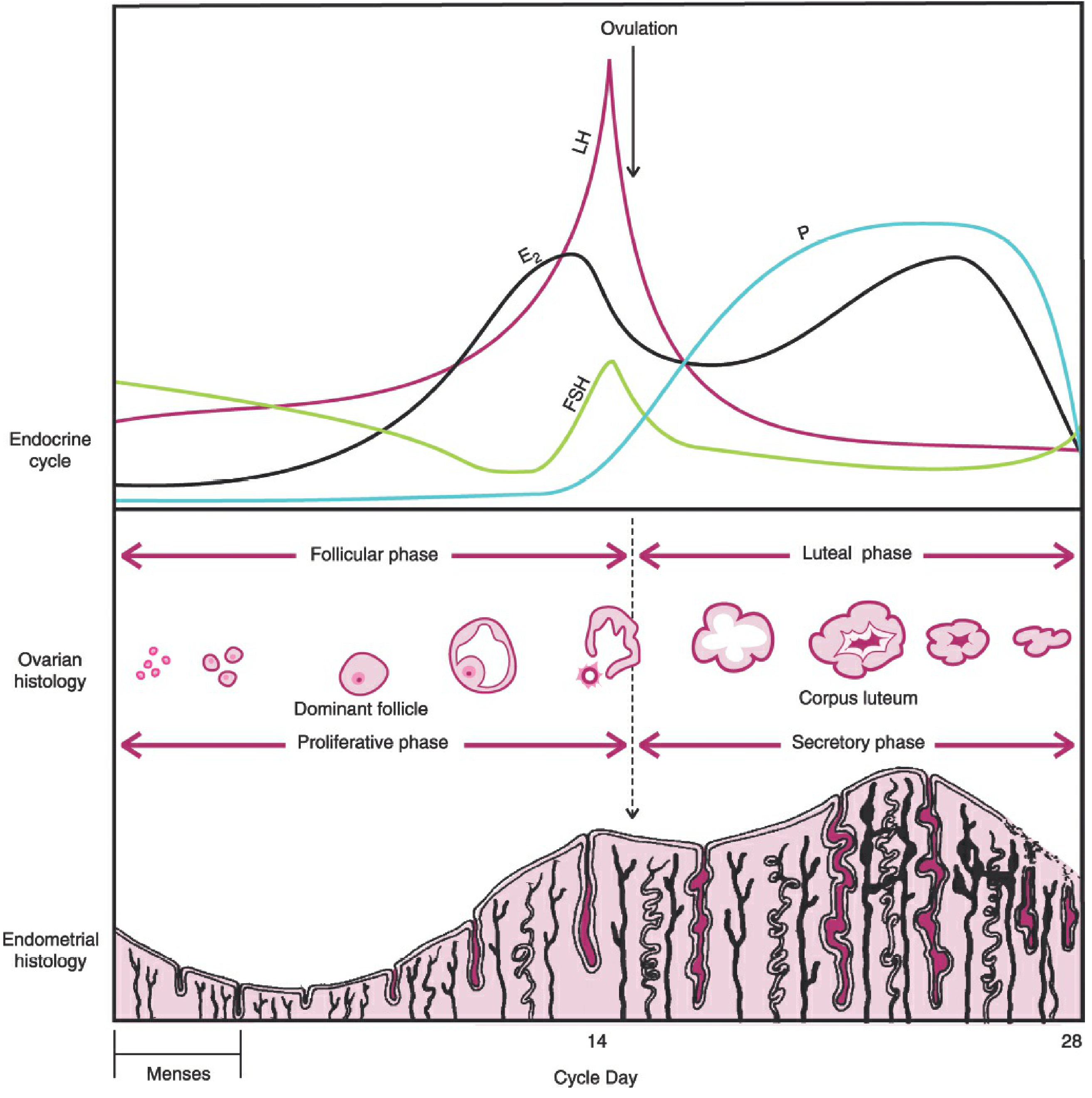
The cycle is divided into:
- Ovarian cycle: Follicular phase + Luteal phase
- Uterine cycle: Proliferative phase + Secretory phase
Normal parameters:
- Cycle length: 21–35 days
- Duration of flow: 2–6 days
- Average blood loss: 20–60 mL
- Follicular phase: 10–14 days (source of most cycle-length variability)
- Luteal phase: consistently ~14 days
Sequential Hormonal Events of the Menstrual Cycle
| Step | Phase | Event |
|---|---|---|
| 1 | Early follicular | Demise of prior corpus luteum → ↓ estrogen & progesterone → FSH begins to rise |
| 2 | Follicular | FSH recruits follicular cohort → follicles secrete ↑ estrogen → endometrial proliferation |
| 3 | Mid-follicular | ↑ Estrogen → negative feedback on FSH (FSH wanes); inhibin B also suppresses FSH |
| 4 | Late follicular | FSH induces LH receptors on granulosa cells → progesterone begins to rise (FSH midcycle surge) |
| 5 | Ovulation | Sufficient estrogen triggers pituitary LH surge → ovulation 24–36 hrs later |
| 6 | Early luteal | Estrogen dips briefly; corpus luteum forms and begins secreting estrogen again + inhibin A |
| 7 | Mid-luteal | Progesterone peaks (presumptive sign of ovulation); endometrium becomes secretory |
| 8 | Late luteal | If no pregnancy: corpus luteum degenerates → ↓ estrogen, progesterone, inhibin A → FSH rises again → new cycle begins |
Cyclic Changes of the Endometrium
- Proliferative phase (corresponds to follicular phase): Estrogen drives glandular and stromal proliferation
- Secretory phase (corresponds to luteal phase): Progesterone transforms endometrium into a receptive state for implantation — glands become tortuous, stromal edema, glycogen accumulates
- Menstruation: Withdrawal of estrogen and progesterone → endometrial shedding
Ovarian Follicular Development (Folliculogenesis)
Folliculogenesis is a dynamic, continuous process from menarche to menopause aimed at monthly recruitment and release of a single dominant follicle.
| Follicle Stage | Size | Key Features | Gonadotropin Dependence |
|---|---|---|---|
| Primordial | 0.03–0.05 mm | Primary oocyte (arrested in prophase I) + 1 layer flattened granulosa cells | Independent |
| Primary | ~0.1 mm | Granulosa cells become cuboidal; zona pellucida forms; FSH receptors develop | Independent |
| Secondary | ~0.2 mm | Multiple cuboidal granulosa layers; theca cells differentiate with LH receptors; neoangiogenesis begins | Transition point |
| Tertiary (antral) | 0.2–20 mm | Antrum forms; theca interna/externa distinct; corona radiata, cumulus oophorus, mural granulosa differentiate | Dependent |
| Preovulatory (Graafian) | >20 mm | Oocyte completes meiosis I → secondary oocyte (arrested in metaphase II); ready for ovulation | Dependent |
Two-cell theory of estrogen synthesis:
- Theca cells (LH-driven) → androgens (androstenedione, testosterone)
- Granulosa cells (FSH-driven) → aromatize androgens → estradiol
Luteal Phase
After ovulation, the ruptured follicle undergoes luteinization → corpus luteum, which secretes progesterone and estradiol. The corpus luteum has a lifespan of approximately 14 days. If pregnancy does not occur, it regresses (luteolysis) → hormone withdrawal → menstruation and the next cycle begins.
PART III: THE GYNECOLOGIC EXAMINATION (per Berek & Novak)
Approach to the Patient
Gynecology requires both medical knowledge and interpersonal skills. The assessment must encompass the "whole patient" — medical status, psychological, social, family, and cultural context. Key principles:
- Non-judgmental communication regarding sexual practices, gender identity, and orientation
- Empathy, attentive listening, and rapport are foundational skills
- Confidentiality is essential for honest disclosure
- Inform the patient of findings immediately after the examination (with patient clothed)
A. History
The complete gynecologic history includes:
- Chief complaint and history of present illness
- Menstrual history: cycle length, duration, flow, last menstrual period (LMP), menarche
- Obstetric history: gravida, para, complications
- Medical and surgical history
- Medications (including OTC and complementary/alternative)
- Family history
- Social history: tobacco, alcohol, recreational drugs, sexual activity, STI exposure, occupation
Technique for present illness (Table 1-5): Use open-ended then directed questions; avoid leading questions; establish negative symptoms to confirm or exclude diagnoses; assess psychosocial response to illness.
B. Physical Examination
A thorough gynecologic examination is performed at initial visit and periodically thereafter. The extent is dictated by the patient's symptoms and clinical situation.
1. General/Abdominal Examination
- Palpate all four quadrants systematically (e.g., clockwise from right upper quadrant)
- Assess for organomegaly (liver, spleen) — auscultate before palpation if organomegaly suspected
- Evaluate for mass effect, omental involvement ("omental cake" in ovarian cancer)
- Assess for rebound tenderness (peritoneal irritation) if acute pain is present
2. Inspection of External Genitalia
- Inspect labia majora, labia minora, clitoris, urethral meatus, introitus, and perineum
- Note skin lesions, pigmentation, edema, atrophy
- Ask patient to bear down (Valsalva): assess for cystocele (anterior wall bulge) or rectocele/enterocele (posterior wall bulge)
3. Speculum Examination (Vagina and Cervix)
- Inspection always precedes palpation
- Warm with tap water; do not lubricate if obtaining Pap smear or cultures
- Insert with blades oblique → rotate to horizontal → open blades to expose cervix
Vaginal inspection — assess for:
- Presence of blood or discharge (culture for gonorrhea, chlamydia; wet prep for trichomonas, BV, candida)
- Mucosal characteristics: color, lesions, vascularity
- Lesion types: inflammatory, neoplastic, vascular, pigmented (Chadwick sign in pregnancy), endometriotic
- Structural abnormalities (congenital and acquired)
Cervical inspection — assess for:
- Unusual bleeding from the os → evaluate for neoplasia
- Mucopurulent discharge → cervicitis
- Polyps (surface or endocervical)
- Carcinoma: may mimic inflammation → biopsy for any suspicious lesion
4. Bimanual Pelvic Examination (Table 1-6)
With one or two lubricated vaginal fingers and the abdominal hand:
- Uterus: Evaluate position, size, shape, symmetry, consistency, tenderness, and mobility; note any masses
- Cervix: Assess position, consistency, tenderness, and cervical motion tenderness (CMT) — rebound tenderness
- Adnexa (bilateral):
- Vaginal fingers in lateral fornix + abdominal hand on ipsilateral lower quadrant
- A normal fallopian tube is not palpable
- Normal ovary (~4 × 2 × 3 cm): sensitive, firm, freely movable — often not palpable
- Any adnexal mass: characterize by location, size, consistency, tenderness, mobility
5. Rectovaginal-Abdominal Examination
- Index finger in vagina + middle finger in rectum + abdominal hand
- Allows higher pelvic exploration (beyond the cul-de-sac limit)
- Used to assess: posterior uterine wall, uterosacral ligaments, posterior cul-de-sac, rectum
6. Rectal Examination
- Inspect perianal/pilonidal/perineal region: color, fissures, fistulae, hemorrhoids
- Ask patient to strain — reveals hidden internal hemorrhoids, polyps, or rectal prolapse
- Palpate with lubricated index finger:
- Note rectal tone, masses, tenderness
- Assess posterior cervix and uterus
- Stool on gloved finger: test for occult blood
Special Populations:
- Adolescents: Pelvic exam not routinely required for healthy teens requesting OCP before initiating intercourse
- Patients with intact hymen: Use rectal-abdominal (rather than bimanual vaginal) technique
- Slender, elderly, or adolescent patients: Single-finger vaginal technique preferred
Summary: Key Teaching Points
| Topic | Core Concept |
|---|---|
| HPO Axis | Hypothalamus → GnRH (pulsatile) → Pituitary → LH/FSH → Ovary → Estrogen/Progesterone |
| GnRH | Must be pulsatile; continuous exposure → downregulation; t½ = 2–4 min |
| FSH | Recruits follicles, drives aromatase, suppressed by estradiol + inhibin B |
| LH surge | Triggered by rising estradiol (positive feedback); causes ovulation 24–36 hrs later |
| Menstrual cycle | 21–35 days; follicular phase variable (10–14 d); luteal phase fixed (~14 d) |
| Folliculogenesis | Primordial → Primary → Secondary (gonadotropin-independent) → Tertiary → Graafian (gonadotropin-dependent) |
| Two-cell theory | Theca (LH) → androgens; Granulosa (FSH) → aromatize to estradiol |
| Corpus luteum | Secretes progesterone + estradiol; lifespan ~14 days; demise triggers next cycle |
| Gynecologic exam | History → Abdominal → External genitalia → Speculum → Bimanual → Rectovaginal |
| Bimanual exam | Assess uterus (position, size, mobility), cervix (CMT), adnexa (masses, tenderness) |
*All content sourced from: Berek & Novak's Gynecology, 16th Edition (ISBN: 9781496380357), Chapter 1 (Initial Assessment and Communication) and Chapter 7 (Reproductive Endocrinology and the Menstrual Cycle).*This comprehensive review covers all three requested topics directly from Berek & Novak's Gynecology (16th Ed.). Here's a quick navigation guide:
What's covered:
Part I — Hormonal Regulation
- The HPO axis (hypothalamus → pituitary → ovary → uterus)
- Hypothalamic anatomy and nuclei (arcuate, supraoptic, paraventricular)
- GnRH: decapeptide structure, pulsatile secretion rules, downregulation vs. upregulation, feedback loops (long/short/ultrashort)
- Anterior pituitary: FSH, LH, prolactin, GH, ACTH roles
- Posterior pituitary: oxytocin (parturition, milk letdown) and vasopressin
Part II — Menstrual Cycle
- Normal parameters (21–35 days, 2–6 days flow, 20–60 mL blood loss)
- 8-step sequential hormonal events from follicular through luteal phase
- Endometrial changes (proliferative vs. secretory phases)
- Folliculogenesis table: 5 stages with sizes, features, and gonadotropin dependence
- Two-cell theory of estrogen synthesis
- Corpus luteum lifespan and luteolysis
Part III — Gynecologic Examination
- Patient-centered approach: communication, confidentiality, cultural sensitivity
- Complete history components (menstrual, obstetric, medical, surgical, social)
- Abdominal exam: systematic palpation, rebound tenderness
- Speculum exam: technique, vaginal and cervical inspection checklist
- Bimanual exam: uterus, cervix (CMT), adnexa assessment
- Rectovaginal exam and rectal exam technique
- Special considerations for adolescents and patients with intact hymen
This is a shared conversation. Sign in to Orris to start your own chat.