
Caspase
Reading File
Caspases are a family of cysteine proteases central to apoptosis (programmed cell death) and inflammation. Let me pull detailed content from the medical library.
Searching sources...
Finding Sources
Finding Sources
Searching PubMed
caspase apoptosis mechanism
Good results. Now let me read the key sections from Robbins (pathology) and Histology textbooks for detailed content, plus the Roitt immunology section.
Reading File
Reading File
Reading File
Excellent. I have very rich content. Let me also fetch the key diagrams and read the extrinsic pathway section from Robbins.
Reading File
Loading Image
I now have comprehensive information to deliver a thorough answer. Let me also quickly look for caspase-1 (inflammasome/pyroptosis) content.
Finding Sources
I now have everything needed for a comprehensive answer. Here it is:
Caspases
Caspases (cysteine-dependent aspartate-specific proteases) are a family of intracellular proteases that serve as the central executors of apoptosis, and also participate in inflammation and pyroptosis. They exist in cells as inactive zymogens (procaspases) and are activated by proteolytic cleavage.
Structure and Biochemistry
- All caspases contain an active-site cysteine residue and cleave substrate proteins at specific aspartate residues.
- They exist as inactive procaspases; activation requires dimerization or proteolytic cleavage.
- Over 600 substrate proteins have been identified as targets for apoptotic caspases (global proteomics analyses) - this "death by a thousand cuts" ensures cells cannot escape once caspases are activated.
Classification
| Class | Members | Role |
|---|---|---|
| Initiator caspases | Caspase-8, -9, -10 | Activated first; trigger the cascade |
| Executioner (effector) caspases | Caspase-3, -6, -7 | Downstream; directly dismantle the cell |
| Inflammatory caspases | Caspase-1, -4, -5 (humans) | Inflammasome-associated; pyroptosis and cytokine processing |
Pathways of Apoptotic Caspase Activation
1. Intrinsic (Mitochondrial) Pathway
Triggered by intracellular stress: DNA damage, growth factor withdrawal, ER stress, p53 signaling, UV radiation.
Key steps:
- BH3-only proteins (BAD, BIM, BID, Puma, Noxa) are upregulated by stress sensors.
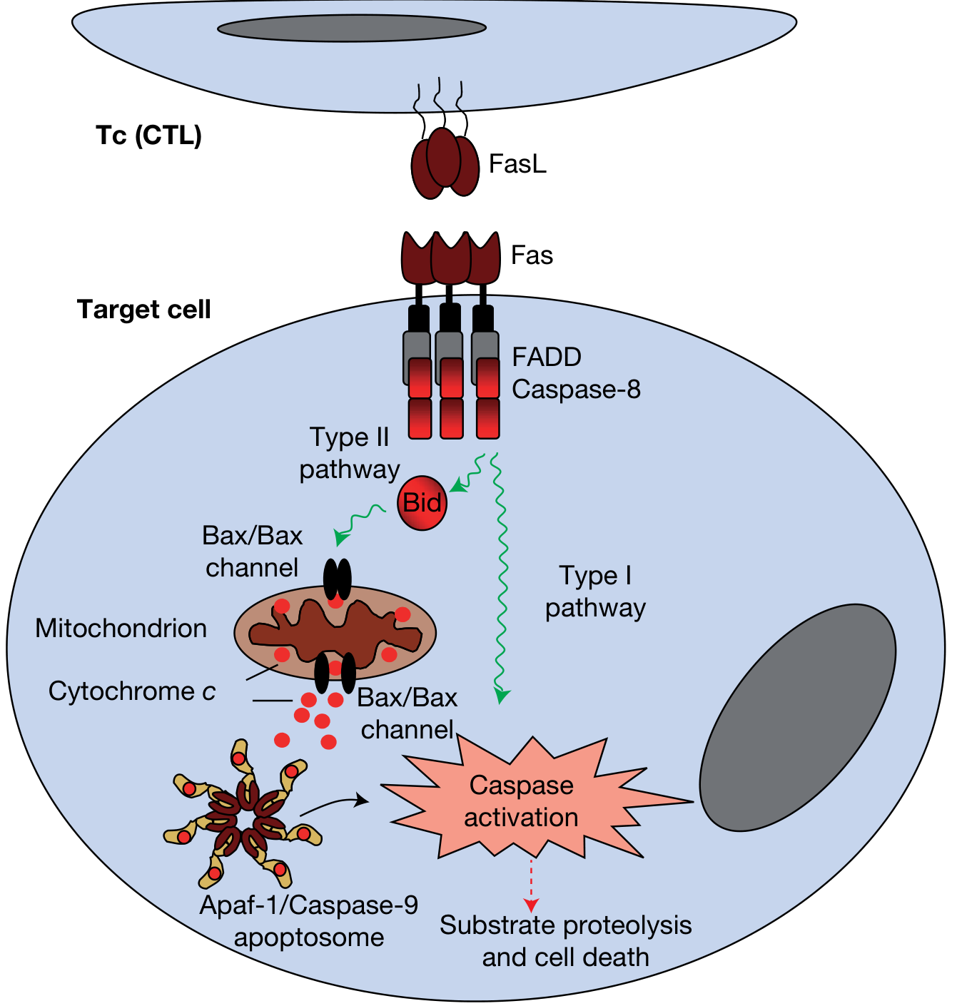
- They activate BAX and BAK, which oligomerize in the outer mitochondrial membrane, forming channels that allow cytochrome c (and SMAC/DIABLO) to leak into the cytoplasm.
- Anti-apoptotic proteins BCL2, BCL-XL, MCL1 normally suppress this by keeping the outer mitochondrial membrane impermeable.
- Cytochrome c binds APAF-1 to form the apoptosome - a multimeric wheel-shaped complex.
- The apoptosome recruits and autocatalytically activates caspase-9 (initiator caspase of this pathway).
- Active caspase-9 activates executioner caspases-3, -6, and -7.
2. Extrinsic (Death Receptor) Pathway
Triggered by engagement of plasma membrane death receptors (members of the TNF receptor family).
Key steps:
- FasL (on CTLs or self-reactive T cells) binds to Fas (CD95), clustering three or more Fas molecules.
- Their cytoplasmic death domains recruit the adaptor FADD (Fas-associated death domain protein).
- FADD recruits and brings together multiple molecules of procaspase-8 (or -10), leading to autocatalytic cleavage and generation of active caspase-8.
- Active caspase-8 either:
- Type I pathway: Directly activates downstream executioner caspases (caspase-3).
- Type II pathway: Cleaves the BH3-only protein BID, which then feeds into the mitochondrial pathway (amplification loop). This cross-talk is especially important in hepatocytes and pancreatic β cells.
- FLIP (a decoy protein produced by some viruses and normal cells) can bind procaspase-8 and block FADD binding, inhibiting this pathway.
Execution Phase (Common Final Pathway)
Both intrinsic and extrinsic pathways converge on caspase-3 (the master executioner). Activated caspase-3:
- Cleaves ICAD (inhibitor of caspase-activated DNase), liberating CAD to degrade chromosomal DNA into oligonucleosomal fragments (the "DNA ladder").
- Proteolytically degrades nuclear lamins, causing nuclear fragmentation.
- Cleaves PARP (poly-ADP-ribose polymerase), disabling DNA repair.
- Triggers cytoskeletal reorganization and membrane blebbing.
- Ultimately produces apoptotic bodies - membrane-bounded fragments containing organelles and nuclear material, which are phagocytosed without triggering inflammation.
Clearance of Apoptotic Cells
Caspases also trigger plasma membrane changes that signal phagocytes:
- Phosphatidylserine flips from the inner to outer membrane leaflet ("eat me" signal for macrophage receptors).
- Apoptotic cells secrete soluble "find me" signals recruiting phagocytes.
- Complement C1q and natural antibodies coat apoptotic bodies for recognition.
- This efficient clearance means apoptotic cells rarely trigger inflammation and are rarely seen on routine H&E (apoptosis occurs >20x faster than mitosis).
Inflammatory Caspases - Caspase-1 and Pyroptosis
Caspase-1 is activated by inflammasomes - large cytoplasmic macromolecular complexes (e.g., NLRP3, NLRC4) that assemble in response to DAMPs, PAMPs, and intracellular danger signals.
Caspase-1 functions:
- Cleaves pro-IL-1β and pro-IL-18 into active, secreted cytokines.
- Cleaves gasdermin-D; the N-terminal fragment forms pores in the plasma membrane, causing pyroptosis - an inflammatory form of cell death characterized by cell swelling and membrane rupture (distinct from apoptosis).
In humans, caspase-4 and caspase-5 serve as noncanonical inflammasome sensors (they also ultimately converge on caspase-1 or gasdermin-D activation).
BCL-2 Family - Regulators of the Intrinsic Pathway
| Group | Members | Mechanism |
|---|---|---|
| Anti-apoptotic | BCL2, BCL-XL, MCL1 | Keep outer mitochondrial membrane impermeable; sequester pro-apoptotic members |
| Pro-apoptotic (pore-formers) | BAX, BAK | Oligomerize to form cytochrome c release channels |
| BH3-only (sensors/activators) | BAD, BIM, BID, Puma, Noxa | Sense cellular stress; activate BAX/BAK and neutralize BCL2/BCL-XL |
BCL2 is frequently overexpressed in B-cell lymphomas (due to t(14;18) translocation), rendering tumor cells resistant to apoptosis.
Granzyme B Pathway (CTL Killing)
In addition to Fas/FasL, cytotoxic T lymphocytes (CTLs) kill targets via perforin/granzyme B:
- Perforin forms pores in the target cell membrane.
- Granzyme B (a serine protease) enters the target cell and directly cleaves and activates caspases (including caspase-3), bypassing the upstream initiator steps.
- This secretory granule pathway is the dominant CTL killing mechanism (evidenced by perforin-knockout mice failing to clear viral infections).
Clinical Significance
| Context | Mechanism |
|---|---|
| Cancer | BCL2 overexpression or caspase mutations allow tumor cells to evade apoptosis |
| Autoimmune disease | Defective Fas/FasL signaling (e.g., ALPS - autoimmune lymphoproliferative syndrome) leads to failure to eliminate self-reactive lymphocytes |
| Ischemia-reperfusion injury | Excessive caspase-3 activation drives neuronal death (caspase inhibitors are neuroprotective in experimental models) |
| Drug targets | IAP inhibitors (smac mimetics), BH3 mimetics (venetoclax targets BCL2), and caspase inhibitors are in clinical development/use |
| Viral evasion | Many viruses encode FLIP homologs or IAPs to block caspase activation and prevent apoptotic clearance |
Sources:
- Robbins & Cotran Pathologic Basis of Disease, pp. 58-62 (Mechanisms of Apoptosis)
- Histology: A Text and Atlas (Ross), pp. 279-281 (Apoptosis, caspase cascades)
- Roitt's Essential Immunology, pp. 272-275 (Caspase activation, CTL killing)
- Goodman & Gilman's Pharmacological Basis of Therapeutics (Apoptosis section)
- Janeway's Immunobiology 10e (Inflammasome and caspase-1)
- Recent reviews: Apoptosis signaling pathways review (2024) (PMID: 39594587); Pyroptosis - mechanisms and clinical perspective (2024) (PMID: 39300122)
This is a shared conversation. Sign in to Orris to start your own chat.