

61847.jpg
Please give the diagnosis difference
I'll analyze the image first to extract all the written content, then provide a differential diagnosis discussion.
analyse_media
Reading File
Now let me search the medical library for the key conditions mentioned and provide a differential diagnosis.
Finding Sources
Finding Sources
Finding Sources
Finding Sources
Reading File
Finding Sources
I now have enough information to provide a thorough differential diagnosis discussion. Here is the full analysis:
Extracted Clinical Information from the Note
Patient Details:
- Diagnosis written: Anemia + CKD (Chronic Kidney Disease)
- Complaints (c/o): Nausea, Vomiting
- Label on note: "For Pancytopenia" (boxed - indicating the workup/reason for evaluation)
CBC (Complete Blood Count):
| Parameter | Value |
|---|---|
| Hb (Hemoglobin) | 4.7 g/dL (severely low; normal: M >13, F >12) |
| WBC | 7000 /μL (normal range) |
| Platelet Count | 7000 /μL (critically low; normal: 1,50,000-4,00,000) |
| Neutrophil | ~42.5% |
| Lymphocyte | ~55.3% |
| Basophil | ~0.5% |
| Monocyte | (mentioned) |
Medications prescribed:
- Inj. Pantoprazole (MSg?) 100cc NS IV TDS
- Inj. Penta CNO? IV BD
- Inj. Emset (Ondansetron) IV TDS
- T. Azith (Azithromycin) 460? 1-0-0
- Neb. Duolin + Budecort
- Inj. Trenefol (A?) IV TDS
- Inj. 100cc NS
Differential Diagnosis Discussion
The key laboratory triad here is:
- Hb: 4.7 (severe anemia)
- Platelets: 7,000 (severe thrombocytopenia)
- WBC: 7,000 (normal total WBC) with relative lymphocytosis (55%)
This is NOT classic pancytopenia (which requires low WBC + low Hb + low platelets). The WBC here is normal. The working label of "Anemia + CKD" is the clinician's primary diagnosis. Below are the key differentials:
1. Anemia of Chronic Kidney Disease (CKD) - PRIMARY DIAGNOSIS
Supports it:
- Labeled explicitly as CKD
- CKD causes anemia due to decreased erythropoietin (EPO) production
- Nausea and vomiting are classic uremic symptoms
- Typically normocytic, normochromic anemia
Against it:
- CKD anemia alone rarely causes Hb this low (4.7 g/dL) without another contributing factor
- CKD does NOT typically cause severe thrombocytopenia (platelets 7,000) - this is a major mismatch that must be explained by a second diagnosis
2. Aplastic Anemia (Bone Marrow Failure)
Supports it:
- Very low Hb + very low platelets
- Relative lymphocytosis (lymphocytes 55%) is characteristic of aplastic anemia where normal marrow elements are replaced by fat/lymphocytes
- Nausea and vomiting may reflect severe anemia
Against it:
- WBC is 7,000 (normal) - true aplastic anemia typically has low WBC/neutropenia
- No mention of absolute neutrophil count (ANC) being low
- Needs bone marrow biopsy to confirm
3. Immune Thrombocytopenic Purpura (ITP) + Separate Cause of Anemia
Supports it:
- Isolated, severe thrombocytopenia (7,000) with near-normal WBC fits ITP
- The severe anemia could be from CKD + chronic disease separately
- Lymphocytosis can be seen in ITP (immune-mediated)
Against it:
- Would require two simultaneous diagnoses
- Less parsimonious explanation
4. Hypersplenism (e.g., from Visceral Leishmaniasis / Kala-Azar, Liver Disease, Portal Hypertension)
Supports it:
- Hypersplenism causes consumption of RBCs and platelets by an enlarged spleen
- Can cause anemia + thrombocytopenia with preserved or even elevated WBC
- Visceral leishmaniasis is a known cause of pancytopenia/hypersplenism in endemic regions
Against it:
- No splenomegaly noted in the record
- No fever/travel history mentioned
5. Megaloblastic Anemia (B12/Folate Deficiency)
Supports it:
- Can cause severe anemia with thrombocytopenia
- CKD patients are prone to nutritional deficiencies
- Nausea and poor oral intake can cause folate deficiency
Against it:
- Typically causes low/normal WBC with hypersegmented neutrophils
- Would expect macrocytic RBCs (MCV not documented)
6. Myelodysplastic Syndrome (MDS)
Supports it:
- Can present with anemia + thrombocytopenia
- Ineffective hematopoiesis; peripheral counts do not reflect bone marrow activity
- More common in older patients
Against it:
- Requires bone marrow examination with dysplasia
- No age specified in the note
Key Missing Workup to Narrow the Differential
| Test | Purpose |
|---|---|
| Peripheral blood smear | Morphology - blasts, hypersegmentation, schistocytes |
| Reticulocyte count | Hypoproliferative vs. hyperproliferative anemia |
| Bone marrow biopsy/aspirate | Gold standard for aplastic anemia, MDS, leukemia |
| Serum B12 and folate | Rule out megaloblastic anemia |
| LFTs + USG abdomen | Splenomegaly, hepatic cause, portal hypertension |
| Serum EPO level | Confirm EPO deficiency in CKD |
| Serum ferritin, iron studies | Rule out iron deficiency anemia |
| Coagulation profile (PT/aPTT) | Rule out DIC |
| ANA, anti-dsDNA | Rule out SLE (can cause cytopenias + renal disease) |
Summary
The clinician's diagnosis of Anemia + CKD is reasonable as the primary working diagnosis. However, the platelets of 7,000 are critically alarming and are not explained by CKD alone. The most important differential to rule out urgently is:
- ITP (isolated immune thrombocytopenia on top of CKD anemia)
- Aplastic anemia (lymphocytosis supports this)
- SLE (can cause renal disease + cytopenias together - unifying diagnosis)
- Megaloblastic anemia (B12/folate deficiency in CKD)
A bone marrow biopsy and peripheral smear are the most critical next steps. The platelet count of 7,000 also puts the patient at high risk of spontaneous bleeding, and the management with IV fluids and antiemetics (Emset/Ondansetron) is appropriate supportive care.
- Frameworks for Internal Medicine, p. 362 (pancytopenia causes)
- Goldman-Cecil Medicine (aplastic anemia diagnosis)
- Brenner and Rector's The Kidney (anemia of CKD)
Correction wbc 1670 in report then give the diagnosis
With WBC = 1,670/μL, this changes everything significantly. Now all three cell lines are low - this is true pancytopenia.
Searching sources...
Finding Sources
Finding Sources
Reading File
Finding Sources
Finding Sources
Finding Sources
Reading File
I now have all the information needed for a complete and corrected analysis.
Corrected CBC - TRUE PANCYTOPENIA
| Parameter | Value | Normal Range | Status |
|---|---|---|---|
| Hb | 4.7 g/dL | 12-16 g/dL | 🔴 Severely LOW |
| WBC | 1,670 /μL | 4,000-11,000 /μL | 🔴 Severely LOW |
| Platelet | 7,000 /μL | 1,50,000-4,00,000 /μL | 🔴 Critically LOW |
| Neutrophil | ~42.5% | 50-70% | Low % |
| Lymphocyte | ~55.3% | 20-40% | Relatively HIGH |
| Monocyte | present | - | - |
ANC (Absolute Neutrophil Count) estimate:
1,670 × 42.5% = ~710/μL → Severe neutropenia (<500 would be very severe)
THIS IS CONFIRMED PANCYTOPENIA
All three cell lines are suppressed: Hb ↓, WBC ↓, Platelets ↓. The relative lymphocytosis (55%) in the context of pancytopenia is a hallmark finding.
Most Likely Diagnosis: Aplastic Anemia (Most Probable)
The textbook finding from Goldman-Cecil Medicine directly describes this picture:
"The peripheral blood in aplastic anemia shows pancytopenia with a relative lymphocytosis, but is otherwise unremarkable." - Goldman-Cecil Medicine, 2-Volume Set
Why Aplastic Anemia fits BEST:
| Finding | Match |
|---|---|
| Hb 4.7 (severe anemia) | ✅ |
| WBC 1,670 (leukopenia) | ✅ |
| Platelets 7,000 (severe thrombocytopenia) | ✅ |
| Lymphocytes 55% (relative lymphocytosis) | ✅ Classic |
| Nausea/vomiting (from severe anemia + uremia) | ✅ |
| CKD label (could be secondary or coincidental) | ✅ |
Severity Classification (Goldman-Cecil):
This patient likely meets Severe Aplastic Anemia (SAA) criteria:
- ANC ~710/μL (approaching the <500/μL threshold)
- Platelet <20,000/μL ✅
- Bone marrow cellularity needs confirmation but counts support it
Differential Diagnosis - Ranked by Likelihood
1. ✅ Aplastic Anemia (MOST LIKELY)
- Pancytopenia + relative lymphocytosis = classic
- Hypocellular bone marrow (need biopsy to confirm)
- Causes: idiopathic/immune-mediated, drugs (NSAIDs, chloramphenicol), infections (EBV, CMV, parvovirus B19, hepatitis), chemical exposure (benzene)
- The CKD may be a separate co-morbidity OR secondary to severe infection that also triggered aplastic anemia
2. Megaloblastic Anemia (B12/Folate Deficiency)
- Can produce pancytopenia + hypersegmented neutrophils
- CKD patients often have dietary deficiency or malabsorption
- Requires: serum B12, folate, MCV (macrocytosis expected)
- Less likely to cause WBC as low as 1,670 alone
3. Myelodysplastic Syndrome (MDS) - Hypoplastic type
- Ineffective hematopoiesis causing all three cell lines to fail
- Dysplastic changes on bone marrow biopsy distinguish from aplastic anemia
- More common in elderly; mutational overlap with aplastic anemia
4. SLE (Systemic Lupus Erythematosus)
- A unifying diagnosis worth considering: SLE causes both lupus nephritis (mimicking CKD) AND immune-mediated pancytopenia
- Patient's renal disease could be lupus nephritis rather than primary CKD
- Requires: ANA, anti-dsDNA, complement levels
5. Bone Marrow Infiltration (Leukemia/Lymphoma/Myelofibrosis)
- Acute leukemia can present with pancytopenia when blasts crowd out normal cells
- Peripheral smear may show blasts
- Lymphoma or metastatic malignancy infiltrating marrow
6. Hypersplenism (Visceral Leishmaniasis / Portal Hypertension)
- Spleen sequesters and destroys all cell lines
- Endemic in many regions; presents with pancytopenia + massive splenomegaly
- Less likely without documented splenomegaly
7. Infections (Sepsis, TB, HIV, Dengue)
- Overwhelming sepsis, miliary TB, HIV, parvovirus B19, EBV, CMV - all can suppress bone marrow
- Should be actively excluded especially in endemic settings
Critical Next Steps (Urgent Workup)
| Test | Purpose |
|---|---|
| Peripheral blood smear | Blasts (leukemia), hypersegmented neutrophils (megaloblastic), schistocytes (TTP/HUS) |
| Bone marrow biopsy + aspirate | Hypocellular (aplastic), dysplasia (MDS), blasts (leukemia) - GOLD STANDARD |
| Reticulocyte count | Hypoproliferative if low (aplastic/MDS) |
| Serum B12 + Folate | Megaloblastic cause |
| ANA, anti-dsDNA, Complement | Rule out SLE |
| HIV, HBsAg, HCV | Viral marrow suppression |
| LFT + USG abdomen | Splenomegaly, hepatic disease |
| Serum LDH, uric acid | Raised in leukemia/hemolysis |
| Renal function (Creatinine, BUN, eGFR) | Confirm CKD severity |
Immediate Clinical Concern
With platelets of 7,000/μL and WBC 1,670/μL, this patient is at high risk for:
- Spontaneous hemorrhage (GI, intracranial)
- Neutropenic sepsis (life-threatening)
The prescribed Emset (Ondansetron) for nausea and IV fluids are appropriate supportive measures. However, platelet transfusion and neutropenic precautions should be considered urgently while bone marrow biopsy is arranged.
- Goldman-Cecil Medicine (aplastic anemia: pancytopenia + relative lymphocytosis, severity criteria)
- Harriet Lane Handbook (aplastic anemia causes and pancytopenia classification)
- Frameworks for Internal Medicine, p. 362 (infections causing bone marrow hypoplasia)
If bone marrow report normal and all other report normal thane diagnosis
If bone marrow is normal and all other investigations are normal, the cause of pancytopenia must come from outside the marrow - meaning destruction or sequestration of blood cells in the periphery.
Searching sources...
Finding Sources
Finding Sources
Reading File
Finding Sources
The textbook gives the exact answer. Here is the diagnosis:
Diagnosis: HYPERSPLENISM
This is the textbook definition, word for word from Schwartz's Principles of Surgery:
"Hypersplenism is defined as a triad of: splenomegaly, pancytopenia, and normocellularity of bone marrow."
When bone marrow is normal AND all other tests are normal, but the patient has pancytopenia - the spleen is the culprit. It is overactive, sequestering and destroying all three blood cell types (RBCs, WBCs, platelets) faster than the marrow (which is healthy and trying to compensate) can replace them.
The Diagnostic Triad of Hypersplenism
| Feature | This Patient |
|---|---|
| 1. Splenomegaly | Must be confirmed on USG abdomen |
| 2. Pancytopenia | ✅ Hb 4.7 + WBC 1,670 + Plt 7,000 |
| 3. Normal bone marrow | ✅ As per your scenario |
All three are present (or presumed) - diagnosis is HYPERSPLENISM.
Why the Spleen Causes This
The spleen acts as a filter. When it is enlarged (from any cause), it becomes overactive and:
- Traps and destroys RBCs → Anemia (Hb 4.7)
- Sequesters WBCs → Leukopenia (WBC 1,670)
- Destroys platelets → Severe thrombocytopenia (Plt 7,000)
The bone marrow responds by trying to produce more cells, but the spleen destroys them faster - hence the marrow looks reactive/normal or even hyperplastic, not hypocellular.
Common Causes of Hypersplenism (to identify the underlying etiology)
| Cause | Clue |
|---|---|
| Liver cirrhosis / Portal hypertension | Most common; LFTs raised, varices on endoscopy |
| CKD with secondary causes | Already labeled CKD in this patient |
| Chronic infections | TB, Malaria, Visceral Leishmaniasis (Kala-Azar) |
| Autoimmune (SLE, Felty's syndrome) | ANA positive, joint disease |
| Haematological | Lymphoma, thalassemia |
| Idiopathic portal hypertension | No cirrhosis, no other cause |
In this patient's context: The pre-existing CKD can lead to portal hypertension-type physiology, or a chronic infection (common in South Asian settings) may be driving the splenomegaly and hypersplenism.
Next Confirmatory Step
USG Abdomen is the single most important test now:
- Will show splenomegaly (confirm the diagnosis)
- Assess liver (cirrhosis, portal hypertension)
- Portal vein diameter (>13mm suggests portal hypertension)
| Test | Purpose |
|---|---|
| USG abdomen | Confirm splenomegaly, portal hypertension |
| LFTs + PT/INR | Liver function, cirrhosis |
| Peripheral smear | Target cells, malaria parasites |
| Malarial antigen / Leishmania serology | Infectious cause |
| Upper GI endoscopy | Esophageal varices if portal HTN suspected |
Summary in One Line
Normal bone marrow + Pancytopenia = Hypersplenism until proven otherwise. Find the spleen on USG, then find why the spleen is enlarged.
- Schwartz's Principles of Surgery, 11th Ed., p. 1559 (hypersplenism triad definition)
In this case vitamin b12 affected
Great clinical addition. If Vitamin B12 is low in this patient, the diagnosis shifts significantly.
Searching sources...
Finding Sources
Finding Sources
Reading File
Reading File
This is the key finding. Let me also fetch the pancytopenia flowchart that appeared in the textbook.
Loading Image
The flowchart perfectly maps this patient's situation. Here is the complete answer:
Diagnosis: Megaloblastic Anemia due to Vitamin B12 Deficiency
This is now the confirmed primary diagnosis explaining ALL findings in this patient.
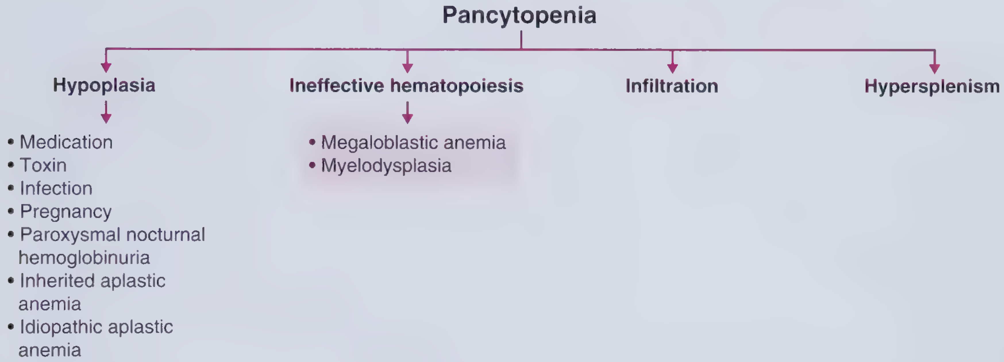
As shown in the flowchart above - when pancytopenia is caused by Ineffective Hematopoiesis, the two causes are Megaloblastic Anemia and Myelodysplasia. With low B12, megaloblastic anemia is the answer.
How Vitamin B12 Deficiency Causes ALL Findings
| Finding | Mechanism |
|---|---|
| Hb 4.7 (severe anemia) | DNA synthesis failure → RBCs cannot divide properly → large, fragile RBCs destroyed in marrow before release (ineffective erythropoiesis) |
| WBC 1,670 (leukopenia) | Same DNA defect → neutrophil precursors fail to mature → hypersegmented neutrophils on smear |
| Platelets 7,000 (thrombocytopenia) | Megakaryocytes affected → large, non-functional platelets |
| Bone marrow NORMAL/HYPERCELLULAR | ✅ Marrow is full of cells trying to divide, but they are abnormal (nuclear-cytoplasmic dissociation) - looks active, NOT empty |
| Nausea, vomiting | Classic GI symptoms of B12 deficiency + severe anemia |
| CKD label | CKD impairs B12 metabolism; or severe megaloblastic anemia may affect renal function |
"Megaloblastic anemia is the most common cause of pancytopenia in the developing world." - Frameworks for Internal Medicine, p. 364
Why the Bone Marrow Appears "Normal" or Non-diagnostic
This is the key teaching point:
"Bone marrow aspiration and biopsy demonstrate hypercellularity with evidence of abnormal proliferation and maturation... large oval-shaped erythroblasts with finely stippled, lacy nuclear chromatin (nuclear-cytoplasmic dissociation)."
In megaloblastic anemia, the marrow is NOT hypoplastic (empty). It is packed with cells but they cannot mature properly - this is called ineffective hematopoiesis. If the biopsy was read as "normal cellularity," megaloblastic changes may have been subtle or missed - but the low B12 confirms the diagnosis without needing marrow.
Complete Clinical Picture - This Patient
Low B12
    ↓
DNA synthesis impaired in ALL rapidly dividing cells
    ↓
RBC precursors → die in marrow → Hb 4.7 (severe anemia)
WBC precursors → fail to mature → WBC 1,670 (leukopenia)
Platelet precursors → fail → Plt 7,000 (thrombocytopenia)
    ↓
Pancytopenia with NORMAL (hypercellular) bone marrow
    ↓
Nausea + vomiting (uremic + anemia symptoms)
    ↓
CKD (co-existing or contributing to B12 depletion)
Classic Peripheral Smear Findings to Confirm
| Finding | Significance |
|---|---|
| Macrocytosis (MCV >100 fL) | Large RBCs, often oval (oval macrocytes) |
| Hypersegmented neutrophils (>5 lobes) | Pathognomonic of B12/folate deficiency |
| Pancytopenia | All three cell lines low |
If MCV on this patient is >100 fL - it seals the diagnosis completely.
Treatment
| Drug | Dose | Route |
|---|---|---|
| Vitamin B12 (Cyanocobalamin) | 1000 mcg daily × 7 days, then weekly × 4, then monthly | IM injection (preferred, especially in CKD where oral absorption may be poor) |
| Folic acid | 5 mg OD | Oral (also give, as often co-deficient) |
Response is rapid - reticulocyte count rises to 20-25% within 5-7 days of B12 replacement. - Textbook of Family Medicine, 9e
Note: Give B12 BEFORE folic acid alone, as giving folate alone in B12 deficiency can worsen neurological damage (subacute combined degeneration of spinal cord).
Final Summary
| Question | Answer |
|---|---|
| Why pancytopenia? | B12 deficiency → ineffective hematopoiesis of ALL cell lines |
| Why bone marrow normal? | Marrow is actually hypercellular but with ineffective, dysmatured cells |
| Why nausea/vomiting? | GI manifestation of B12 deficiency + severe anemia |
| Primary diagnosis | Megaloblastic Anemia (B12 Deficiency) |
| Treatment | Vitamin B12 IM injections + Folic acid |
- Frameworks for Internal Medicine, p. 364 (most common cause of pancytopenia in developing world)
- Textbook of Family Medicine, 9e, p. 2709-2713 (megaloblastic pancytopenia, treatment response)
- Tintinalli's Emergency Medicine (B12 deficiency: hematologic + neurologic effects)
Vitamin b12 deficiency causes
Vitamin B12 deficiency has several well-known causes across dietary, gastric, intestinal, and other categories.
Searching sources...
Finding Sources
Finding Sources
Reading File
Reading File
Here is a complete, organized breakdown of all causes of Vitamin B12 deficiency:
Causes of Vitamin B12 Deficiency
First, understand the normal absorption pathway - B12 from food binds to Intrinsic Factor (IF) secreted by gastric parietal cells → complex absorbed in distal ileum → stored in liver. Any disruption along this path causes deficiency.
1. DIETARY INSUFFICIENCY (Inadequate Intake)
| Cause | Details |
|---|---|
| Strict Vegan / Vegetarian diet | B12 is found ONLY in animal products (meat, fish, eggs, dairy). Vegans eating no meat or dairy become deficient - common in South Asia |
| Malnutrition / Poverty | Inadequate intake of animal-protein foods |
| Elderly ("tea and toast" diet) | Poor dietary variety, reduced intake |
| Prolonged breastfeeding | Infant of a B12-deficient vegan mother |
2. GASTRIC CAUSES (Lack of Intrinsic Factor)
| Cause | Details |
|---|---|
| Pernicious Anemia (Most Common overall) | Autoimmune destruction of gastric parietal cells → no Intrinsic Factor secreted → B12 cannot be absorbed. Anti-IF antibodies are diagnostic |
| Total / Subtotal Gastrectomy | Parietal cells removed surgically → no IF production |
| Partial gastrectomy | Reduced parietal cell mass → reduced IF |
| Chronic Atrophic Gastritis | Gradual loss of parietal cells with aging |
| H. pylori infection | Damages gastric mucosa → impairs IF and acid secretion |
| Gastric bypass surgery | Bypasses parietal cell-rich stomach segments |
3. INTESTINAL CAUSES (Malabsorption in Distal Ileum)
| Cause | Details |
|---|---|
| Crohn's Disease (Ileitis) | Inflammation of distal ileum - the ONLY site of B12-IF complex absorption |
| Ileal resection / surgical removal | Loss of absorption site |
| Tropical Sprue | Mucosal damage in small bowel |
| Celiac Disease | Villous atrophy affects absorption broadly |
| Bacterial overgrowth (blind loop syndrome) | Gut bacteria consume B12 before it can be absorbed |
| Fish tapeworm (Diphyllobothrium latum) | Parasite competes for and consumes B12 in the gut |
| Radiation enteritis | Ileal mucosal damage |
4. DRUG-INDUCED
| Drug | Mechanism |
|---|---|
| Metformin (very common) | Interferes with calcium-dependent IF-B12 receptor in ileum; long-term use causes B12 depletion |
| Proton Pump Inhibitors (PPIs) e.g., Pantoprazole | Reduce gastric acid → impair release of B12 from food protein |
| H2 blockers (ranitidine, famotidine) | Same mechanism as PPIs |
| Nitrous oxide (N2O) anesthesia | Irreversibly oxidizes B12 → functional deficiency acutely |
| Colchicine, Neomycin | Impair ileal absorption |
| Oral contraceptive pills | Mild reduction in B12 levels |
Clinically relevant: This patient is on Pantoprazole IV TDS - long-term PPI use is a recognized cause of B12 deficiency!
5. INCREASED DEMAND / UTILIZATION
| Cause | Details |
|---|---|
| Pregnancy | Increased fetal demand depletes maternal stores |
| Hyperthyroidism | Increased metabolic demand |
| Haematological malignancy | Rapid cell turnover consumes B12 |
6. CHRONIC KIDNEY DISEASE (Relevant to This Patient)
| Mechanism | Details |
|---|---|
| Dietary restriction | CKD patients are often on restricted diets with less animal protein |
| Dialysis | B12 is water-soluble and dialyzed out |
| Reduced absorption | Uremic enteropathy impairs gut absorption |
| Drug interactions | CKD patients on multiple drugs including PPIs, metformin |
The "Five P's" of Vitamin B12 Deficiency (Memory Aid)
From Swanson's Family Medicine Review:
| P | Manifestation |
|---|---|
| Pancytopenia | Low Hb + WBC + Platelets |
| Peripheral neuropathy | Tingling, numbness in hands/feet |
| Posterior spinal column neuropathy | Loss of vibration + proprioception (subacute combined degeneration) |
| Papillary (tongue) atrophy | Beefy red, smooth, sore tongue (glossitis) |
| Pyramidal tract signs | Spasticity, increased reflexes |
Connecting to This Patient
This patient is on Pantoprazole IV TDS (written on the note) - this is a PPI which itself contributes to B12 malabsorption. Combined with:
- Likely vegetarian/vegan diet (common in South Asia)
- CKD (poor dietary intake, reduced absorption)
- Nausea/vomiting (poor oral intake, further depleting B12)
All of these converge to produce severe B12 deficiency → Megaloblastic anemia → Pancytopenia.
Quick Summary by Category
DIETARY      → Vegan diet, malnutrition, elderly
GASTRIC      → Pernicious anemia (#1), gastrectomy, H. pylori, atrophic gastritis
INTESTINAL   → Crohn's, ileal resection, blind loop, tapeworm, celiac
DRUGS        → Metformin (#1 drug cause), PPIs, H2-blockers, N2O
RENAL        → CKD, dialysis
INCREASED DEMAND → Pregnancy, malignancy
- Katzung's Basic & Clinical Pharmacology, 16th Ed. (causes and treatment of B12 deficiency)
- Swanson's Family Medicine Review (five P's of B12 deficiency)
- Creasy & Resnik's Maternal-Fetal Medicine (pernicious anemia as most common cause)
This is a shared conversation. Sign in to Orris to start your own chat.