
Ascending tract physiology notes
Reading File
Now let me search the medical library for ascending tract content in parallel.
Finding Sources
Finding Sources
Finding Sources
Finding Sources
Finding Sources
Reading File
Finding Sources
Reading File
Finding Sources
Reading File
Reading File
Reading File
Finding Sources
Loading Image
Loading Image
Finding Sources
I now have comprehensive material to compile detailed notes. Let me also grab the sensory fiber type table and pain modulation section.
Reading File
Reading File
Loading Image
I now have excellent material to write comprehensive ascending tract physiology notes. Let me compile everything.
Ascending Tract Physiology - Complete Notes
Sources: Neuroanatomy through Clinical Cases, 3rd Ed. | Morgan & Mikhail's Clinical Anesthesiology, 7e | Adams & Victor's Principles of Neurology, 12th Ed.
Overview
Somatosensation - bodily sensations of touch, pain, temperature, vibration, and proprioception - is carried to the cortex via two principal ascending systems. They differ in fiber type, spinal cord location, level of decussation, and modalities conveyed.
| Pathway | Modalities | Decussation Level |
|---|---|---|
| Posterior column-medial lemniscal (PCML) | Vibration, joint position, fine/discriminative touch | Internal arcuate fibers - lower medulla |
| Anterolateral pathways (spinothalamic + others) | Pain, temperature, crude touch | Anterior commissure - spinal cord (2-3 segments above entry) |
| Lateral corticospinal tract (motor, for reference) | Motor | Pyramidal decussation - cervicomedullary junction |
Key point: Because touch is partially carried by both pathways, isolated lesions of either one do not fully abolish touch.
1. Sensory Neuron Fiber Types
All primary sensory neurons have their cell bodies in the dorsal root ganglia (DRG). Each DRG cell has a bifurcating stem axon - one branch goes to the periphery, the other enters via the dorsal root.
| Fiber | Alternate Name | Diameter (µm) | Myelinated | Sensory Modality |
|---|---|---|---|---|
| A-α | Type I | 13-20 | Yes (heavily) | Proprioception (muscle spindle Ia, Golgi tendon Ib) |
| A-β | Type II | 6-12 | Yes | Fine touch, pressure, vibration |
| A-δ | Type III | 1-5 | Lightly | Sharp/fast pain, cold temperature |
| C | Type IV | 0.2-1.5 | No (unmyelinated) | Slow/burning pain, warm temperature, crude touch |
- Large-diameter myelinated fibers (A-α, A-β) enter the medial dorsal root entry zone → posterior columns
- Small-diameter and unmyelinated fibers (A-δ, C) enter the lateral dorsal root entry zone → synapse immediately in dorsal horn gray matter
2. Posterior Column - Medial Lemniscal (PCML) Pathway
Modalities: Proprioception, vibration sense, fine/discriminative touch
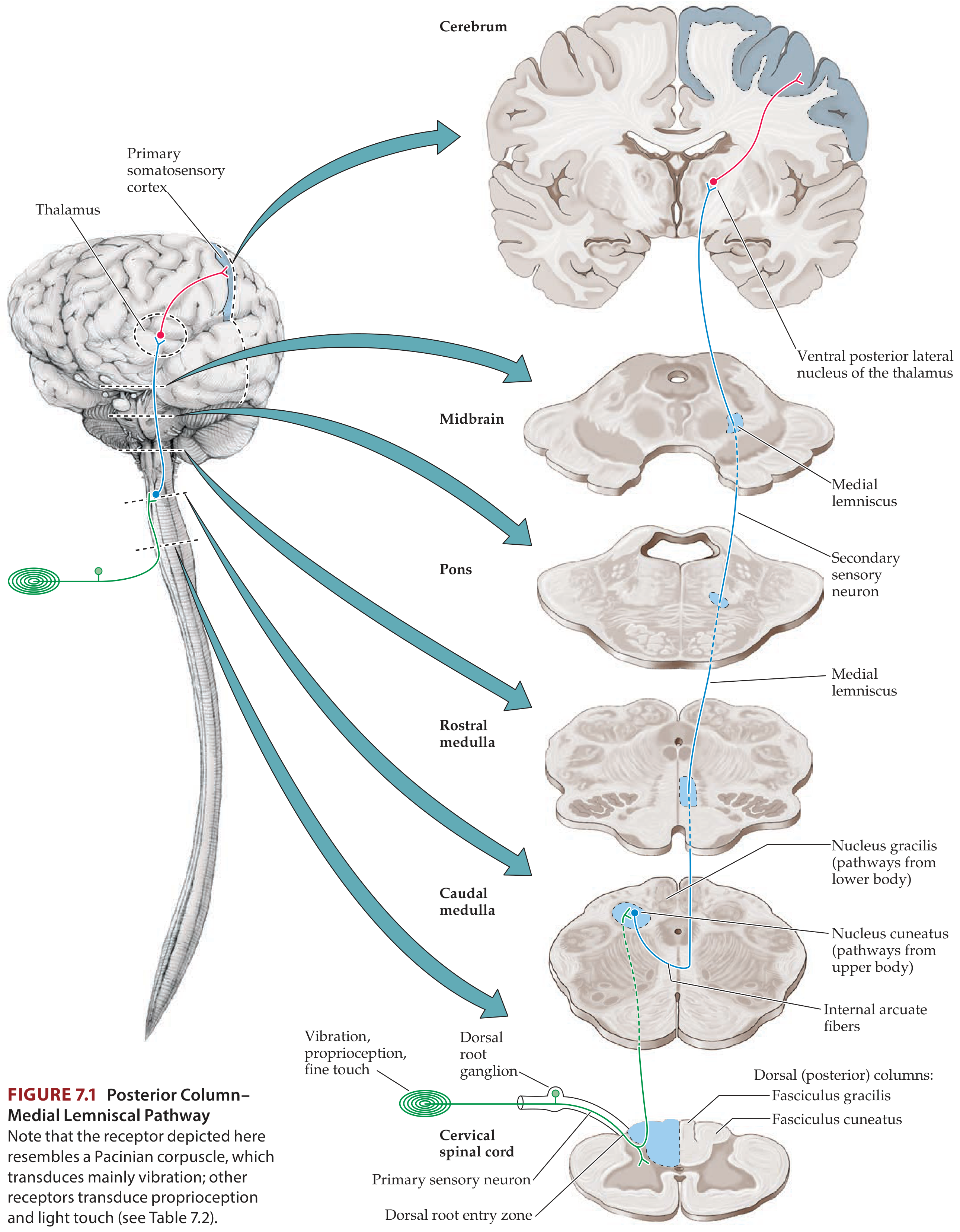
Step-by-Step Course
-
1st-order neuron - DRG cell body; peripheral receptor (Pacinian corpuscle for vibration, muscle spindles/Golgi tendon organs for proprioception). Axon enters spinal cord via medial dorsal root entry zone.
-
Ascent in posterior (dorsal) columns - Fiber travels ipsilaterally all the way to the lower medulla without synapsing.
- Fasciculus gracilis (medial): lower trunk and leg fibers; from below ~T6
- Fasciculus cuneatus (lateral): upper trunk, arm, neck fibers; from above ~T6
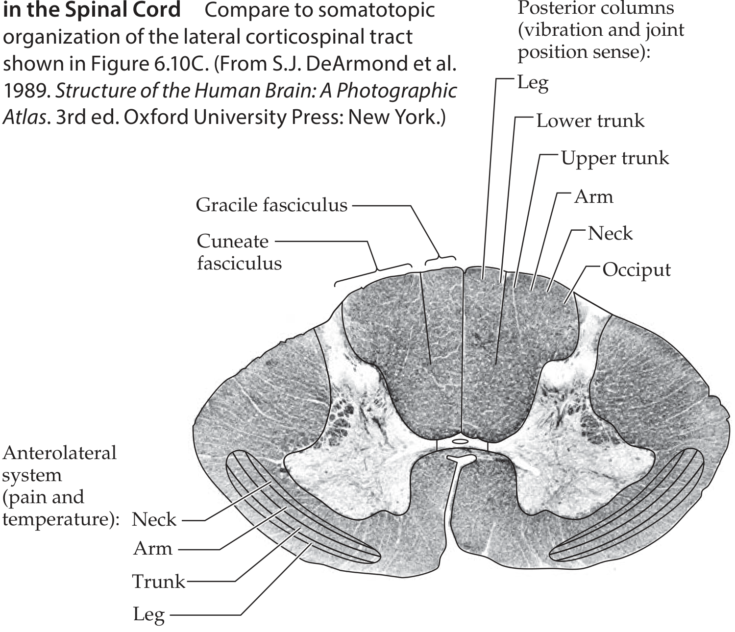
- Somatotopic rule: fibers add on laterally as you ascend - so legs are most medial in the posterior columns.
-
2nd-order neuron - Synapse in the posterior column nuclei of the lower medulla:
- Fasciculus gracilis → Nucleus gracilis
- Fasciculus cuneatus → Nucleus cuneatus
-
Decussation - Axons of 2nd-order neurons cross as internal arcuate fibers in the lower medulla.
-
Medial lemniscus - After crossing, fibers form the medial lemniscus on the contralateral side. Somatotopic orientation shifts as it ascends:
- Medulla: vertical orientation; legs represented ventrally ("the little person stands up")
- Pons/midbrain: inclined position; legs move laterally, arms medially ("the little person lies down")
-
Thalamic relay - Medial lemniscus terminates in the Ventral Posterior Lateral (VPL) nucleus of the thalamus.
-
3rd-order neuron - Thalamic radiations project to primary somatosensory cortex (postcentral gyrus, Brodmann areas 3, 1, 2).
Diagram - PCML Pathway:
3. Anterolateral Pathways
Modalities: Pain, temperature, crude touch
This system comprises three tracts: spinothalamic, spinoreticular, and spinomesencephalic.
Step-by-Step Course
-
1st-order neuron - Small-diameter A-δ and C fibers enter via the lateral dorsal root entry zone. Some axon collaterals ascend/descend 1-2 segments in Lissauer's tract before synapsing.
-
Synapse in dorsal horn - 1st-order neurons synapse onto 2nd-order neurons mainly in:
- Lamina I (dorsal horn marginal zone)
- Lamina V (deeper dorsal horn)
-
Decussation in spinal cord - 2nd-order axons cross via the anterior (ventral) commissure to the contralateral anterolateral white matter. Important: it takes 2-3 spinal segments for fibers to cross - so a lateral cord lesion causes contralateral pain/temperature loss a few segments below the lesion level.
-
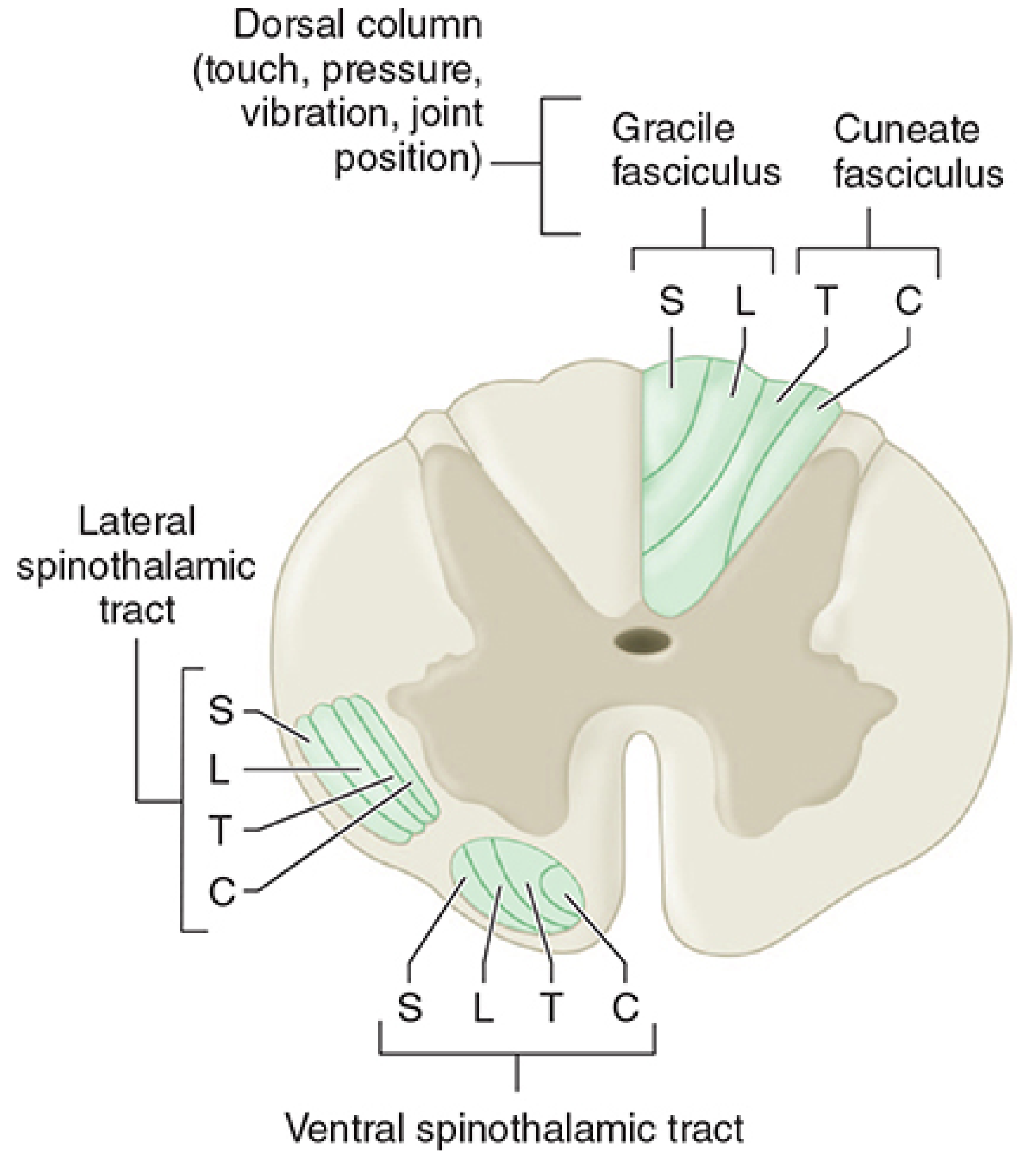
Ascent in anterolateral white matter - Somatotopic organization:
- Legs most lateral, arms more medial (fibers from anterior commissure add on medially as they ascend)
- This lateral-to-medial arrangement is preserved throughout the brainstem
- In the medulla: located laterally, in the groove between the inferior olives and inferior cerebellar peduncles
- In pons/midbrain: just lateral to the medial lemniscus
-
Thalamic relay - Primary relay in VPL (separate neurons from PCML fibers). Additional projections to:
- Intralaminar thalamic nuclei (central lateral nucleus)
- Medial thalamic nuclei (mediodorsal nucleus)
-
3rd-order neuron - From VPL → primary somatosensory cortex (postcentral gyrus). Medial thalamic projections → frontal and cingulate cortex (emotional aspects of pain).
-
Face pain - Carried by the analogous trigeminothalamic tract (not the spinothalamic tract).
Spinal cord cross-section showing somatotopic organization:
Detailed somatotopic map (cadaveric cross-section):
The Three Anterolateral Tracts
| Tract | Projection | Function |
|---|---|---|
| Spinothalamic (neospinothalamic) | VPL thalamus → somatosensory cortex | Discriminative pain/temperature (location, intensity, duration) |
| Spinoreticular | Medullary/pontine reticular formation → intralaminar thalamus | Arousal and autonomic responses to pain; emotional aspects |
| Spinomesencephalic | Periaqueductal gray (PAG), superior colliculus | Activates antinociceptive descending pathways |
Additionally:
- Lateral spinothalamic (neo) - VPL - discriminative pain quality
- Medial spinothalamic (paleo) - Medial thalamus - autonomic and unpleasant emotional pain perception
- Spinohypothalamic/spinotelencephalic - Hypothalamus; evoke emotional behavior
- Spinocervical tract - Lateral cervical nucleus → contralateral thalamus; major alternative pain pathway (ascends uncrossed)
4. Somatosensory Cortex
- Primary somatosensory cortex - Postcentral gyrus, Brodmann areas 3, 1, 2
- Somatotopic (homuncular) organization: face most laterally, leg most medially (same as motor cortex)
- Receives from VPL (body) and VPM (face via trigeminothalamic tract)
- Projects to secondary somatosensory cortex - in the Sylvian fissure/parietal operculum (also somatotopic)
- Further processing in superior parietal lobule association cortex (areas 5 and 7)
Lesions of somatosensory cortex → cortical sensory loss: impaired two-point discrimination, graphesthesia, stereognosis, but relatively preserved crude pain/touch (subcortical processing intact).
5. Central Modulation of Pain
Gate Control Theory
- A-β fiber (large, non-pain) activation in dorsal horn inhibits pain transmission
- Clinical application: TENS (transcutaneous electrical nerve stimulation) activates A-β fibers to reduce chronic pain; rubbing an injured area does the same
Descending Modulatory Pathways
- Periaqueductal gray (PAG) - Receives inputs from hypothalamus, amygdala, cortex → key "off switch" for pain
- PAG → Rostral Ventromedial Medulla (RVM) relay → spinal cord dorsal horn
- Two key descending neurotransmitters:
- Serotonin (5-HT) - Raphe nuclei neurons project to dorsal horn
- Norepinephrine (NE) - Locus ceruleus (stimulated by RVM via substance P) projects to dorsal horn
Endogenous Opioid System
- Opiate receptors distributed throughout - peripheral nerves, dorsal horn, PAG, RVM
- Enkephalin and dynorphin - concentrated in PAG, RVM, dorsal horn
- β-endorphin - concentrated in hypothalamic regions projecting to PAG
- Exogenous opioids (morphine) mimic these at all these sites
6. Key Clinical Correlations
Lesion Localization Rules
| Lesion Location | PCML Deficit | Anterolateral Deficit | Rule |
|---|---|---|---|
| Lateral half of spinal cord (Brown-Séquard) | Ipsilateral vibration/position loss below lesion | Contralateral pain/temp loss 2-3 levels below lesion | Pathways cross at different levels |
| Anterior cord | Spared | Bilateral pain/temp loss | Anterior commissure or both anterolateral columns |
| Medulla (lateral) | Spared | Ipsilateral face + contralateral body pain/temp loss | Lateral medullary syndrome |
| Left cerebral cortex lesion | Right-sided all modalities | Right-sided all modalities | Both pathways already crossed before cortex |
Syringomyelia
- Cavitation of the central cord damages fibers crossing in the anterior commissure → bilateral loss of pain and temperature at the segmental levels of the cavity ("cape distribution"), with preserved posterior column function ("dissociated sensory loss")
7. Quick Summary Table
| Feature | PCML Pathway | Spinothalamic (Anterolateral) |
|---|---|---|
| Modalities | Vibration, proprioception, fine touch | Pain, temperature, crude touch |
| Fiber types | A-α, A-β (large myelinated) | A-δ, C (small/unmyelinated) |
| 1st synapse | Posterior column nuclei (medulla) | Dorsal horn (laminae I, V) |
| Decussation | Internal arcuate fibers, lower medulla | Anterior commissure, spinal cord |
| Spinal cord location | Posterior (dorsal) columns | Anterolateral white matter |
| Somatotopy in cord | Legs medial, arms lateral | Legs lateral, arms medial |
| Thalamic relay | VPL | VPL (+ medial/intralaminar nuclei) |
| Cortex | Postcentral gyrus (areas 3, 1, 2) | Postcentral gyrus + frontal/cingulate |
| Ipsilateral deficit | Ipsilateral cord lesion → ipsilateral loss | Ipsilateral cord lesion → contralateral loss |
Sources: Neuroanatomy through Clinical Cases, 3rd Ed., Ch. 7 | Morgan & Mikhail's Clinical Anesthesiology, 7e, Ch. 47 | Adams & Victor's Principles of Neurology, 12th Ed.
This is a shared conversation. Sign in to Orris to start your own chat.