
A 30 Year old male met with road traffic accident and had massive haemorrhage. The patient was pale, pulse was rapid and thread skin was cold and clammy. (2+3+10) (2023P1)
I'll read the medical library skill first, then research this comprehensively.
Reading File
Now let me search the medical library for hemorrhagic shock, its classification, pathophysiology, and management - all in parallel.
Finding Sources
Finding Sources
Finding Sources
Excellent results. Now let me read the most relevant sections in parallel - Schwartz's signs/symptoms table, Miller's pathophysiology, Guyton's physiology, and Sabiston's management.
Reading File
Reading File
Reading File
Reading File
Excellent content. Now let me read more on management (Sabiston treatment section) and Guyton's progressive/irreversible shock, plus the Rosen's hemorrhagic shock section.
Reading File
Reading File
Reading File
Now let me get the management section from Schwartz's and Guyton's irreversible/positive feedback mechanisms section:
Reading File
Reading File
I now have comprehensive content. Let me fetch the key Guyton figure on positive feedback loops to include visually:
Loading Image
Loading Image
I now have all the information needed for a thorough answer. The question is marked (2+3+10) suggesting:
- 2 marks: Diagnosis / identify the condition (hemorrhagic/hypovolemic shock)
- 3 marks: Classification (ATLS Classes I-IV)
- 10 marks: Pathophysiology + Management
Let me now compose the full answer.
Hemorrhagic (Hypovolemic) Shock Following Road Traffic Accident
PART 1 (2 marks) - Diagnosis
The clinical picture - pallor, rapid thready pulse, cold and clammy skin following massive hemorrhage after RTA - is classic hemorrhagic (hypovolemic) shock.
Definition: Hemorrhagic shock results from rapid reduction in intravascular blood volume following hemorrhage, leading to an imbalance between systemic oxygen delivery and oxygen consumption, causing inadequate tissue perfusion.
- Pallor = peripheral vasoconstriction diverting blood from skin
- Rapid thready pulse = compensatory tachycardia + reduced stroke volume
- Cold and clammy skin = sympathetic-mediated vasoconstriction + sweat gland activation
This is a "cold shock" characterized by increased systemic vascular resistance (SVR) and decreased cardiac output (CO) - in contrast to septic/distributive shock which is warm. (Sabiston Textbook of Surgery)
PART 2 (3 marks) - Classification (ATLS Classes)
Based on ATLS (Advanced Trauma Life Support), hemorrhagic shock is classified into four classes based on blood loss:
| Parameter | Class I | Class II | Class III | Class IV |
|---|---|---|---|---|
| Blood loss (mL) | Up to 750 | 750-1500 | 1500-2000 | >2000 |
| Blood loss (% BV) | Up to 15% | 15-30% | 30-40% | >40% |
| Pulse rate | <100 | >100 | >120 | >140 |
| Blood pressure | Normal | Normal | Decreased | Decreased |
| Pulse pressure | Normal/Increased | Decreased | Decreased | Decreased |
| Respiratory rate | 14-20 | >20-30 | 30-40 | >35 |
| Urine output (mL/hr) | >30 | 20-30 | 5-15 | Negligible |
| CNS/Mental status | Slightly anxious | Mildly anxious | Anxious & confused | Confused & lethargic |
(Schwartz's Principles of Surgery, 11th ed.)
The patient described (massive hemorrhage, rapid thready pulse, cold/clammy skin) fits Class III-IV shock.
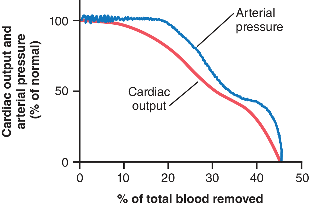
Key note: Only 10% of total blood volume can be removed with almost no effect on arterial pressure or cardiac output. Greater blood loss diminishes cardiac output first, then arterial pressure - both fall to zero when ~40-45% of total blood volume is removed. (Guyton and Hall Medical Physiology)
PART 3 (10 marks) - Pathophysiology and Management
PATHOPHYSIOLOGY
A. Initial Hemodynamic Response
Hemorrhage reduces intravascular volume → decreased venous return → decreased cardiac preload → decreased cardiac output → decreased arterial blood pressure.
The cardiac output falls first; arterial pressure is maintained longer via sympathetic reflexes (see Guyton graph below):
B. Neuroendocrine (Sympathoadrenal) Compensation
Baroreceptors in the carotid sinus and aortic arch detect the fall in blood pressure and trigger a powerful sympathetic response:
- Arteriolar vasoconstriction - increases total peripheral resistance (maintains BP)
- Venous constriction - increases venous return to the heart
- Tachycardia - heart rate can rise to 160-180 bpm
- Adrenal medulla releases epinephrine and norepinephrine
- Renin-angiotensin-aldosterone system (RAAS) activation - renal vasoconstriction + sodium and water retention
- ADH (vasopressin) release from posterior pituitary - peripheral vasoconstriction + free water retention
- Cortisol and glucagon release - mobilizes glucose
Blood is preferentially redirected to the heart, brain, and kidneys at the expense of the skin (explaining cold, clammy skin), gut, and muscle.
C. Microcirculatory and Cellular Response
- Ischemic cells cannot maintain aerobic metabolism → switch to anaerobic glycolysis → lactic acid accumulation → metabolic acidosis
- Base deficit becomes progressively more negative even while BP and pH appear normal (base deficit is the earliest marker of clinically significant hemorrhage)
- Individual ischemic cells take up interstitial fluid → further depleting intravascular volume
- Cellular edema can restrict adjacent capillary flow - the "no-reflow" phenomenon - preventing reversal of ischemia even after macroperfusion is restored
D. Inflammatory & Organ-level Response
- Ischemic cells produce and release: lactate, free radicals, prostacyclin, thromboxane, prostaglandins, leukotrienes, endothelin, complement, interleukins, TNF, and damage-associated molecular patterns (DAMPs)
- These compounds cause direct cellular damage and amplify the systemic inflammatory response
- Multiple organ failure (MOF) can result from this inflammatory cascade
Organ-specific effects:
- CNS: Reflexes and cortical activity depressed; irreversible damage with prolonged ischemia
- Kidney: Initially compensates by vasoconstriction; prolonged hypotension → acute tubular necrosis (ATN)
- Lung: Capillary leak → ARDS (Adult Respiratory Distress Syndrome)
- Liver & gut: Mucosal barrier breakdown → bacterial translocation
E. Progressive vs. Compensated Shock
There is a critical threshold of blood loss. Below this threshold, the body's compensatory mechanisms (baroreceptors, RAAS, ADH, reverse stress-relaxation of vessels) can restore circulation = compensated (nonprogressive) shock.
Beyond this threshold, shock becomes progressive through vicious positive-feedback cycles as shown in the diagram below from Guyton and Hall:
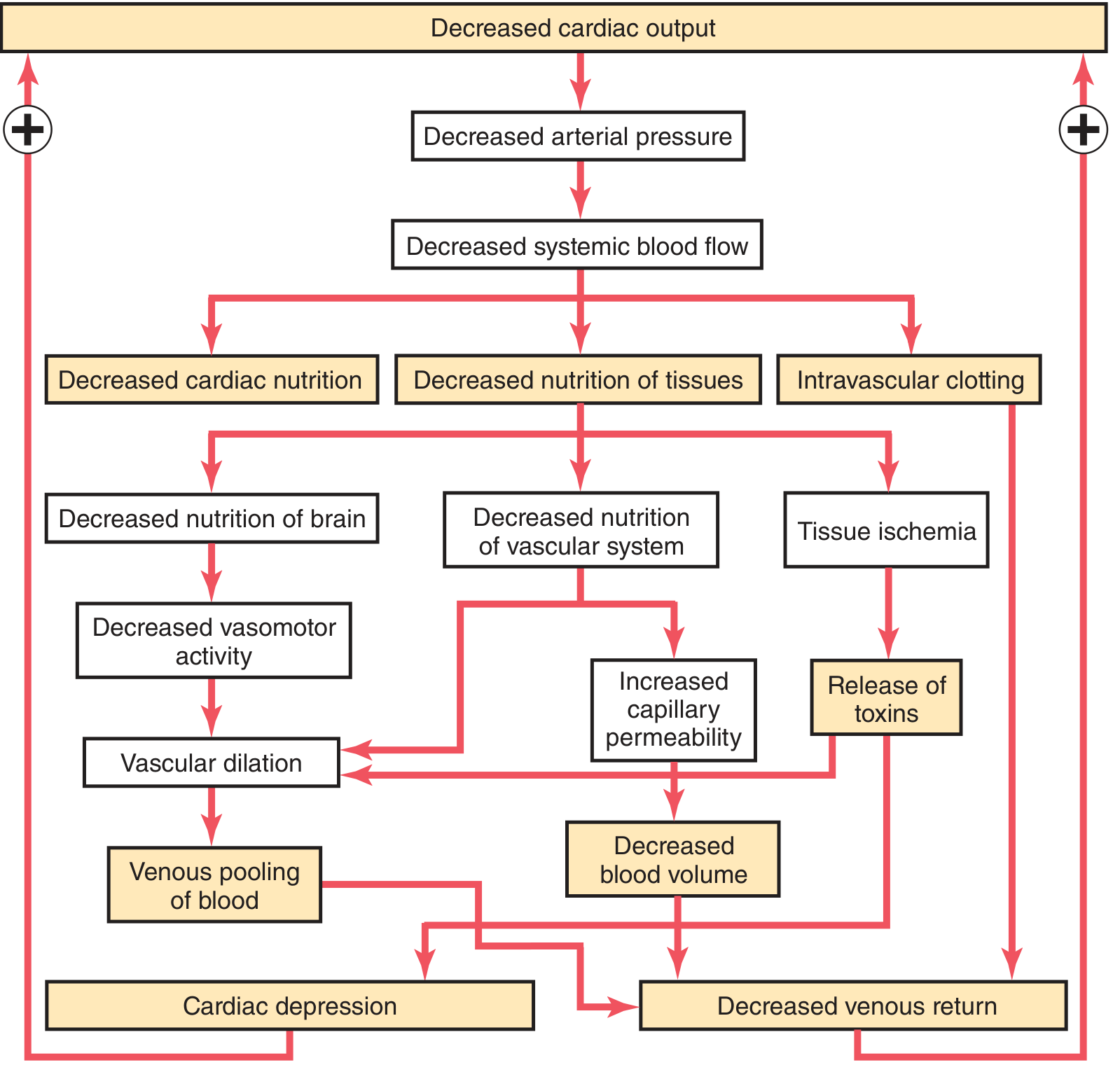
Key positive-feedback cycles include:
- Decreased cardiac output → decreased coronary flow → cardiac depression → further decreased output
- Decreased systemic flow → decreased vasomotor center nutrition → vasomotor failure → vascular dilation → venous pooling
- Tissue ischemia → increased capillary permeability → further blood volume loss
- Intravascular clotting → tissue ischemia → toxin release → further cardiac depression
Once irreversible shock is reached, massive ischemic damage, cell membrane failure (Na-K ATPase failure), and widespread intravascular coagulation make survival impossible even with blood transfusion.
MANAGEMENT
Management follows the ATLS primary survey (ABCDE) principle.
1. Immediate - Control of Hemorrhage
- Direct pressure on external bleeding wounds
- Tourniquet for extremity hemorrhage
- Pelvic binder for pelvic fractures (can lose >2000 mL into pelvis)
- Emergency surgical hemorrhage control (damage control surgery) if internal hemorrhage
2. Airway and Breathing
- Maintain airway; administer high-flow oxygen (100%)
- Intubate if GCS < 8 or airway at risk
3. Intravenous Access and Resuscitation
- Establish two large-bore peripheral IV lines (16G or larger)
- Draw blood for: FBC, crossmatch, coagulation studies, ABG, base deficit, lactate
4. Fluid Resuscitation Strategy
Hemorrhagic shock = blood products are the resuscitative fluid of choice. Crystalloid alone leads to anemia and dilutional coagulopathy. (Sabiston Textbook of Surgery)
- Massive Transfusion Protocol (MTP): Packed Red Blood Cells (pRBCs) : Fresh Frozen Plasma (FFP) : Platelets in 1:1:1 ratio
- Recent evidence suggests whole blood may be superior to component therapy
- Tranexamic acid (TXA): Should be given within 3 hours of injury (CRASH-2 trial evidence) - reduces fibrinolysis and mortality
- Viscoelastic testing (TEG/ROTEM) can guide targeted component replacement, especially when transfusion exceeds 10 units pRBCs
- Avoid hypothermia (use warmed fluids) - hypothermia worsens coagulopathy
The lethal triad of trauma must be prevented:
Hypothermia + Acidosis + Coagulopathy = irreversible death
5. Response to Resuscitation (classify patient)
| Category | Description | Action |
|---|---|---|
| Responder | Vital signs normalize | Investigate for occult injury |
| Transient responder | Initial improvement then deteriorates | Urgent surgical evaluation |
| Non-responder | No response despite resuscitation | Immediate surgical intervention |
6. Vasopressors
- Early vasopressor use in hemorrhagic shock has minimal evidence but may be used as a temporizing bridge while blood products are given
- Evidence supports vasopressin in a "vasopressin-deficient state" seen in prolonged shock
7. Permissive Hypotension (Damage Control Resuscitation)
- For penetrating vascular injuries: target SBP ~90 mmHg (to prevent dislodging an endogenous clot)
- Exception: TBI patients require SBP >100 mmHg to maintain cerebral perfusion pressure
- This concept is NOT appropriate for blunt trauma with TBI
8. Endpoints of Resuscitation
- Normalize: HR, BP, urine output (>0.5 mL/kg/hr)
- Clearance of lactate
- Improvement in base deficit (toward 0)
- Normalization of coagulation
9. Monitoring
- Urine output (Foley catheter)
- ABG with lactate and base deficit
- CVP / IVC ultrasound to distinguish hemorrhagic from cardiogenic shock
- eFAST (extended Focused Assessment with Sonography for Trauma) to identify source
10. Definitive Hemorrhage Control
- Damage control surgery: abbreviated initial operation to stop bleeding and contamination; definitive repair after resuscitation in ICU
- Interventional radiology: angioembolization for pelvic/hepatic/splenic hemorrhage
Summary
| Aspect | Key Points |
|---|---|
| Diagnosis | Hemorrhagic shock - rapid pulse, pallor, cold/clammy = Class III-IV |
| Mechanism | Hypovolemia → ↓CO → sympathetic activation → vasoconstriction → ischemia → acidosis |
| Classification | ATLS Classes I-IV based on % blood volume lost |
| Management | Stop bleeding, O2, MTP (1:1:1), TXA within 3 hrs, damage control surgery |
| Goals | Correct lethal triad (hypothermia, acidosis, coagulopathy) |
Sources: Guyton and Hall Textbook of Medical Physiology | Schwartz's Principles of Surgery, 11th ed. | Sabiston Textbook of Surgery | Miller's Anesthesia, 10th ed. | Rosen's Emergency Medicine
This is a shared conversation. Sign in to Orris to start your own chat.