
Enzymes classification
enzyme classification IUB system oxidoreductases transferases hydrolases
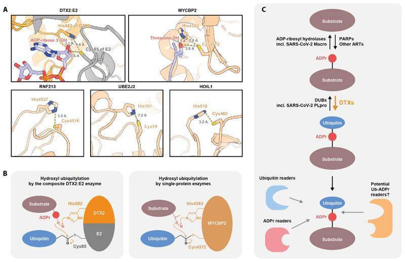
Educational medical graphic illustrating the molecular structural mechanisms of hydroxyl-ubiquitylation. Panel A presents high-resolution structural models (PDB and AlphaFold2) of five enzymes: DTX2:E2, MYCBP2, RNF213, UBE2J2, and HOIL1. These models highlight the spatial proximity between a catalytic Histidine (His) residue and a Cysteine (Cys) residue, with labeled inter-residue distances ranging from 3.1 to 3.8 Angstroms, demonstrating the conserved arrangement for ubiquitin transfer. Panel B provides schematic diagrams of hydroxyl ubiquitylation, distinguishing between composite enzyme complexes (DTX2:E2) and single-protein enzymes (MYCBP2). It illustrates the proposed catalytic mechanism where His (e.g., His582 or His4583) acts as a general base to deprotonate the hydroxyl group on an ADP-ribose (ADPr) or Threonine substrate, facilitating nucleophilic attack on the E2/E3~Ubiquitin thioester. Panel C depicts a signaling pathway for the synthesis, removal, and recognition of the composite ADPr-Ubiquitin modification. This flowchart identifies PARPs/ARTs for initial ADP-ribosylation and DTX enzymes for subsequent ubiquitylation, alongside reversing hydrolases like SARS-CoV-2 Macro and PLpro. The pathway concludes with the recognition of this hybrid modification by specific Ubiquitin, ADPr, and potential dual-modality reader domains.
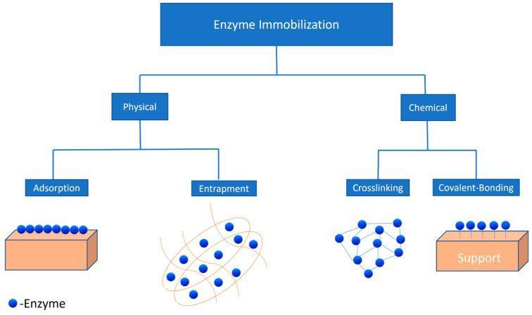
This educational flowchart illustrates the primary methodologies for enzyme immobilization, categorized into physical and chemical techniques. The diagram is organized hierarchically, beginning with 'Enzyme Immobilization' at the top level. The physical category branches into Adsorption and Entrapment. Adsorption is shown as enzymes (blue circles) resting on the surface of a support material, while Entrapment depicts enzymes confined within a fibrous matrix. The chemical category branches into Crosslinking and Covalent-Bonding. Crosslinking is represented by enzymes interconnected by molecular bridges in a lattice structure, and Covalent-Bonding shows enzymes directly attached to a support surface via chemical bonds. This classification is clinically and biotechnologically relevant for enhancing enzyme stability and reusability in biomedical applications such as diagnostic assays and pharmacological production.
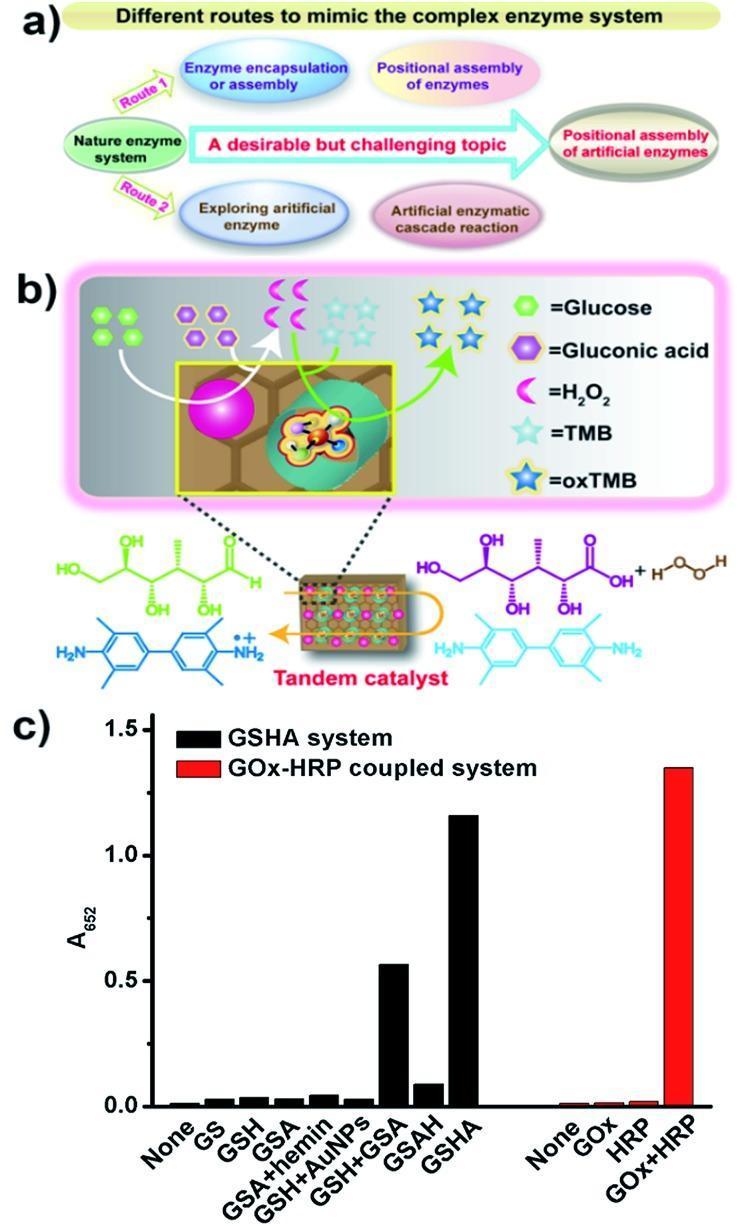
This composite educational graphic illustrates the development of artificial enzyme systems for tandem catalysis. Panel (a) is a flowchart depicting two research routes to mimic natural enzyme systems: Route 1 focuses on encapsulation/assembly of natural enzymes, while Route 2 explores artificial enzymes to achieve cascade reactions. Panel (b) provides a schematic and molecular-level illustration of a GSHA (graphene-mesoporous silica-hemin-AuNPs) tandem catalyst. It demonstrates a two-step reaction where Glucose is oxidized to Gluconic acid, producing H2O2 as an intermediate, which then facilitates the oxidation of TMB to oxTMB. Chemical structures show the transformation of glucose (green) and TMB (light blue) into their respective products. Panel (c) is a bar graph comparing the catalytic activity (measured by absorbance at 652 nm) of the GSHA system components versus the natural GOx-HRP coupled system. The data highlights that the integrated GSHA construct exhibits significantly higher activity than its individual components (GS, GSH, GSA) and approaches the performance of the natural GOx+HRP enzyme pair, demonstrating the effectiveness of spatial positional assembly in biomimetic catalysts.
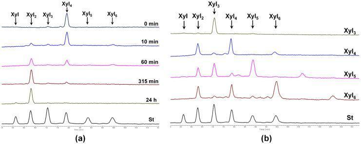
This image consists of two panels (a and b) showing High-Performance Liquid Chromatography (HPLC) chromatograms related to the enzymatic hydrolysis of xylooligosaccharides (XOs) by SlXyn30A. Panel (a) illustrates a time-course analysis of xylotetraose (Xyl4) hydrolysis over 24 hours (0 min, 10 min, 60 min, 315 min, and 24 h). The chromatograms show a progressive decrease in the Xyl4 peak intensity, coinciding with the emergence and growth of products like xylobiose (Xyl2). Panel (b) compares the hydrolysis products generated from various substrates (Xyl3, Xyl4, Xyl5, and Xyl6) after 30 minutes of reaction. Both panels include a standard (St) chromatogram for peak identification. The peaks are labeled as Xyl1 through Xyl6, representing the degree of polymerization of the xylooligosaccharides. This diagnostic data demonstrates the enzyme's xylobiohydrolase activity and transglycosylation capabilities, which are relevant in the study of glycoside hydrolases and their potential applications in human gut health through prebiotic fiber metabolism.
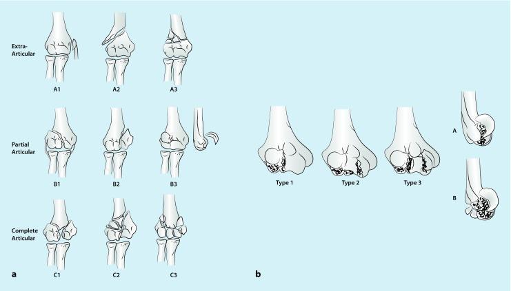
Educational orthopedic diagrams illustrating classification systems for distal humerus fractures. Section (a) displays the AO classification system in three rows: Extra-articular (Type A), showing fractures proximal to the joint line; Partial Articular (Type B), showing fractures extending into the joint but leaving part of the articular surface attached to the shaft; and Complete Articular (Type C), showing complete dissociation of the articular surface from the shaft. Section (b) illustrates the Dubberley classification for coronal shear fractures (AO Type B3). Frontal views categorize fractures into Type 1 (capitellum), Type 2 (continuous capitellum and trochlea), and Type 3 (separate capitellum and trochlea fragments). Lateral profiles differentiate between Type A (absence of dorsal comminution) and Type B (presence of posterior/dorsal comminution). These diagrams serve as clinical guides for orthopedic surgical planning, helping determine the necessity of headless compression screws or locking plate osteosynthesis based on fracture morphology and joint involvement.
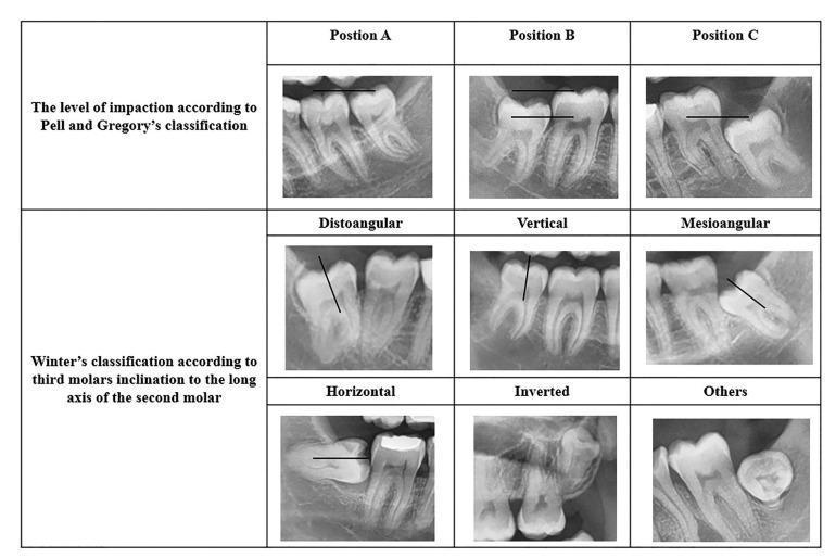
A composite image displaying nine panoramic dental radiographs categorized by two standard systems for classifying impacted mandibular third molars: the Pell and Gregory classification and Winter's classification. The top row illustrates the Pell and Gregory system based on the vertical depth of the impacted tooth relative to the second molar: Position A (at the occlusal plane), Position B (between the occlusal plane and cementoenamel junction), and Position C (below the CEJ). The bottom two rows demonstrate Winter's classification based on the angulation of the long axis of the third molar relative to the second molar or occlusal plane. Visualized categories include distoangular (tilted away from the second molar), vertical (parallel), mesioangular (tilted toward the second molar), horizontal (90-degree tilt), inverted (crown facing downward), and other atypical positions. These radiographic examples serve as an educational diagnostic guide for oral and maxillofacial surgery planning and risk assessment.
Enzyme Classification
The 6 IUB Classes
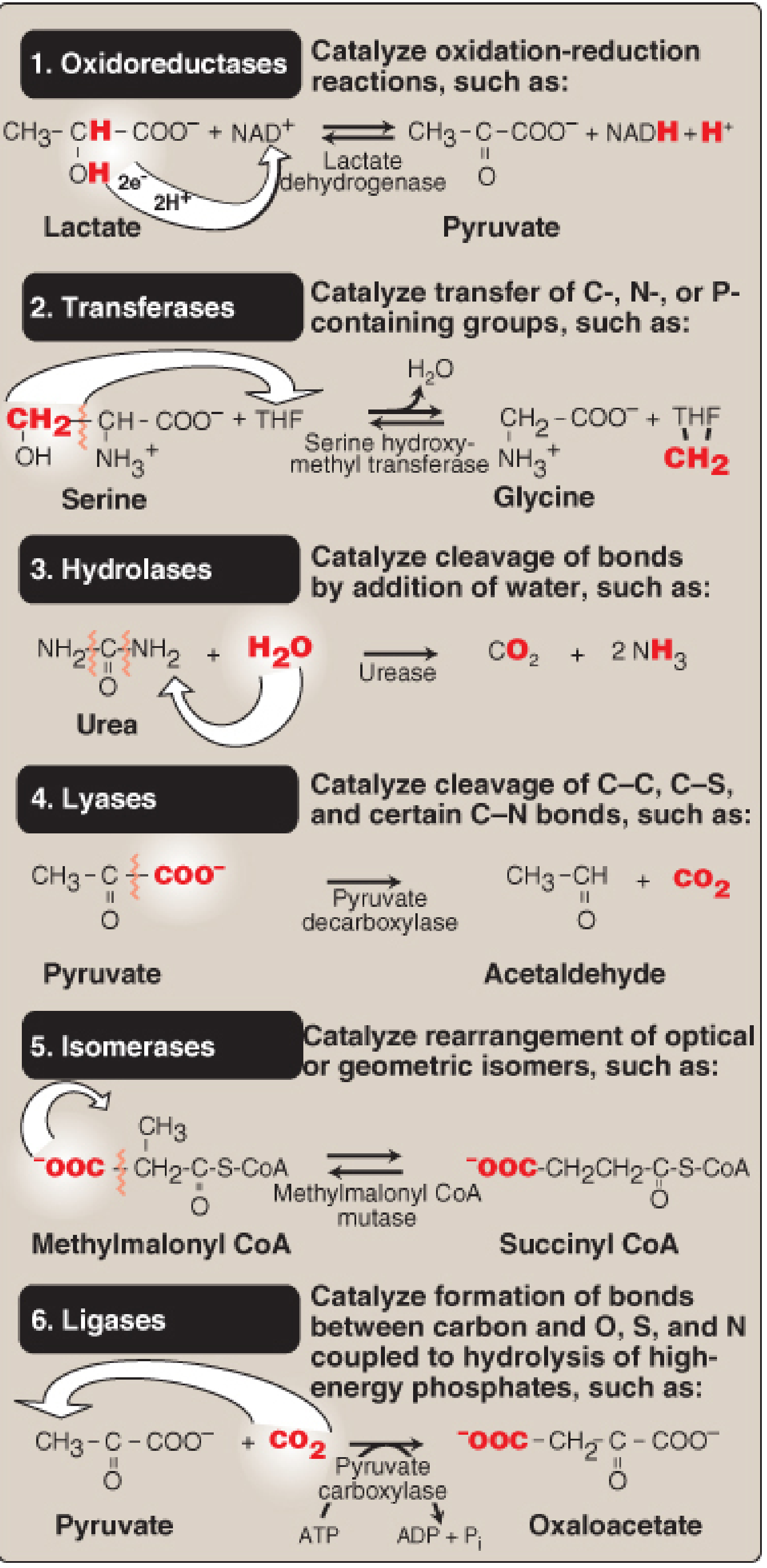
1. Oxidoreductases (EC 1)
- Reaction: Catalyze oxidation-reduction reactions - transfer of electrons (hydrogen atoms or oxygen)
- Key reaction: AH₂ + B → A + BH₂
- Subgroups: Dehydrogenases, oxidases, peroxidases, reductases, oxygenases
- Examples:
- Lactate dehydrogenase (LDH): Lactate + NAD⁺ → Pyruvate + NADH
- Succinate dehydrogenase
- Cytochrome oxidase
2. Transferases (EC 2)
- Reaction: Catalyze transfer of functional groups (methyl, amino, phosphate, acyl, etc.) from donor to acceptor
- Key reaction: A-X + B → A + B-X
- Subgroups: Aminotransferases (transaminases), kinases, methyltransferases, glycosyltransferases
- Examples:
- Serine hydroxymethyl transferase: Serine + THF → Glycine + methylene-THF
- Hexokinase (phosphate group transfer)
- Alanine aminotransferase (ALT), Aspartate aminotransferase (AST)
3. Hydrolases (EC 3)
- Reaction: Catalyze cleavage of bonds by addition of water (hydrolysis)
- Key reaction: A-B + H₂O → A-OH + B-H
- Subgroups: Proteases, lipases, phosphatases, nucleases, glycosidases
- Examples:
- Urease: Urea + H₂O → CO₂ + 2 NH₃
- Trypsin, chymotrypsin (peptide bonds)
- Alkaline phosphatase
4. Lyases (EC 4)
- Reaction: Catalyze cleavage or formation of C-C, C-O, C-S, and certain C-N bonds by means other than hydrolysis or oxidation (often without water)
- Key reaction: A-B → A + B (elimination/addition forming double bonds)
- Subgroups: Decarboxylases, aldolases, dehydratases, synthases
- Examples:
- Pyruvate decarboxylase: Pyruvate → Acetaldehyde + CO₂
- Aldolase (splits fructose-1,6-bisphosphate)
- Carbonic anhydrase (CO₂ + H₂O ⇌ HCO₃⁻ + H⁺)
5. Isomerases (EC 5)
- Reaction: Catalyze rearrangement of atoms within a molecule - interconversion of optical or geometric isomers
- Key reaction: A → B (isomer of A)
- Subgroups: Racemases, epimerases, mutases, isomerases
- Examples:
- Methylmalonyl-CoA mutase: Methylmalonyl-CoA → Succinyl-CoA
- Glucose-6-phosphate isomerase
- Phosphoglucose isomerase
6. Ligases (EC 6)
- Reaction: Catalyze formation of new bonds between two molecules, coupled to the hydrolysis of a high-energy phosphate (usually ATP)
- Key reaction: A + B + ATP → A-B + ADP + Pᵢ
- Subgroups: Synthetases, carboxylases (require biotin), amino-acyl-tRNA synthetases
- Examples:
- Pyruvate carboxylase: Pyruvate + CO₂ + ATP → Oxaloacetate + ADP + Pᵢ
- Acetyl-CoA carboxylase
- DNA ligase
Note (2018 update): A 7th class, Translocases (EC 7), was officially added by the IUPAC-IUBMB to cover enzymes that catalyze the movement of ions or molecules across membranes (e.g., ion pumps and transporters). Many older textbooks still list only 6 classes.
EC Numbering System
| Position | Meaning |
|---|---|
| EC 1.x.x.x | Class (e.g., 1 = Oxidoreductase) |
| EC 1.1.x.x | Subclass (type of bond/group involved) |
| EC 1.1.1.x | Sub-subclass (coenzyme used, etc.) |
| EC 1.1.1.27 | Serial number (specific enzyme) |
Quick Summary Table
| Class | EC # | Reaction Type | Memory Aid | Example |
|---|---|---|---|---|
| Oxidoreductases | EC 1 | Redox (e-⁻ transfer) | Oxygen changes | LDH, Cytochrome oxidase |
| Transferases | EC 2 | Group transfer | Transport groups | Kinases, Aminotransferases |
| Hydrolases | EC 3 | Bond cleavage + H₂O | Hydro = water | Lipase, Urease, Trypsin |
| Lyases | EC 4 | Bond cleavage/addition (non-hydrolytic) | Leave a double bond | Aldolase, Decarboxylases |
| Isomerases | EC 5 | Structural rearrangement | Isomer interconversion | Mutases, Racemases |
| Ligases | EC 6 | Bond formation + ATP | Link with energy | Synthetases, Carboxylases |
| Translocases | EC 7 | Membrane transport | Transport across membrane | Na⁺/K⁺-ATPase |
Enzyme Nomenclature
- Recommended (trivial) name: Adds "-ase" to the substrate (e.g., urease, glucosidase) or describes the action (e.g., lactate dehydrogenase). Some retain old trivial names with no hint of function (e.g., trypsin, pepsin).
- Systematic name: Full description of the reaction catalyzed, including all substrates, followed by "-ase". Example: Lactate:NAD⁺ oxidoreductase (EC 1.1.1.27).