
Fistula
I'll start by reading the medical library skill, then search for comprehensive information on fistula.
Reading File
Now let me search the medical library for fistula content in parallel:
Finding Sources
Finding Sources
Good results. Now let me read the key sections from Bailey & Love (general surgery overview), Sabiston (fistula definition + anal fistula), and the GI textbook - in parallel:
Reading File
Reading File
Reading File
Excellent content. Let me now read more of the anal fistula section and also get content on enterocutaneous fistula and arteriovenous fistula - key types:
Reading File
Finding Sources
Finding Sources
Let me read the enterocutaneous fistula content from Yamada's GI textbook and the AV fistula content from Sabiston, plus fetch the key images:
Reading File
Reading File
Loading Image
Let me get the AV fistula hemodialysis access content and the Pye's Surgical Handicraft content on intestinal fistula, plus the MRI image:
Reading File
Reading File
Loading Image
I now have comprehensive content to compile a thorough answer. Let me compose the full response.
Fistula - Comprehensive Overview
Definition
A fistula is an abnormal communication between two epithelium-lined surfaces. The tract is usually lined by granulation tissue but may become epithelialised in chronic cases. Fistulae may be congenital (e.g., tracheo-oesophageal, branchial) or acquired (e.g., enterocutaneous from Crohn's disease or surgery, arteriovenous for dialysis access).
- Bailey and Love's Short Practice of Surgery, 28th Ed.
Classification by Type
| Type | Connection | Common Examples |
|---|---|---|
| Enterocutaneous | Bowel to skin | Post-operative, Crohn's |
| Rectovaginal | Rectum to vagina | Obstetric trauma, IBD, radiation |
| Vesicovaginal | Bladder to vagina | Obstetric injury, surgery |
| Arteriovenous (AV) | Artery to vein | Congenital, dialysis access, trauma |
| Tracheoesophageal | Trachea to oesophagus | Congenital, malignancy |
| Pharyngocutaneous | Pharynx to skin | Post-laryngectomy complication |
| Anal (fistula-in-ano) | Anal canal to perianal skin | Cryptoglandular abscess, Crohn's |
| Obstetric/Genitourinary | Genitourinary tract | Prolonged obstructed labour |
| Aortoenteric | Aorta to GI tract | Post-aortic graft, erosion |
Fistula-in-Ano (Anal Fistula)
Pathogenesis
Fistula-in-ano results from persistent communication between the cryptoglandular complex in the anal canal (internal opening) and the perianal skin (external opening) after spontaneous or surgical drainage of a perianal abscess. Approximately 50% of patients with a perianal abscess will ultimately develop a fistula. - Sabiston Textbook of Surgery
Parks Classification
The standard classification system divides anal fistulae into four types based on their relationship to the anal sphincter complex:
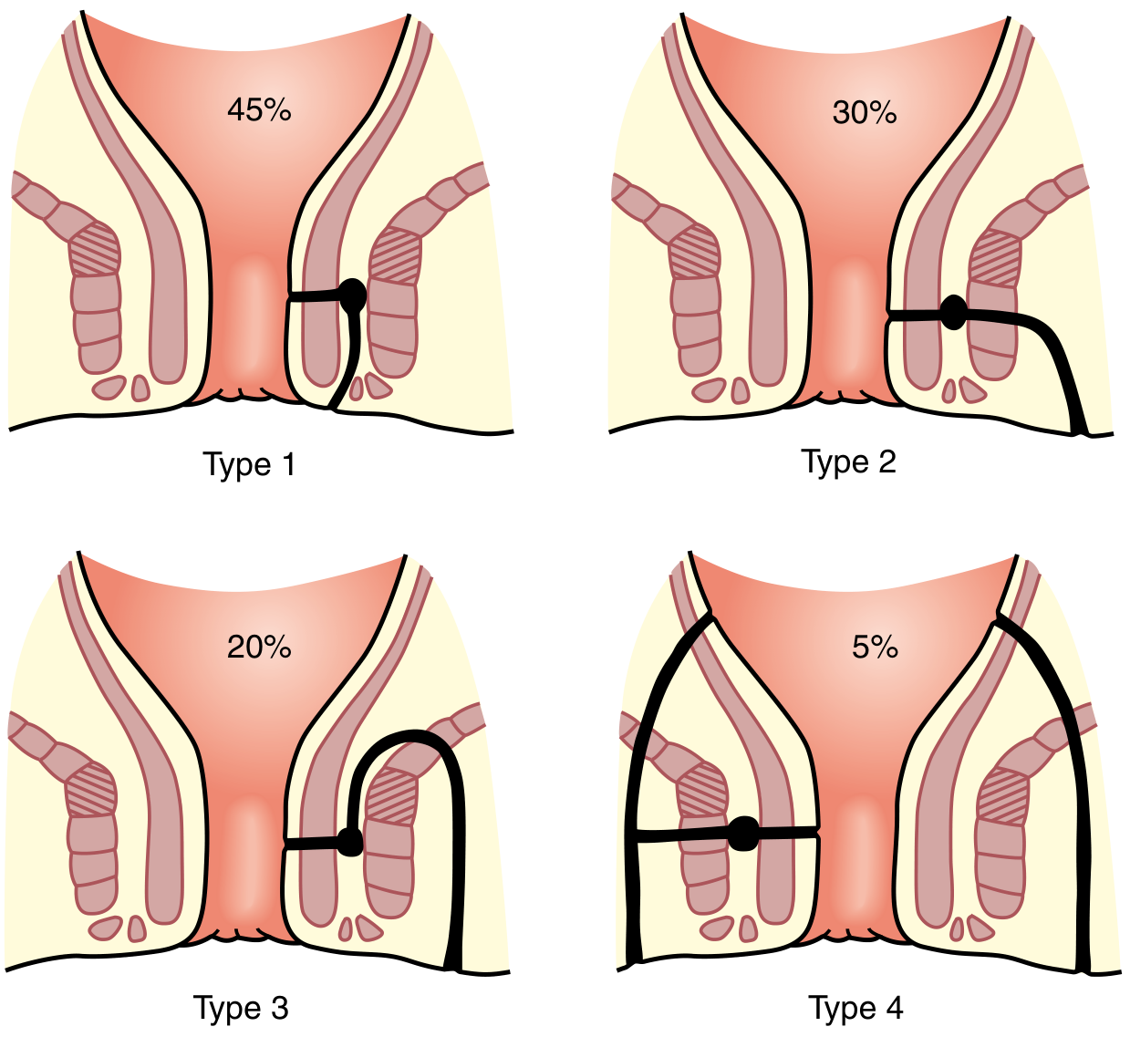
| Type | Name | Frequency | Description |
|---|---|---|---|
| Type 1 | Intersphincteric | 45% | Tracks between internal and external sphincters |
| Type 2 | Transsphincteric | 30% | Crosses the external sphincter |
| Type 3 | Suprasphincteric | 20% | Goes above the puborectalis |
| Type 4 | Extrasphincteric | 5% | Entirely outside the sphincter mechanism |
Goodsall's Rule
- External opening anterior to transverse anal line → track runs radially to the internal opening
- External opening posterior → track curves to a posterior midline internal opening
- Exception: anterior openings >3 cm from anal verge may curve posteriorly (horseshoe fistula)
Diagnosis
- Clinical: chronic perianal drainage, cyclical pain/swelling relieved by discharge
- Physical exam: cordlike subcutaneous structure, external opening(s)
- "Watering can perineum" (multiple openings) suggests perianal Crohn's disease
- MRI with anal fistula protocol is the gold standard for complex anatomy
MRI of anal fistulae - axial T2-weighted images:
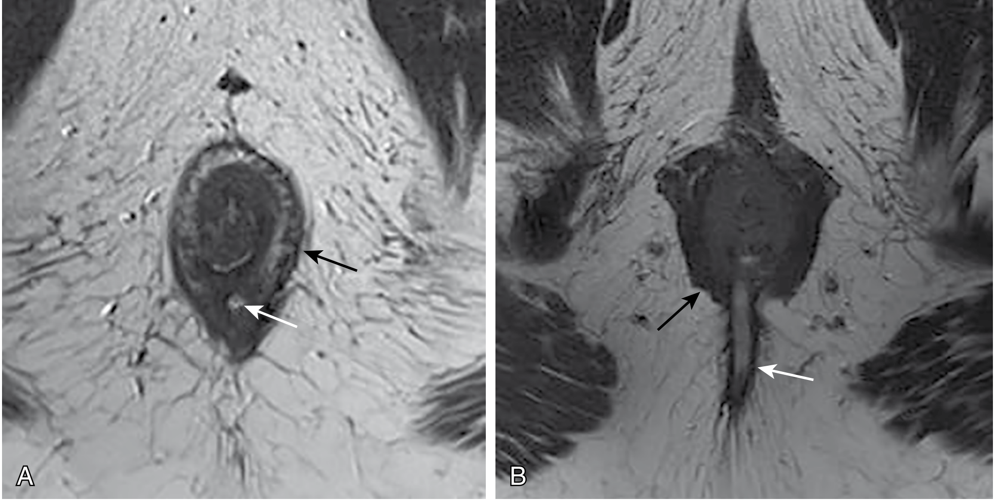
(A) Intersphincteric fistula (white arrow) lying within the intersphincteric plane. (B) Transsphincteric fistula (white arrow) penetrating the external sphincter boundary.
Treatment Goals (Anal Fistula)
- Treat any undrained infection
- Define fistula anatomy
- Remove or ablate epithelialised tracts
- Avoid or minimise risk of fecal incontinence
- Prevent recurrence
Surgical options:
-
Fistulotomy (lay-open): For low-lying fistulae involving <1/3 of the external sphincter (intersphincteric/low transsphincteric). Fecal incontinence risk <5% with normal preoperative sphincter function; recurrence rate 2-8%.
-
Seton: A suture/drain placed through the tract; used as a cutting or draining seton for complex/high fistulae to protect continence
-
Advancement flap: Mucosal or full-thickness flap to close the internal opening
-
LIFT procedure (Ligation of Intersphincteric Fistula Tract)
-
Bioprosthetic plug, fibrin glue, video-assisted anal fistula treatment (VAAFT)
-
Sabiston Textbook of Surgery
Enterocutaneous Fistula (ECF)
Causes
- 75-80% are post-surgical complications (anastomotic leak, inadvertent bowel injury)
- Pre- or post-surgical malnutrition is a major risk factor
- Crohn's disease, radiation, cancer, foreign body
Output Classification
| Class | Output | Clinical significance |
|---|---|---|
| Low output | <200 mL/day | More likely to close spontaneously |
| Medium output | 200-500 mL/day | Variable |
| High output | >500 mL/day | Significant fluid/electrolyte loss, skin excoriation from digestive enzymes, difficult to manage |
Conservative Management ("SNAP" principles)
- Skin care - protect surrounding skin from corrosive secretions
- Nutrition - parenteral or enteral nutrition is critical; high-output fistulae worsen malnutrition
- Anatomy - contrast sinogram/fistulogram, CT scan to delineate tract
- Plan - identify spontaneous closure potential vs. need for surgery
Conservative measures:
- Nil by mouth, nasogastric drainage
- IV fluid and electrolyte replacement (especially for high-output)
- Total parenteral nutrition (TPN)
- Somatostatin analogues (octreotide) to reduce output
- Wound/stoma care with collection devices
Contraindications to spontaneous closure (mnemonic FRIEND):
-
Foreign body in tract
-
Radiation damage
-
Inflammation (IBD, abscess)
-
Epithelialisation of tract
-
Neoplasm
-
Distal obstruction
-
Pye's Surgical Handicraft; Yamada's Textbook of Gastroenterology
Arteriovenous Fistula (AVF)
Types
- Congenital AV fistulae - developmental malformations
- Acquired AV fistulae - trauma (penetrating injury causing artery-to-vein communication), iatrogenic (post-catheterisation), neoplastic erosion
- Surgically created AVF - for hemodialysis access
Hemodialysis AVF
The surgically created AVF is the preferred vascular access for hemodialysis ("fistula first" principle from KDOQI guidelines). Common configurations:
- Radiocephalic (Brescia-Cimino) - wrist, first choice
- Brachiocephalic - antecubital
- Brachiobasilic - requires transposition
AVF advantages over AV graft (AVG):
- Lower infection risk
- Better long-term patency
- No prosthetic material
However, AVF requires 6 months maturation time before use. Factors negatively affecting AVF maturation and patency: female sex, coronary artery disease, diabetes, obesity, elderly patients.
Key complications of AVF:
-
Stenosis/thrombosis (most common)
-
Steal syndrome causing distal ischemia
-
Venous hypertension
-
Aneurysm/pseudoaneurysm
-
Infection
-
High-output cardiac failure (rare, with large AV connections)
-
Sabiston Textbook of Surgery, 25th Ed.
Head & Neck Fistulae (Pharyngocutaneous)
Context
A common complication after laryngectomy, glossectomy, neck dissection, or other ablative head/neck surgery. Forms an abnormal connection between the pharynx/aerodigestive tract and the surrounding neck/skin.
Risk Factors
- Prior radiation therapy
- Malnutrition
- Postoperative hypothyroidism
Consequences
- Prolonged open wounds
- Severe infections
- Carotid blowout (most devastating - saliva and bacteria pooling near great vessels)
Management
- Conservative: Local wound care, debridement, strict NPO with enteral feeding (NG or gastrostomy), dry packing. Barium swallow to confirm closure before resuming oral intake.
- Surgical: Required for uncontrolled infection, systemic sepsis, or proximity to carotid. Pectoralis major muscle flap is the gold standard for large fistula closure.
Prevention: Use of vascularized tissue (pedicled pectoralis, radial forearm or anterolateral thigh free flap) to reinforce pharyngeal closure significantly reduces pharyngocutaneous fistula risk.
- Sabiston Textbook of Surgery
Factors Preventing Fistula Closure (General)
Any fistula is unlikely to close spontaneously if:
- There is distal obstruction to flow
- A foreign body is present in the tract
- The tract is epithelialised
- There is active disease (malignancy, active IBD, radiation necrosis)
- There is an abscess cavity that has not been drained
- The fistula has a short, straight, wide-bore tract
Summary Table: Common Fistulae
| Fistula | Aetiology | Key Investigation | Treatment |
|---|---|---|---|
| Fistula-in-ano | Cryptoglandular abscess, Crohn's | MRI pelvis | Fistulotomy, seton, advancement flap |
| Enterocutaneous | Post-operative, IBD | CT/sinogram | SNAP, surgery if no spontaneous closure |
| Rectovaginal | Obstetric, IBD, radiation | Examination, MRI | Advancement flap, colostomy |
| Vesicovaginal | Obstetric, gynaecologic surgery | Cystoscopy, CT urogram | Surgical repair (open/laparoscopic) |
| AVF (dialysis) | Surgical creation | Duplex USS | Maturation procedures, angioplasty if stenotic |
| Pharyngocutaneous | Post-laryngectomy | Barium swallow, CT neck | Conservative / pectoralis flap |
| Tracheoesophageal | Congenital, malignancy | Bronchoscopy, endoscopy | Stenting, surgical repair |
This is a shared conversation. Sign in to Orris to start your own chat.