
Create a case of moderate pneumonia not severe pneumonia
chest x-ray lobar pneumonia consolidation
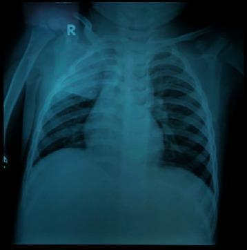
This diagnostic image is a posterior-anterior (PA) pediatric chest X-ray. The primary finding is a dense, homogeneous opacity located in the right upper lobe, characterized by a well-defined inferior border along the minor fissure, which is highly consistent with lobar consolidation. Bilateral perihilar and parenchymal spotting (reticulonodular opacities) are also visible, suggesting an associated bronchopneumonia component. The cardiac silhouette and mediastinal structures appear within normal limits for a pediatric patient, though the bony structures of the rib cage and vertebrae are visible with moderate clarity. This imaging demonstrates typical radiological signs of community-acquired or atypical pneumonia, such as right superior lobar pneumonia. Clinically, this serves as an educational example of differentiating focal lobar consolidation from generalized patchy bronchopneumonia patterns in a pediatric context.
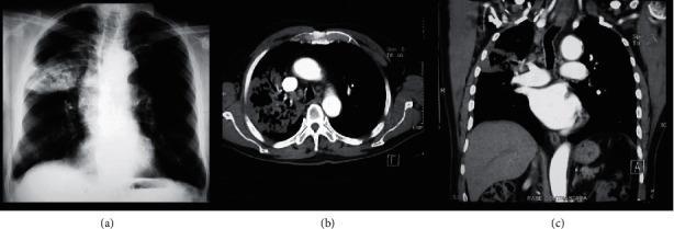
A multimodal diagnostic image set featuring a posteroposterior (PA) chest X-ray (a), an axial chest CT scan with contrast (b), and a coronal chest CT reconstruction (c) demonstrating necrotizing pneumonia. The images highlight extensive pathology in the right lung. Image (a) shows a heterogeneous opacification and lobar consolidation within the right middle or upper zone containing radiolucent lucencies. The CT images (b and c) provide high-resolution details of a dense lobar consolidation specifically in the right upper lobe, characterized by internal cavitation or ‘caverns’ representing tissue necrosis. Furthermore, the CT scans reveal significant parahilar lymphadenopathy and patchy consolidation. This comparative imaging illustrates the superior sensitivity of CT in identifying pulmonary cavitation and hilar adenopathy compared to conventional radiography. The visual evidence is consistent with fungal infections such as Histoplasma capsulatum, emphasizing clinical significance in identifying complications like necrosis and lymph node involvement in infectious pulmonary diseases.
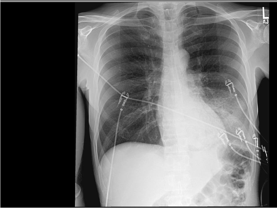
This diagnostic image is an anteroposterior (AP) chest X-ray demonstrating a significant dense consolidation in the left lower lobe. The primary finding is an area of increased opacity in the left paracardiac and retrocardiac region, which partially obscures the left hemidiaphragm and lower mediastinal border, consistent with lobar pneumonia or an inflammatory infiltrate. The right lung field appears clear without evidence of effusion or focal consolidation. Multiple medical devices are visible overlying the thorax, including radiopaque ECG leads and associated wires distributed across both lung fields. The cardiac silhouette appears within normal limits for an AP projection, and the bony structures, including the ribs and clavicles, are intact. This radiographic presentation is typical for acute infectious processes such as Legionella pneumonia, as suggested by the clinical context. The image serves as a key educational example of lobar consolidation and the visual identification of indwelling monitoring equipment in an acute clinical setting.
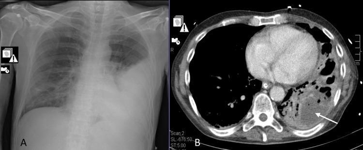
The composite image displays two diagnostic modalities for the chest. Image A is a frontal chest X-ray (radiography) demonstrating extensive opacification and consolidation in the left lower lung field, consistent with lobar pneumonia. The heart border is obscured, and the left costophrenic angle is obliterated. Image B is an axial computed tomography (CT) scan of the chest at the level of the heart, showing the evolution of the pathology. It reveals a large area of consolidation in the left posterior lung containing multiple low-density, hypodense regions. A white arrow points to a well-defined pulmonary abscess and surrounding cavitation within the necrotizing lung tissue. The right lung remains relatively clear on both modalities. These images illustrate the clinical progression from initial community-acquired pneumonia to necrotizing pneumonia and abscess formation, a significant complication often associated with specific bacterial pathogens or co-infections.
Case: Moderate Community-Acquired Pneumonia (CAP)
Patient Presentation
Examination Findings
| Parameter | Finding |
|---|---|
| Temperature | 38.6°C |
| Heart rate | 104 bpm |
| Respiratory rate | 24/min |
| Blood pressure | 128/78 mmHg |
| SpO2 | 93% on room air |
| Mental status | Alert and oriented, no confusion |
| Chest auscultation | Right lower lobe crepitations with bronchial breathing; dullness to percussion over the right base |
| Other systems | Unremarkable, no signs of shock or peripheral cyanosis |
Investigations
- CBC: WBC 14,200/mm3 with neutrophilic predominance
- Renal function: Urea 6.2 mmol/L (below the 7 mmol/L CURB-65 threshold), creatinine within normal limits
- CRP: Elevated at 96 mg/L
- Blood gas (room air): Mild hypoxemia, PaO2 68 mmHg, no hypercapnia or acidosis
- Blood cultures: Pending (drawn before antibiotics)
- Sputum culture: Sent, pending
- Chest X-ray: Right lower lobe homogeneous consolidation with air bronchograms, no effusion, no cavitation
Severity Assessment: Why This Is Moderate, Not Severe
- Confusion: Absent (0)
- Urea >7 mmol/L: Absent, 6.2 mmol/L (0)
- Respiratory rate ≥30/min: Absent, 24/min (0)
- Blood pressure criteria: Absent, hemodynamically stable (0)
- Age ≥65: Present, 68 years (1)
Diagnosis
Management Plan
- Admission: General ward, not ICU; continuous SpO2 monitoring and vital sign checks every 4-6 hours
- Oxygen therapy: Supplemental oxygen via nasal cannula to maintain SpO2 ≥94% (titrate down as tolerated)
- Empirical antibiotics (per standard CAP guidance for hospitalized non-ICU patients): a respiratory fluoroquinolone (e.g., levofloxacin) or combination beta-lactam (e.g., ceftriaxone) plus a macrolide (e.g., azithromycin), pending culture results and adjusted for local resistance patterns and comorbidities
- Supportive care: IV fluids if oral intake reduced, antipyretics for fever, glycemic monitoring given diabetes, incentive spirometry/chest physiotherapy, DVT prophylaxis given reduced mobility
- Monitoring for deterioration: Watch for any new confusion, rising respiratory rate, falling blood pressure, or worsening hypoxemia that would reclassify this as severe pneumonia and prompt ICU referral
- De-escalation: Switch to oral antibiotics once clinically stable (afebrile, improving respiratory status, tolerating oral intake) for a total course of 5-7 days
- Follow-up: Repeat chest X-ray at 6 weeks if smoking history or persistent symptoms, to exclude underlying malignancy or non-resolving pneumonia
Expected Course
Answer
- Alert, no confusion
- Urea 6.2 mmol/L (below the 7 mmol/L threshold)
- Respiratory rate 24/min (below the ≥30/min severe threshold)
- Blood pressure stable (128/78 mmHg)
- Single risk factor present: age ≥65
- Single-lobe consolidation, no septic shock, no need for mechanical ventilation or vasopressors - so it fails to meet ATS/IDSA severe CAP major or minor criteria