
Femur tens steps and procedure
Reading File
Finding Sources
Finding Sources
Searching PubMed
pediatric femur elastic nailing
Searching the Web
pediatric femoral shaft fracture titanium elastic nail surgical technique entry point procedure
Finding Sources
Reading Web Page
https://pmc.ncbi.nlm.nih.gov/articles/PMC7851225
Reading Article
PMID: 38447564
Reading File
Reading File
Assuming you mean TENS/TEN: titanium elastic nailing for a pediatric femoral shaft fracture. This is an operative outline for training and should be performed by an orthopedic surgeon with fluoroscopy.
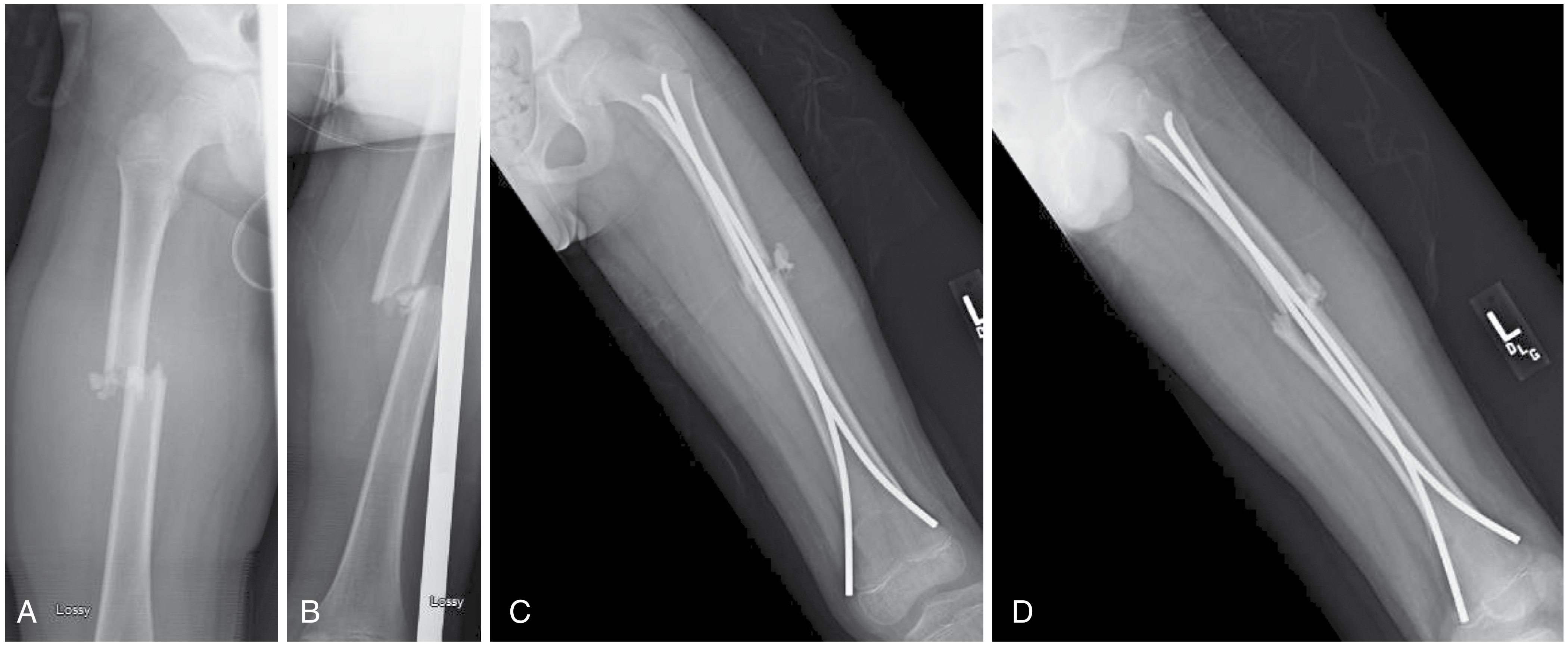
Femur TENS procedure: 10 steps
-
Confirm indication and plan
- Obtain AP and lateral radiographs of the entire femur.
- Best suited to a skeletally immature child with a length-stable, transverse or short-oblique diaphyseal fracture.
- Measure the canal and select two equal-diameter nails, usually each approximately 40% of the narrowest canal diameter.
-
Anaesthesia, antibiotics, and positioning
- Give general anaesthesia and perioperative antibiotic prophylaxis.
- Position supine on a radiolucent table, with the complete femur visible on image intensifier in AP and lateral planes.
-
Closed reduction
- Apply gentle traction and correct length, angulation, translation, and rotation under fluoroscopy.
- Open reduction is reserved for an irreducible fracture or interposed soft tissue.
-
Mark distal entry sites
- For the usual retrograde technique, make medial and lateral entry points in the distal femoral metaphysis.
- They should be about 2-3 cm proximal to the distal femoral physis and must not injure the physis.
-
Make skin incisions and create cortical openings
- Make small medial and lateral incisions.
- Protect soft tissues, especially medially.
- Use an awl or drill to make an oblique cortical entry hole directed into the medullary canal.
-
Prepare the two nails
- Pre-bend both nails into smooth, similar curves.
- The apex of each curve should be planned near the fracture level so opposing nails create balanced three-point fixation.
-
Insert the first nail
- Insert the first nail through one distal entry point and advance it up the canal to just below the fracture.
- Keep fluoroscopic control and avoid cortical perforation.
-
Insert the second nail and cross the fracture
- Insert the second nail from the opposite side to the fracture level.
- Reduce the fracture and advance each nail across it under fluoroscopy.
- Confirm restoration of length, coronal and sagittal alignment, and rotation.
-
Advance and seat nail tips
- Advance the medial and lateral nails to divergent proximal positions, avoiding the proximal femoral physis.
- Cut the distal ends short enough to reduce irritation but leave enough length for later removal.
- Bend and recess the nail ends appropriately.
-
Final imaging, closure, and postoperative care
- Obtain AP and lateral fluoroscopic views of the entire femur to verify reduction, nail position, and absence of physeal penetration.
- Irrigate and close the small wounds.
- Provide analgesia and an individualized plan for weight bearing, physiotherapy, and follow-up radiographs. Nail removal is commonly considered after union, depending on local practice and symptoms.
Key technical points
- Use two equal-sized, symmetrically prebent nails for balanced fixation.
- The nails should be inserted from opposite sides and their tips should diverge proximally.
- Avoid both distal and proximal physes.
- Confirm rotation clinically and fluoroscopically before finishing.
- Nail-end prominence can cause painful skin irritation and is a common minor complication.
Important limitations
TENS is less reliable for length-unstable, comminuted, long-oblique/spiral, subtrochanteric fractures, and in older or heavier children. Increased complications are reported with age over 11 years, weight above about 49 kg, open injury, marked comminution, and unstable patterns. In such cases, submuscular plating, a rigid trochanteric-entry nail in appropriate adolescents, or other fixation may be preferable. Campbell's Operative Orthopaedics, 15th ed., p. 1852.
A 2024 systematic review of flexible intramedullary nailing for length-unstable pediatric femoral fractures found a notable minor-complication rate, particularly nail prominence, so fracture selection and postoperative monitoring matter (systematic review, PMID 38447564).
This is a shared conversation. Sign in to Orris to start your own chat.