
Diplopia evaluation
Reading File
Now let me search the medical library for diplopia evaluation content, running parallel searches across headings and paragraphs.
Finding Sources
Finding Sources
Excellent finds. Let me read the most comprehensive sources in parallel - Rosen's binocular diplopia section, Neuroanatomy Through Clinical Cases, and Harrison's.
Reading File
Reading File
Reading File
Now let me read more from Harrison's (CN palsies) and Rosen's (CN IV, VI, and special tests), and fetch the key diagnostic figures.
Reading File
Reading File
Excellent. Now let me read the CN IV and CN VI palsy details from Harrison's, plus the Adams & Victor section on Diplopia & Strabismus for the Park's 3-step test and additional clinical details.
Reading File
Reading File
I have rich content from multiple textbooks. Let me fetch a couple of key diagnostic images now.
Loading Image
Loading Image
Excellent images retrieved. Now let me get the INO image and read Harrison's CN IV/VI section.
Loading Image
Reading File
I now have comprehensive information from multiple authoritative textbooks. Let me compile the full, structured response.
Diplopia: Clinical Evaluation
1. First Step - Monocular vs. Binocular
The most important initial question: does the diplopia resolve when either eye is covered?
- Monocular diplopia - persists with one eye closed. Almost always an ocular (non-neurological) cause: corneal abnormality, uncorrected refractive error, cataract, lens dislocation, foveal traction, or rarely malingering/psychiatric disease. Test with a pinhole: diplopia from refractive error resolves; diplopia from retinal or lenticular pathology does not.
- Binocular diplopia - resolves when either eye is covered. This means ocular misalignment is present and requires systematic neurological evaluation.
- Harrison's Principles of Internal Medicine 22E, p. 278; Neuroanatomy Through Clinical Cases 3e, p. 599
2. Key Historical Features in Binocular Diplopia
Ask about:
- Direction of diplopia: purely horizontal, purely vertical, or torsional
- Gaze position that worsens it: e.g., worse looking left, worse reading/looking down
- Mode of onset: acute vs. gradual
- Intermittency and diurnal variation: worse at end of day = myasthenia gravis
- Head tilt: compensatory head posture suggests CN IV palsy
- Associated symptoms: ptosis, pain, proptosis, headache, jaw claudication, fever, weakness, ataxia
- Medical history: diabetes, hypertension, thyroid disease, cancer, MS
- Harrison's 22E, p. 278; Rosen's Emergency Medicine, p. 206
3. Structural Causes (Orbital/Restrictive Diplopia)
These are recognized by local orbital signs and a pattern that does NOT follow classic CN palsy stereotypes:
| Cause | Key Features |
|---|---|
| Thyroid eye disease (Graves) | Inferior + medial recti fibrosis; restriction of elevation and abduction; proptosis, eyelid retraction, injection |
| Orbital myositis / pseudotumor | Painful proptosis, periorbital swelling, restricted motility; mimics CN palsy but pattern atypical |
| Orbital fracture (blowout) | Trauma history; muscle entrapment (inferior rectus); worsens on upgaze |
| Orbital tumor / abscess | Gradual onset; mass effect; proptosis; may have fever if infectious |
| Giant cell arteritis | Age >50; headache, scalp tenderness, jaw claudication; ESR/CRP elevated |
Dedicated orbital imaging (MRI with fat saturation + gadolinium) is useful when the cause is not evident clinically.
- Harrison's 22E, p. 278; Rosen's Emergency Medicine, p. 206
4. Neuromuscular Cause: Myasthenia Gravis
A major cause of painless, variable, binocular diplopia - can mimic any CN palsy.
- Diplopia fluctuates, worsens with fatigue/at end of day, improves with rest
- Not confined to a single CN distribution
- Pupils always normal
- May have fatigable ptosis (unilateral or bilateral)
- ~50% of patients present with purely ocular symptoms
- Bedside ice test: apply ice to closed eyelid for 5 minutes; improvement of ptosis ~5 mm = positive (cold mitigates acetylcholinesterase inhibition)
- Labs: anti-AChR antibodies (may be negative in purely ocular form); anti-MuSK antibodies
- Beware: Botulism from food or wound poisoning can mimic ocular myasthenia
- Harrison's 22E, p. 278; Rosen's Emergency Medicine, p. 206
5. CN Palsy Patterns
Once restrictive orbitopathy and myasthenia are excluded, a cranial nerve lesion is the most likely cause.
Extraocular muscle actions by gaze position
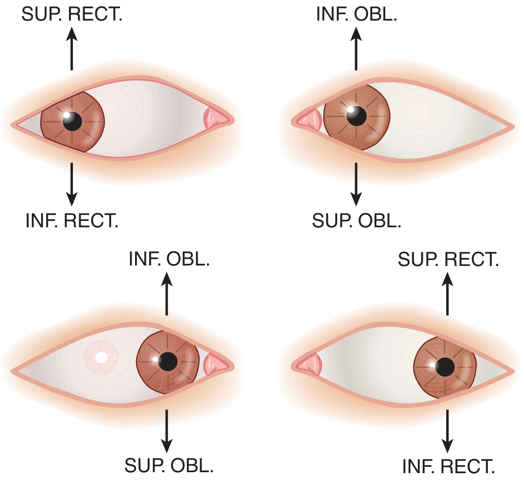
Figure: Muscles chiefly responsible for vertical movements in different gaze positions - Adams & Victor's Principles of Neurology, 12e
CN III (Oculomotor) Palsy
- Muscles affected: medial rectus, superior rectus, inferior rectus, inferior oblique, levator palpebrae, iris sphincter
- Eye position: down and out (unopposed lateral rectus + superior oblique)
- Signs: ptosis, dilated pupil (parasympathetic fibers run on the outside of the nerve = vulnerable to compression)
- Diplopia: in all directions except lateral gaze to the affected side
- Pupil-involving CN III: urgent neuroimaging + CT/MR angiogram to exclude posterior communicating artery aneurysm
- Pupil-sparing CN III: typically microvascular ischemia (diabetes, hypertension); spontaneous recovery in months
- Aberrant regeneration (lid elevates on downgaze/adduction): indicates compressive or traumatic etiology - NOT seen with microvascular CN III
Brainstem/midbrain syndromes with CN III:
- Nothnagel's: CN III + cerebellar ataxia (superior cerebellar peduncle)
- Benedikt's: CN III + contralateral tremor/chorea/athetosis (red nucleus)
- Weber's: CN III + contralateral hemiparesis (cerebral peduncle)
- Harrison's 22E, pp. 278-279
CN IV (Trochlear) Palsy
- Muscle affected: superior oblique (depresses and intorts the globe)
- Result: hypertropia + excyclotorsion of the affected eye
- Complaint: vertical diplopia, especially on reading or looking down
- Bielschowsky head tilt test (cardinal feature): diplopia worsens on tilting head toward the side of palsy, improves on tilting away
- Common causes: closed head trauma (nerve crosses at dorsal midbrain, vulnerable to tentorial impact), microvascular ischemia, congenital (review old photos for habitual head tilt)
- No aneurysmal cause expected (unlike CN III)
- Management: base-down prism; if persistent, inferior oblique weakening surgery
- Harrison's 22E, p. 279
CN VI (Abducens) Palsy
- Muscle affected: lateral rectus
- Result: failure of abduction; eye esotropic at rest
- Complaint: horizontal diplopia, worst on gaze to the side of the lesion
- Important distinction: CN VI nucleus lesion vs. fascicle lesion
- Nuclear lesion = complete ipsilateral gaze palsy (both ipsilateral LR and contralateral MR fail via MLF)
- Fascicle lesion = isolated lateral rectus weakness only
Brainstem syndromes with CN VI:
- Foville's (dorsal pons): lateral gaze palsy + ipsilateral facial palsy + contralateral hemiparesis
- Millard-Gubler (ventral pons): lateral rectus weakness only + ipsilateral facial palsy + contralateral hemiparesis
- Gradenigo's syndrome (petrous apex): mastoiditis + deafness + pain + CN VI palsy
CN VI has the longest intracranial course and is a false localizing sign in raised ICP (stretches over petrous apex).
- Harrison's 22E, p. 279
6. Internuclear Ophthalmoplegia (INO)
A supranuclear cause of diplopia from a medial longitudinal fasciculus (MLF) lesion:
- Finding: failure of adduction of one eye on horizontal gaze (with preserved adduction on convergence) + nystagmus in the abducting eye
- Cause: most commonly MS (bilateral INO = virtually pathognomonic for MS), also brainstem infarct
- In patients with MS, diplopia may be the presenting (isolated) syndrome
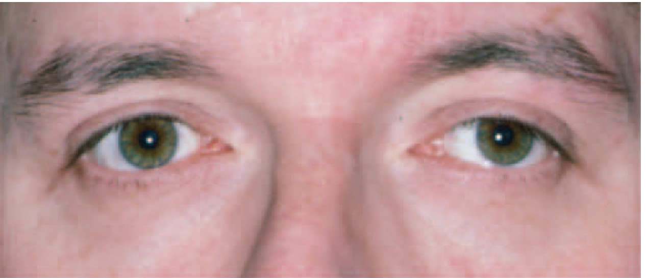
Figure: Left internuclear ophthalmoplegia - Harrison's Principles of Internal Medicine 22E
7. Other Important Neurological Causes
| Condition | Clue |
|---|---|
| Wernicke encephalopathy | Alcohol/malnutrition/bariatric surgery; triad of ophthalmoplegia (usually CN VI), ataxia, altered mentation |
| Miller-Fisher syndrome | Classic triad: ophthalmoplegia + ataxia + areflexia (no limb weakness) |
| Basilar meningitis | Diplopia + headache, photophobia, meningism, fever |
| Raised ICP | False-localizing CN VI palsy |
| Brainstem lacunar stroke | Crossed findings: CN palsy ipsilateral, motor/sensory deficits contralateral |
| Cavernous sinus syndrome | CN III, IV, V1/V2, VI all affected; Horner syndrome possible; causes: carotid aneurysm, cavernous sinus thrombosis, pituitary adenoma, meningioma, Tolosa-Hunt, herpes zoster |
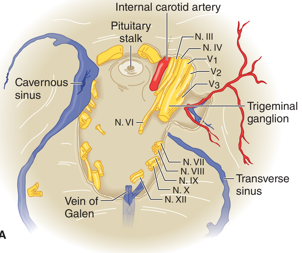
Figure: Cavernous sinus and cranial nerve relationships - Adams & Victor's Principles of Neurology, 12e
8. Bedside Examination Tools
Cover Test
- More sensitive than motility testing for subtle misalignment
- Perform in primary gaze, then with head turned/tilted in each direction fixating a distant target
- Detects comitant deviations (equal in all gaze positions = strabismus) vs. incomitant (CN palsy, restrictive)
Red Glass Test
- Place red glass over one eye (typically right); patient follows a white light in 9 gaze positions
- Reports positions of red (right eye) and white (left eye) images
- Image further from midline in the direction of attempted gaze = from the abnormal eye
Corneal Light Reflex (Hirschberg Test)
- Shine flashlight from directly in front; symmetric corneal reflections = normal
- Displaced reflection = eye misalignment
Ice Test (for Myasthenia)
- Ice pack on closed eyelid for 5 minutes
- Improvement in ptosis (~5 mm) = positive for myasthenia
- Neuroanatomy Through Clinical Cases 3e, pp. 599-600; Rosen's Emergency Medicine, p. 206
9. Ancillary Testing
| Indication | Test |
|---|---|
| Suspected myasthenia | Anti-AChR, anti-MuSK antibodies; ice test; repetitive nerve stimulation |
| Suspected thyroid eye disease | TSH, free T4, thyroid antibodies |
| Suspected GCA (age >50, headache, jaw claudication) | ESR, CRP; temporal artery biopsy if ESR >50 or CRP >20 |
| Suspected MS | Brain/spinal cord MRI with gadolinium |
| CN III with pupil involvement | CT/MR angiogram (exclude PComA aneurysm) |
| Orbital/restrictive cause | MRI orbit with fat saturation + gadolinium |
| Suspected stroke/brainstem lesion | MRI brain with DWI |
| Metabolic causes | CBC, BMP, glucose, HbA1c, B1 (thiamine if Wernicke suspected) |
- Harrison's 22E, p. 278; Rosen's Emergency Medicine, pp. 206-208
10. Diagnostic Algorithm Summary
Diplopia
   |
   ├─ Monocular (persists with one eye closed)
   │     → Ocular cause (cornea, lens, retina)
   │     → Pinhole test (resolves = refractive; persists = structural)
   │
   └─ Binocular (resolves covering either eye)
         |
         ├─ Signs of orbital disease (proptosis, pain, restricted motility atypical pattern)
         │     → Restrictive: Thyroid, pseudotumor, fracture, tumor, GCA
         │     → MRI orbit
         │
         ├─ Variable, fatigue-related, pupils normal
         │     → Myasthenia gravis
         │     → Ice test, AChR antibodies, EMG
         │
         └─ Follows CN distribution
               |
               ├─ CN III: Down-out eye + ptosis ± dilated pupil
               │     Pupil involved → aneurysm workup (CTA/MRA)
               │     Pupil spared + microvascular risk → ischemic CN III
               │
               ├─ CN IV: Vertical diplopia, worse reading, head tilt
               │     Bielschowsky test; closed head trauma common
               │
               ├─ CN VI: Horizontal diplopia, esotropia, worse ipsilateral gaze
               │     Gaze palsy → nuclear; isolated LR → fascicle
               │
               └─ Supranuclear / other
                     INO (MLF) → MS or stroke
                     Gaze palsy → frontal/pontine
                     Cavernous sinus → multiple CN (III+IV+V1+VI)
Sources: Harrison's Principles of Internal Medicine 22E; Rosen's Emergency Medicine; Adams & Victor's Principles of Neurology 12e; Neuroanatomy Through Clinical Cases 3e
This is a shared conversation. Sign in to Orris to start your own chat.