
white painful skin erosion b/w fingers
interdigital candidiasis white erosion between fingers
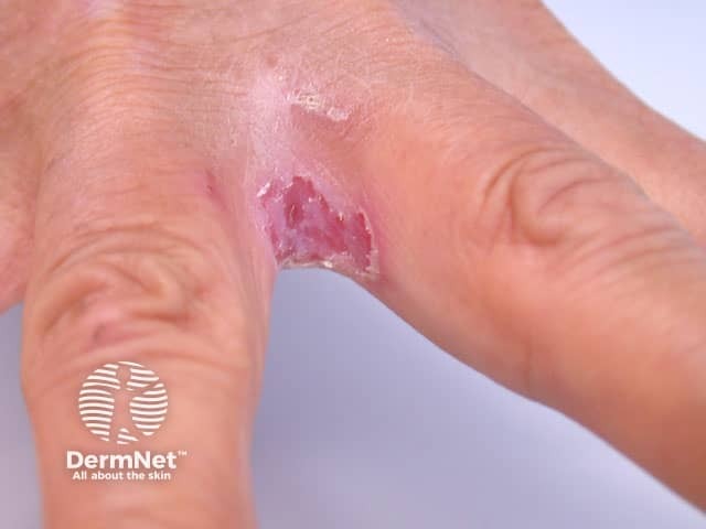
A high-resolution clinical photograph of the palmar aspect of a hand showing an interdigital erosive lesion consistent with Erosio interdigitalis blastomycetica. The interdigital web space, inferred between two adjacent digits, exhibits shallow, superficial erosions with erythematous, macerated margins and a moist surface. White crusting or scale accents the lesion border, and surrounding skin demonstrates mild desquamation and edema from moisture exposure. The appearance is classic for candidal intertrigo involving interdigital folds, though the differential includes dermatophytic infection and bacterial intertrigo. Clinically, this finding supports a fungal etiology in moist skin creases; appropriate management includes topical antifungal therapy and moisture control, with consideration of barrier protection to reduce maceration. Risk factors such as sweating, frequent hand immersion, diabetes mellitus, or immunosuppression may predispose to interdigital candidiasis. In practice, treatment choices include azole or allylamine antifungals, alongside hygiene measures and drying of interdigital spaces. The image is valuable for dermatology, infectious diseases, and primary care education to recognize interdigital candidiasis, to distinguish it from tinea manuum or contact dermatitis, and to illustrate the characteristic morphology of a superficial interdigital erosion. This image also serves as a reference for trainees evaluating intertriginous hand infections and for documenting treatment response over time and outcome.
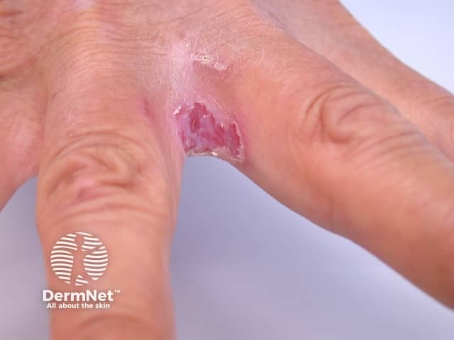
Clinical photography of an interdigital lesion on the hand consistent with candidal infection. Modality: Dermatology clinical photograph; view: external dorsal/palmar aspect of the fingers with the affected interdigital web space between the index and middle fingers. The lesion presents as a moist, erythematous macerated plaque with shallow erosions and peripheral crusting at the web space, sometimes accompanied by fissuring and mild edema. Surface is shiny and friable with possible satellite pustules; consistent with candidiasis of intertriginous skin. The appearance may mimic eczema or bacterial intertrigo, but the moist, red-to-pink tissue with erosions and a history of maceration favors Candida species. Diagnostic significance: photodocumented interdigital candidiasis; correlation with clinical symptoms (pruritus, burning) and risk factors such as occlusive skin folds, sweating, diabetes, immunosuppression; confirm with KOH preparation showing yeast and pseudohyphae or fungal culture; differential diagnoses include dermatophytosis (tinea) of the hand, irritant contact dermatitis, and atopic dermatitis. Potential clinical use cases: educational dermatology case, image-based teaching of intertrigo etiologies, differential for interdigital infections, radiology not relevant; management implications include topical azole antifungals (clotrimazole, miconazole), topical allylamines (terbinafine), or systemic azoles in extensive disease; emphasis on moisture control and barrier repair. Photographic features support clinical teaching, remote diagnosis, and standardized image datasets.
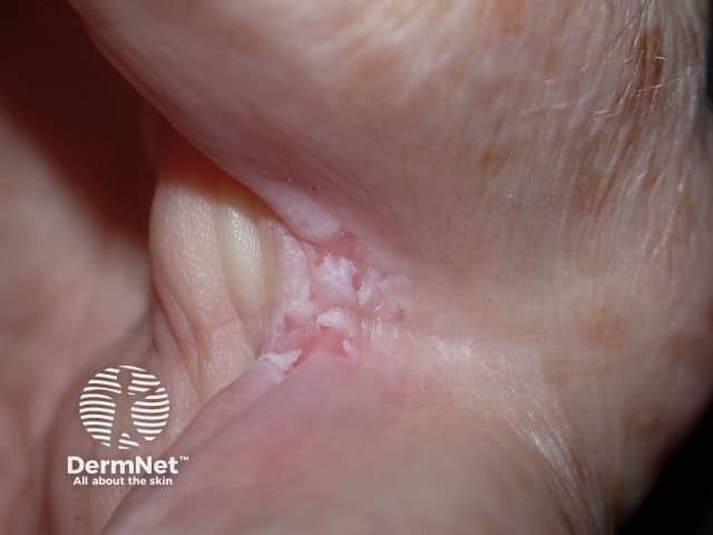
This is a high-resolution clinical photograph of cutaneous candidiasis presenting as intertrigo in an interdigital skin fold. The image was captured using digital photography with close-up macro settings under natural lighting to preserve true color and detail. The affected area shows moist, erythematous maceration with fissuring and a slightly shiny surface, characteristic of moisture-associated dermatitis. Superficial white curd-like plaques are evident along the skin folds, with associated mild edema and desquamation. The surrounding skin exhibits mild erythema but no deep ulceration. The appearance is consistent with Candida species infection, most commonly Candida albicans, in an intertriginous region; alternative Candida species may be involved. This pattern—moist, red plaques with fissures and satellite lesions in adjacent folds—helps distinguish candidal intertrigo from other causes of intertrigo such as dermatophyte infection (tinea intertrigo) or irritant dermatitis. Clinically, this image is relevant for diagnosis, educational purposes, and differential diagnosis deliberations in dermatology and infectious disease. It supports antifungal therapy selection, such as azole or nonazole antifungals, and monitoring of treatment response. It may be used in teaching about risk factors (diabetes, obesity, humid environments), symptomatology, and the importance of keeping skin folds dry to prevent recurrence.
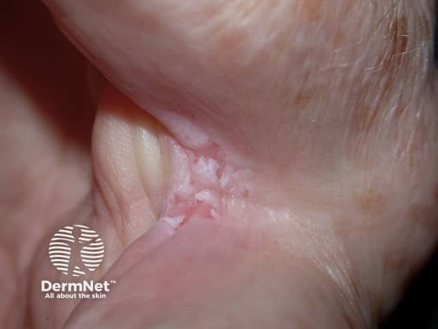
This clinical dermatology photograph depicts a close-up view of a moist, erythematous interdigital skin lesion consistent with candidal infection. Modality: Clinical photography in high-resolution color; close-up macro view; ambient lighting with color fidelity; no magnification metadata. The anatomic location is the distal interdigital fold of a digit, with involvement of periungual skin and adjacent lateral nail fold. Visual features include erythematous maceration, fissuring, and a thin whitish curd-like scale on an eroded surface; minimal variegated erythema; slight edema; no crusting. The appearance is characteristic of cutaneous candidiasis or candidal intertrigo: moist, reddish plaques in warm, moist folds with possible satellite pustules, fissures, and discomfort. Differential considerations include tinea cruris/corporis or irritant contact dermatitis; however, the presence of white curd-like material favors Candida albicans infection. Diagnostic significance: prompt antifungal therapy (azoles or polyenes) often yields rapid improvement; evaluate for risk factors such as occlusion, sweating, obesity, diabetes, immunosuppression. Potential clinical uses: educational reference for dermatology training, differential diagnosis practice, AI-assisted image retrieval for dermatology intertrigo/candida cases, decision support for antifungal treatment planning, and patient education about hygiene and moisture control. Contextual cues such as duration, accompanying itching, and secondary bacterial colonization should guide culture or microscopy when lesions fail to respond.
White Painful Skin Erosion Between Fingers
Clinical Appearance

Most Likely Cause: Cutaneous Candidiasis
- Moist, macerated, white-to-bright-red erosions
- Pain and burning sensation
- Surrounding collarette of scale
- Possible satellite papules or pustules peripherally (characteristic of Candida)
- Fissures in the depths of the web space
Differential Diagnosis
| Condition | Key Features |
|---|---|
| Candidal intertrigo (most likely) | White maceration, satellite pustules, burning pain, KOH shows pseudohyphae |
| Tinea manuum (dermatophyte) | Dry scaly lesions, less macerated, KOH shows septate hyphae |
| Interdigital bacterial intertrigo | Foul smell, fiery-red, moist; caused by Staphylococcus, Pseudomonas, Corynebacterium |
| Contact/irritant dermatitis | History of chemical/allergen exposure, no satellite lesions |
| Scabies | Intense nocturnal itch, burrows, involves multiple people in household |
| Herpetic whitlow | Vesicles/ulcers, often on a single finger, HSV-positive; consider in healthcare workers |
Diagnosis
- KOH preparation from the roof of a pustule or lesion: reveals pseudohyphae and budding yeast in Candida; septate hyphae in dermatophytes
- Fungal culture if KOH is inconclusive
- Wood's lamp: coral-red fluorescence suggests erythrasma (Corynebacterium)
- Skin swab for bacterial culture if superinfection is suspected
Treatment
-
Topical imidazole - first line:
- Clotrimazole 1% cream, twice daily for 4 weeks
- Miconazole, ketoconazole, or sulconazole are alternatives
-
Systemic antifungals for extensive disease:
- Fluconazole 100 mg orally daily for 2 weeks
- Itraconazole 100 mg orally daily for 2 weeks
-
Moisture control - keep the web space dry; separate skin surfaces with gauze or antifungal powder
Key Points
- The white appearance is due to maceration in the moist web space; Candida thrives here
- Satellite pustules confirm Candida over other etiologies
- Always check for diabetes - recurrent or resistant interdigital candidiasis is a red flag for uncontrolled blood sugar
- Barrier protection and drying after hand-washing prevent recurrence
deep flesh wound always reoccurs in winter times for my mother treatment
Where exactly does the deep flesh wound appear? · How would you describe the wound? · Does your mother have any known medical conditions?
diabetic heel fissures deep cracks treatment
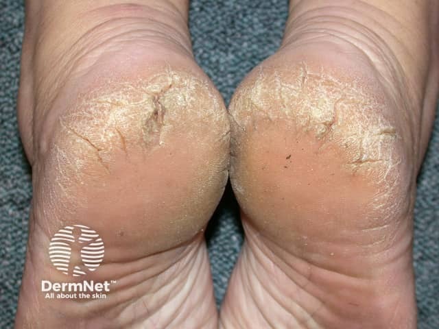
Imaging modality: Clinical photography of skin in the posterior heel region. The subject shows bilateral fissured, hyperkeratotic plaques on the posterior aspect of both heels with surrounding dryness and desquamation. The skin is thickened with a yellow-tan hue and rough texture, consistent with chronic callus formation. Cracks align with weight-bearing areas and extend transversely across the posterior calcaneal border. This appearance is typical of severe xerosis with hyperkeratosis and fissuring; differential includes irritant dermatitis, psoriasis, fungal infection, and mechanical trauma. The image demonstrates a non-inflammatory, non-pustular pattern with fissures confined to the plantar/posterior heel. Clinically relevant findings include risk of secondary infection through fissures, pain on ambulation, and impaired mobility. Notable features are bilateral symmetry, well-defined keratotic plaques, and mild surrounding erythema. In practice, this image supports diagnoses such as heel fissures due to dry skin (xerosis) or chronic callosity; it is useful for educational demonstrations, dermatology outreach, triage, and treatment planning. Potential use cases include patient education on moisturization regimens, evaluation of response to emollients and keratolytics, and monitoring of hyperkeratotic lesion evolution in at-risk populations (elderly, diabetic, or sedentary individuals). Additional imaging notes may include peri-heel hygiene assessment and footwear evaluation.

This sequence of grayscale clinical photographs serves as a comparison chart illustrating the dermatological progression of a human foot undergoing topical treatment for xerosis and hyperkeratosis. Image A (Day 0) demonstrates the baseline clinical condition of the plantar surface, characterized by deep skin fissures (heel cracks), prominent epidermal ridges, and a dry, rough texture indicative of compromised barrier function. Image B (after 4 weeks) shows intermediate progression, with a visible reduction in the depth and severity of the fissures and a softening of the skin texture. Image C (after 8 weeks) displays the therapeutic outcome, showcasing a significant resolution of the cracks and restoration of a smooth, hydrated appearance with reduced visibility of lines. This visual timeline is representative of clinical efficacy studies in podiatry and dermatology, focusing on skin regeneration, emolliency, and the repair of cracked heels using therapeutic foot care formulations.
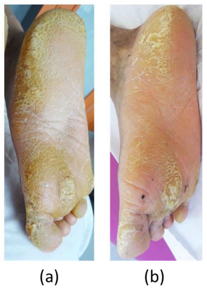
This clinical photograph exhibits a comparison of the plantar surface of the foot in a patient with pachyonychia congenita (specifically KRT6A-associated palmoplantar keratoderma), showing treatment progression over 11 months. Panel (a) illustrates the baseline condition characterized by severe, focal hyperkeratosis with thick, yellowish, and fissured plaques on the heel, lateral border, and metatarsal heads. The skin texture is markedly rough, and deep cracks are visible in weight-bearing areas. Panel (b) shows the result after 11 months of oral erlotinib therapy, demonstrating a significant reduction in the thickness and extent of the keratoderma. The yellowish hyperkeratotic plaques have noticeably thinned, the surface appears smoother, and the deep fissures are largely resolved. The skin tone is more uniform, and the inflammatory borders are less prominent. This comparison serves as a clinical demonstration of the efficacy of EGFR inhibitors in managing the hyperkeratotic symptoms of keratin-related disorders.
Deep Heel Fissures in a Diabetic Patient - Recurring Every Winter
Why This Happens (The Mechanism)
- Autonomic neuropathy - destroys the sweat glands in the foot, causing severely dry skin that cannot self-lubricate. Cold, dry winter air makes this far worse.
- Sensory neuropathy - your mother may not feel minor trauma or pressure building up, so callus forms unnoticed over bony pressure points (especially the heel).
- Motor neuropathy - causes foot muscle changes and abnormal weight distribution, increasing pressure on the heel.
Why This Is Serious - Do Not Ignore
- Infected ulcers (cellulitis, abscess)
- Osteomyelitis (bone infection) - develops in up to 50-60% of moderate-to-severe infections
- Limb-threatening complications - up to 15% of diabetic foot ulcers lead to amputation
"You cannot examine the feet of your diabetic patients too often, and you cannot examine them with their shoes on!" - Symptom to Diagnosis, 4th Ed
Treatment Plan
Immediate (Active Fissure Care)
| Step | What to Do |
|---|---|
| 1. Soak | Warm water soak for 10-15 min to soften thick skin |
| 2. Gentle debridement | Use a pumice stone or foot file after soaking to gently reduce thick callus - never cut aggressively |
| 3. Moisturize deeply | Apply a urea-based cream (25-40% urea) or salicylic acid cream to the heel immediately after soaking while skin is still damp |
| 4. Seal fissures | Use a medical-grade liquid bandage or wound closure adhesive over open fissures |
| 5. Cover & protect | Wear cotton socks to bed after applying cream |
- If there are signs of infection (redness, warmth, swelling, discharge, odor, fever): see a doctor immediately for antibiotics - mild infections: oral cephalexin or amoxicillin-clavulanate; deeper infections: hospitalization may be needed
Ongoing Daily Maintenance
- Moisturize heels every single day, year-round - do not wait for winter
- Use a thick emollient (petroleum jelly, urea cream, or shea butter) at night with socks
- Keep feet clean and dry; check between toes daily
- Never walk barefoot - always use soft-soled, well-cushioned footwear
- Change socks daily; wear breathable cotton or moisture-wicking socks
Prevention Before Winter
- Start intensive moisturizing 4-6 weeks before winter begins (around October)
- Get the callus professionally trimmed by a podiatrist once every 3 months
- Use a humidifier indoors during winter to reduce ambient dryness
Most Important: Blood Sugar Control
When to See a Doctor (Urgent Red Flags)
- The fissure is deep enough to bleed
- There is redness, warmth, or swelling around the wound
- The wound has a bad smell or discharge
- She has fever or chills
- The wound is not healing within 2 weeks
Recommended Products
| Product Type | Examples |
|---|---|
| Urea cream (25-40%) | Carmol 40, Eucerin Urea 30%, Flexitol Heel Balm |
| Emollient for nightly use | Vaseline, CeraVe Healing Ointment |
| Fissure sealant | New-Skin Liquid Bandage, 3M Cavilon |
| Cushioned insoles | Diabetic-grade orthotic insoles |
A practical daily routine beats any single treatment. The fissures return every winter because the skin is never maintained adequately in summer. Year-round daily heel cream is the most effective prevention.