
Edrophrium drug
edrophonium drug mechanism uses myasthenia gravis Tensilon test pharmacology
edrophonium Tensilon test myasthenia gravis neuromuscular junction acetylcholinesterase
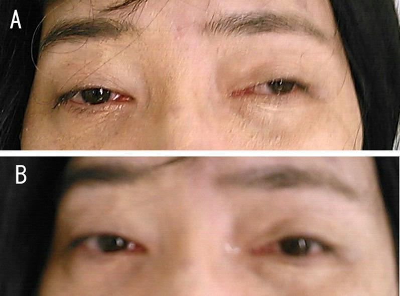
This clinical photograph is a side-by-side comparison illustrating a positive Tensilon (edrophonium) test in a patient with Myasthenia Gravis (MG). Panel A shows the patient's orbital region at baseline, demonstrating significant bilateral asymmetrical ptosis (drooping of the upper eyelids). The left eyelid is more severely affected, covering a substantial portion of the visual axis. Panel B shows the same patient after the administration of edrophonium, an acetylcholinesterase inhibitor. There is a marked improvement in the bilateral ptosis, characterized by an increased palpebral fissure width and elevation of the upper lid margins, exposing more of the iris and pupil. This visual change is a classic diagnostic indicator for disorders of the neuromuscular junction, specifically MG associated with anti-acetylcholine receptor (AchR) antibodies. The image serves as a clinical teaching tool for neurology and ophthalmology to demonstrate the transient reversal of fatiguable muscle weakness through pharmacological intervention.
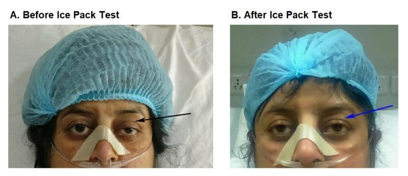
Two side-by-side clinical photographs labeled A and B demonstrate a bedside ice pack test in a patient with suspected myasthenia gravis. Image A (Before Ice Pack Test) shows significant left-sided blepharoptosis, with the upper eyelid drooping to partially cover the pupil (indicated by a black arrow). Image B (After Ice Pack Test) demonstrates a positive result, where the left eyelid shows temporary elevation and resolution of ptosis (indicated by a blue arrow) following local cooling. The patient is shown in a clinical setting wearing a blue surgical cap, a nasal cannula for oxygen delivery, and an adhesive nasal strip for stabilization. This comparative series illustrates the clinical utility of the ice pack test, which inhibits acetylcholinesterase activity at the neuromuscular junction to temporarily improve muscle strength in myasthenic patients. The images highlight key diagnostic physical exam findings in neuro-ophthalmology and neuromuscular medicine.
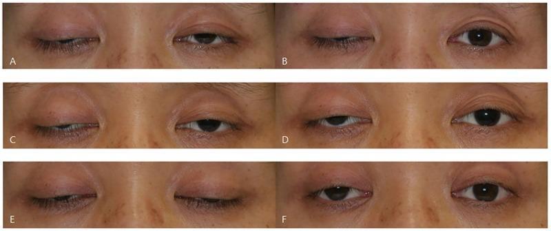
This set of six clinical photographs (A-F) illustrates the variability of the ice test in a patient with myasthenia gravis presenting with bilateral asymmetric ptosis. The images are arranged in pairs representing three different days, showing the patient's eyes before and after a 2-minute ice application. Frames A, C, and E show baseline bilateral blepharoptosis with varying degrees of upper eyelid drooping obscuring the pupil and iris. Frames B, D, and F show the post-intervention state. The series demonstrates three distinct clinical responses in the right eye: (B) a negative result with no significant change, (D) an equivocal result showing subtle 1.0 mm elevation, and (F) a positive result with a marked 2.0 mm increase in the palpebral fissure. This comparison serves as an educational tool for neurology and ophthalmology students to understand the fluctuating nature of neuromuscular junction disorders and the diagnostic utility (and inconsistency) of the ice pack test, which temporarily improves ptosis by inhibiting acetylcholinesterase at the motor endplate.
Edrophonium (Tensilon)
Drug Class & Mechanism of Action
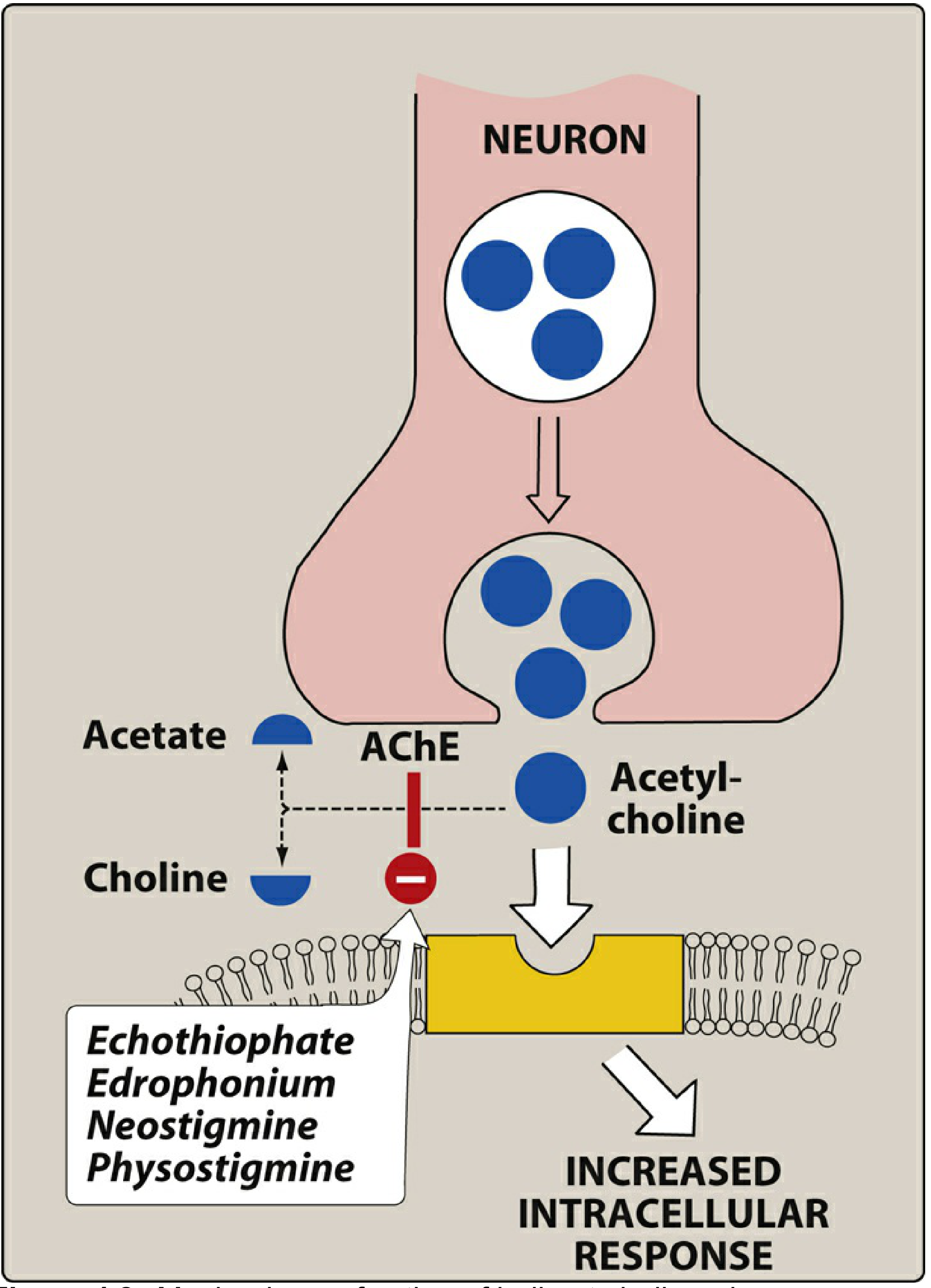
- Chemical structure: Quaternary amine - charged and polar, so it cannot cross the blood-brain barrier. Its actions are therefore limited to the periphery with no CNS effects.
- Elimination: Rapid renal elimination accounts for its very short duration of action.
Pharmacokinetics
| Parameter | Value |
|---|---|
| Onset | 30-60 seconds (IV) |
| Peak effect | < 2 minutes |
| Duration | 5-20 minutes |
| Route | Intravenous (IV) |
| Elimination | Renal |
Clinical Uses
1. Diagnosis of Myasthenia Gravis (MG) - "Tensilon Test"
- Give 2 mg IV as a test dose; wait 45 seconds
- If no response, give an additional 8 mg IV
- Positive result: Visible improvement in muscle strength (e.g., resolution of ptosis, fusion of diplopia) lasting ~5 minutes = confirms MG
Important note: The edrophonium test has lost favor as a primary diagnostic tool due to the widespread availability of autoantibody testing (anti-AChR, anti-MuSK antibodies). Edrophonium is also increasingly difficult to obtain globally and has not been available in the United States since 2018. (Barash's Clinical Anesthesia, 9e; Bradley & Daroff's Neurology)
2. Differentiating Myasthenic Crisis vs. Cholinergic Crisis
| Feature | Myasthenic Crisis | Cholinergic Crisis |
|---|---|---|
| Cause | Undermedication / disease exacerbation | Overmedication (excess ACh) |
| After edrophonium | Muscle strength improves | Weakness worsens; fasciculations, respiratory depression |
| Treatment | Increase anticholinesterases (pyridostigmine) | Atropine; ventilatory support |
- Dose used: 1-2 mg IV (small doses are used for this differentiation to avoid precipitating worsening in cholinergic crisis)
- Caution: Equipment for ventilatory support must be available due to the risk of respiratory arrest
3. Reversal of Neuromuscular Blockade (Anesthesia)
- Dose: 0.5-1.0 mg/kg IV
- Co-administer with atropine (not glycopyrrolate) because atropine's fast onset matches that of edrophonium - 0.014 mg atropine per 1 mg edrophonium
- Preferred for mivacurium reversal: Unlike neostigmine, edrophonium does not inhibit plasma cholinesterase (which is required to metabolize mivacurium), making it uniquely useful here
4. Pediatric Dosing (Myasthenia Diagnosis)
- Total edrophonium IV dose in children: 0.15 mg/kg, not to exceed 10 mg
- Give 1/10th of the total dose as a test dose first to assess for adverse cholinergic effects
Adverse Effects / Cholinergic Toxicity
- Salivation, lacrimation, urination, defecation
- Bradycardia, hypotension
- Increased bronchial secretions, bronchospasm
- Miosis, abdominal cramps, nausea, vomiting
- Muscle fasciculations
- Skeletal muscle weakness/paralysis (at high doses - depolarizing blockade)
- Respiratory paralysis (most dangerous)
Comparison with Other Anticholinesterases
| Drug | Duration | CNS Entry | Key Use |
|---|---|---|---|
| Edrophonium | 5-20 min (short) | No (quaternary) | Diagnosis of MG, crisis differentiation, reversal |
| Neostigmine | 30 min - 2 hr | No (quaternary) | Reversal of NMB, MG management |
| Pyridostigmine | 3-6 hr | No (quaternary) | Long-term MG management |
| Physostigmine | 30 min - 2 hr | Yes (tertiary) | Anticholinergic toxidrome reversal |
Key Points Summary
- Prototype short-acting AChE inhibitor - reversible, quaternary amine
- Onset 30-60 sec, duration 5-20 min - fastest onset of all anticholinesterases
- Primary use: Tensilon test for MG diagnosis and crisis differentiation
- Anesthesia: Reversal of non-depolarizing blockade; preferred for mivacurium reversal (does not inhibit plasma cholinesterase)
- Antidote for cholinergic crisis: Atropine (muscarinic blocker)
- No CNS effects due to quaternary structure
- Availability: Withdrawn from the US market in 2018; increasingly unavailable worldwide
Curb
What are you referring to by 'Curb'?
CURB-65 score pneumonia criteria management 2024 guidelines
CURB-65 Score - Community-Acquired Pneumonia (CAP) Severity
What is CURB-65?
The 5 Criteria (1 point each)
| Letter | Parameter | Threshold |
|---|---|---|
| C | Confusion | New confusion (abbreviated Mental Test score ≤8, or disorientation to person, place, or time) |
| U | Blood Urea nitrogen | BUN >7 mmol/L (>19 mg/dL) |
| R | Respiratory rate | ≥ 30 breaths/min |
| B | Blood pressure | Systolic < 90 mmHg OR diastolic ≤ 60 mmHg |
| 65 | Age | ≥ 65 years |
Score Interpretation & Management
| Score | Mortality Risk | Management |
|---|---|---|
| 0 | ~1.5% (30-day) | Outpatient treatment |
| 1 | Low risk | Outpatient (consider admission if age ≥65 is the only point) |
| 2 | Intermediate (3-15%) | Hospital admission OR virtual ward/same-day emergency care |
| 3-5 | High (>15-22%) | Inpatient ± ICU consideration |
Key note: Scores of ≥3 carry a 22% overall 30-day mortality and may require ICU-level admission.
CRB-65 (Simplified Version - No Labs Needed)
| CRB-65 Score | Action |
|---|---|
| 0 | Manage in primary care (low risk) |
| 1 | Consider referral to hospital or virtual ward |
| ≥2 | Refer to hospital |
CURB-65 vs. PSI - Quick Comparison
| Feature | CURB-65 | PSI |
|---|---|---|
| Variables | 5 | 20 |
| Complexity | Simple, bedside | Complex, calculator needed |
| Best for | Identifying high-risk patients | Identifying low-risk patients |
| Preferred by | British Thoracic Society, NICE | IDSA-ATS guidelines |
| Validation | Good | More robust |
IDSA/ATS Criteria for ICU Admission (Separate from CURB-65)
- Septic shock requiring vasopressors
- Acute respiratory failure requiring mechanical ventilation
- Respiratory rate ≥30 breaths/min
- PaO2/FiO2 ratio ≤250
- Multilobar infiltrates
- Confusion/disorientation
- Uremia (BUN ≥20 mg/dL)
- Leukopenia (WBC <4,000 cells/mm³)
- Thrombocytopenia (platelets <100,000/mm³)
- Hypothermia (core temp <36°C)
- Hypotension requiring aggressive fluid resuscitation
Important Caveats
- Clinical judgment always takes priority - scores support but do not replace clinical assessment
- CURB-65 does not account for comorbidities, oxygenation (SpO2), ability to take oral medications, or social factors (e.g., homelessness, cognitive impairment)
- If O2 saturation on room air is <92%, hospitalization is generally needed regardless of score
- A low CURB-65 score does not exclude severe pneumonia, especially in patients with significant comorbidities or frailty
- The score can be misleading in pregnancy