
Structure of haemoglobin
Haemoglobin is a tetrameric protein made of four globin chains, each carrying a haem group. The most common adult form (HbA) has two alpha and two beta chains. Each haem contains a porphyrin ring with a central iron (Fe²+) atom that binds one oxygen molecule, giving a total O2-binding capacity of four per haemoglobin molecule.
Searching sources...
Reading File
Finding Sources
Finding Sources
Finding Sources
Finding Sources
Good hits. Now reading the most relevant sections in parallel - Ganong's physiology, Harrison's, Guyton & Hall, Thompson & Thompson Genetics, and Histology textbook.
Reading File
Reading File
Reading File
Reading File
Reading File
Excellent content retrieved. Now fetching the key structural images from Guyton & Hall and the Histology/Thompson textbooks.
Loading Image
Loading Image
Loading Image
Loading Image
All images and content retrieved. Here is the comprehensive answer.
Structure of Haemoglobin
Overview
Haemoglobin (Hb) is a globular, tetrameric protein found in red blood cells. It is the primary oxygen-carrying molecule in vertebrates. Each haemoglobin molecule consists of four subunits, with each subunit made up of a globin polypeptide chain bound to a haem prosthetic group.
- Ganong's Review of Medical Physiology - p.557
- Guyton and Hall Textbook of Medical Physiology - p.445
1. Quaternary Structure - The Tetramer
Haemoglobin is a tetramer of two pairs of unlike globin polypeptide chains. In normal adult haemoglobin (HbA), these are:
- 2 alpha (α) chains + 2 beta (β) chains → designated α₂β₂
The four chains fold and fit together to form a compact globular molecule. The arrangement is not symmetric - the alpha and beta chains interact at specific interfaces (α₁β₁ and α₁β₂ contacts), and cooperative oxygen binding depends critically on these inter-subunit contacts.
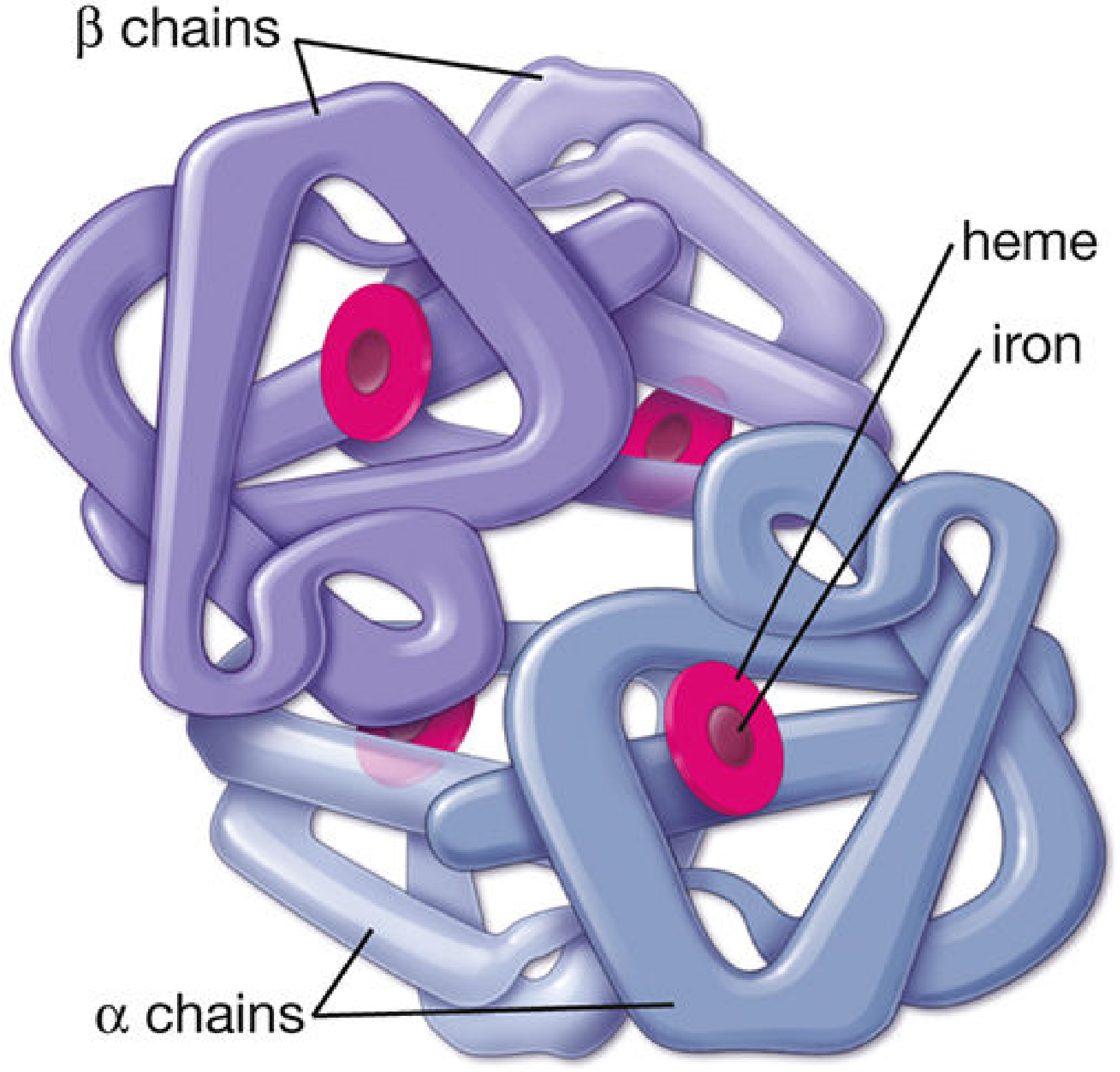
Structural diagram of the haemoglobin molecule - two α-chains (light blue) and two β-chains (purple), each carrying a heme group (pink) with a central iron atom.
2. The Haem Group
Each globin subunit contains one haem prosthetic group embedded in a hydrophobic cleft (pocket) of the polypeptide chain. The folding of the globin chain places haem near the surface, where it is accessible to oxygen.
Haem structure:
- A tetrapyrrole porphyrin ring (protoporphyrin IX) with four pyrrole rings (A, B, C, D) joined by methene bridges
- A central Fe²+ (ferrous) iron atom coordinated to all four nitrogen atoms of the porphyrin ring
- The iron also forms two additional coordination bonds:
- One to the proximal histidine (His F8 / β-His92) - a covalent link anchoring haem to globin
- One to O₂ (or a water molecule in deoxyHb) on the distal side
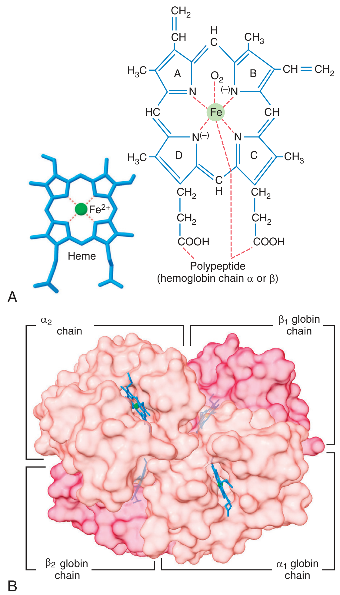
Figure A: The heme moiety showing the four pyrrole rings (A-D), central iron atom coordinated to nitrogen atoms of the porphyrin, and attachment to the globin polypeptide. Figure B: The complete haemoglobin tetramer (α₂β₂).
Because there are four haem groups per molecule (one per chain), and each iron can bind one O₂, a single haemoglobin molecule can carry four molecules of O₂ (eight oxygen atoms total). Oxygen binds loosely and reversibly via a coordination bond with iron - it is carried as molecular O₂ (not ionic) and released unchanged in the tissues.
- Guyton and Hall Textbook of Medical Physiology - p.445
3. Tertiary Structure - The Globin Chain
Each individual globin chain has a remarkably conserved tertiary structure - virtually all globins (regardless of species or chain type) adopt the same folded conformation called the globin fold:
- 7-8 helical regions labelled A through H
- The haem is held in a hydrophobic pocket between helices E and F
- Proximal histidine (His F8 / β-His92) - covalently links haem iron to the protein
- Phenylalanine (Phe CD1 / β-Phe42) - wedges the porphyrin ring into the haem pocket; critical for haem retention
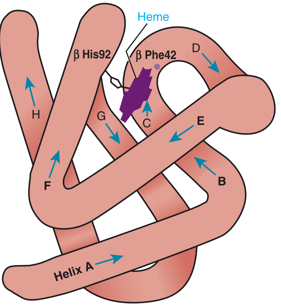
The tertiary structure of a globin subunit: eight helical regions (A-H), with heme (purple) nestled in the pocket between helices. His92 provides the iron-protein covalent link; Phe42 locks the porphyrin in place.
Variants that disrupt helical structure, replace highly conserved residues, or introduce charged groups into the hydrophobic interior invariably produce haemoglobinopathies.
- Thompson & Thompson Genetics and Genomics in Medicine - p.252
4. Types of Normal Haemoglobin
All normal human haemoglobins share the α₂X₂ tetrameric plan, but differ in their non-alpha chains. These change during development in a process called globin switching:
| Haemoglobin | Chains | Normal Adults | Notes |
|---|---|---|---|
| HbA | α₂β₂ | ~96% | Predominant adult form |
| HbA₂ | α₂δ₂ | 1.5-3% | β and δ differ in only 10/146 amino acids |
| HbF | α₂γ₂ | <1% | Fetal form; higher O₂ affinity |
| HbH | β₄ | Pathologic | Forms in severe α-thalassaemia |
| Embryonic Hbs | ε, ζ chains | Absent after birth | Earliest embryonic haemoglobins |
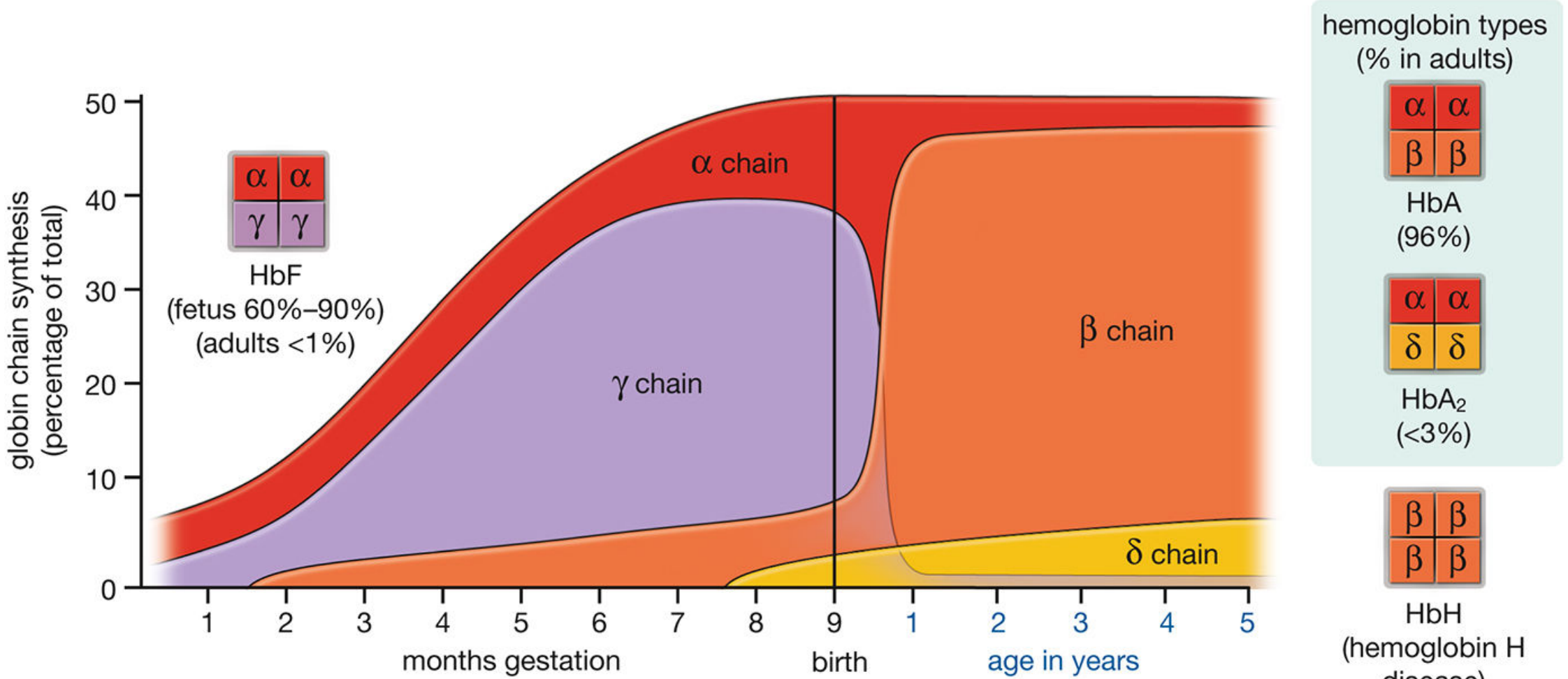
Globin switching: In the fetus, γ-chain synthesis produces HbF (60-90%). After birth, β-chain synthesis rises sharply while γ-chains decline, resulting in predominantly HbA (96%) and HbA₂ (<3%) in adults.
- Histology: A Text and Atlas - p.685
5. Globin Gene Locations
- α-globin genes (α1 and α2) - clustered on chromosome 16 (two copies per haplotype)
- β-globin gene cluster (β, δ, γ, ε) - on chromosome 11; genes are arranged in the same order as their developmental expression
6. Clinically Relevant Points
| Feature | Significance |
|---|---|
| Fe²+ state | Must remain ferrous (Fe²+) for O₂ binding; Fe³+ (methaemoglobin) cannot carry O₂ |
| Cooperative binding | Binding of O₂ to one subunit increases affinity of remaining subunits (sigmoid O₂ dissociation curve) |
| HbA1c | Glucose attached to N-terminal valine of β chains; marker of long-term glycaemic control |
| Sickle cell (HbS) | Val→Glu substitution at position 6 of β chains; deoxyHbS polymerises into crystals that deform and lyse RBCs |
| Unstable haemoglobins | Mutations weakening heme-globin linkage, disrupting α-helical structure, or introducing polar residues into the hydrophobic core |
Sources: Ganong's Review of Medical Physiology (26th ed.) | Guyton and Hall Textbook of Medical Physiology | Thompson & Thompson Genetics and Genomics in Medicine (9th ed.) | Histology: A Text and Atlas (Pawlina) | Harrison's Principles of Internal Medicine 22E | Robbins, Cotran & Kumar Pathologic Basis of Disease
This is a shared conversation. Sign in to Orris to start your own chat.