
Explain thyroid in full detail as per surgery
thyroid gland anatomy surgical diagram lobes isthmus
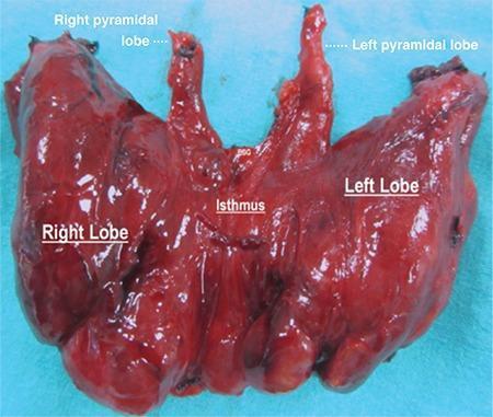
Anatomical specimen photograph of a human thyroid gland post-total thyroidectomy, set against a blue surgical background. The specimen is labeled to demonstrate a rare anatomical variation: bilateral pyramidal lobes. Centrally, the isthmus connects the voluminous right and left lateral lobes. Arising superiorly from the junction points between the isthmus and each lateral lobe are two distinct, elongated, conical structures identified as the right and left pyramidal lobes. The entire gland exhibits a reddish-brown, glistening parenchyma with a multinodular texture consistent with toxic multinodular goiter. The anatomical landmarks are labeled with white text: 'Right Lobe', 'Left Lobe', 'Isthmus', 'Right pyramidal lobe', and 'Left pyramidal lobe'. This clinical photograph is highly relevant for endocrine surgery and gross anatomy education, highlighting developmental remnants of the thyroglossal duct and the necessity of complete dissection during thyroidectomy to ensure total removal of all functional thyroid tissue.
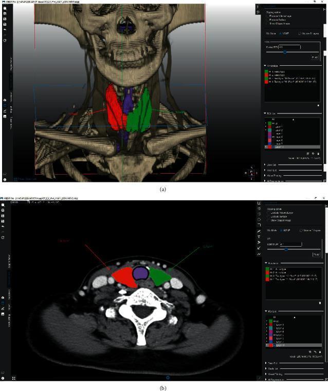
This medical visual demonstrates volumetric segmentation of the thyroid gland using specialized medical imaging software. Image (a) presents a 3D reconstruction overlay on a CT-derived skeletal model of the neck and upper thorax. The segmentation highlights the anatomical lobes: the right thyroid lobe is colored green, the left lobe is red, and the isthmus/central region is purple. Image (b) shows a corresponding axial CT slice at the level of the thyroid. In this cross-sectional view, the colored segmentation masks (red for left lobe, green for right lobe, and purple for the central isthmus) are superimposed on the soft tissue structures anterior to the trachea. The image illustrates the application of semi-automated graph-cut algorithms for preoperative planning in endocrine surgery, specifically for thyroid lobectomy. Key anatomical landmarks visible include the trachea, carotid arteries, jugular veins, and cervical vertebrae. This content is designed for intermediate to advanced medical training in radiology and surgical oncology, focusing on thyroid anatomy and computer-assisted volumetric measurement.
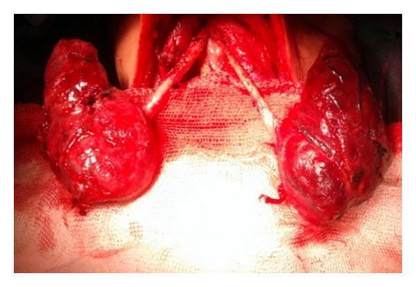
This clinical photograph captures an intraoperative view of the neck during a surgical thyroid exploration, illustrating a rare congenital anomaly: agenesis of the thyroid isthmus. The image displays two distinct, enlarged, reddish-brown thyroid lobes situated laterally within the surgical field. Crucially, there is a complete absence of the central isthmus that typically connects the two lobes across the midline. Between the separated lobes, the cartilaginous rings of the trachea are clearly exposed and visible medially. The thyroid lobes exhibit a glistening, irregular surface texture with focal areas of hemorrhage, consistent with surgical manipulation in a patient with Graves' disease. The surgical field is stabilized with medical gauze, providing high contrast for the anatomical structures. This visual serves as a key educational example for endocrine surgeons and students to recognize anatomical variations of the thyroid gland that may be encountered during total thyroidectomy or other cervical procedures.
thyroidectomy surgical technique recurrent laryngeal nerve parathyroid
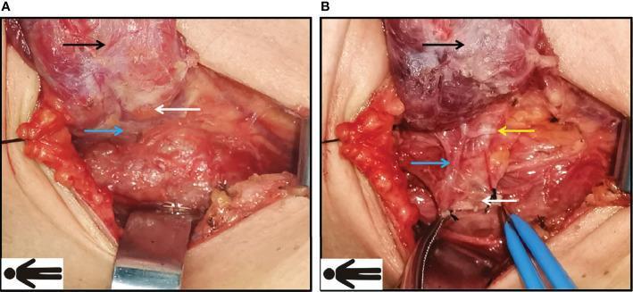
Two-panel clinical photograph illustrating the preservation of the inferior right parathyroid gland and recurrent laryngeal nerve (RLN) during a thyroidectomy surgical procedure using the Fang’s capillary fascia preservation technique. Panel A shows the operative field after raising the right thyroid lobe (black arrow), revealing the inferior parathyroid gland (white arrow) and its associated blood supply (blue arrow) in situ. Panel B demonstrates the further dissection and mobilization of tissue to protect vital structures. The sternothyroid ligament is manipulated to expose a fan-like surgical plane. Key landmarks identified include the superiorly positioned thyroid lobe (black arrow), the inferior parathyroid gland (white arrow), its distinct vascular pedicle (blue arrow), and the recurrent laryngeal nerve (yellow arrow) running in close proximity within the tracheoesophageal groove. The images emphasize the importance of identifying and maintaining the microvascular supply to the parathyroid glands and avoiding mechanical or electrical injury to the RLN during Level VI lymph node dissection. This material is intended for endocrine surgery and surgical anatomy education.
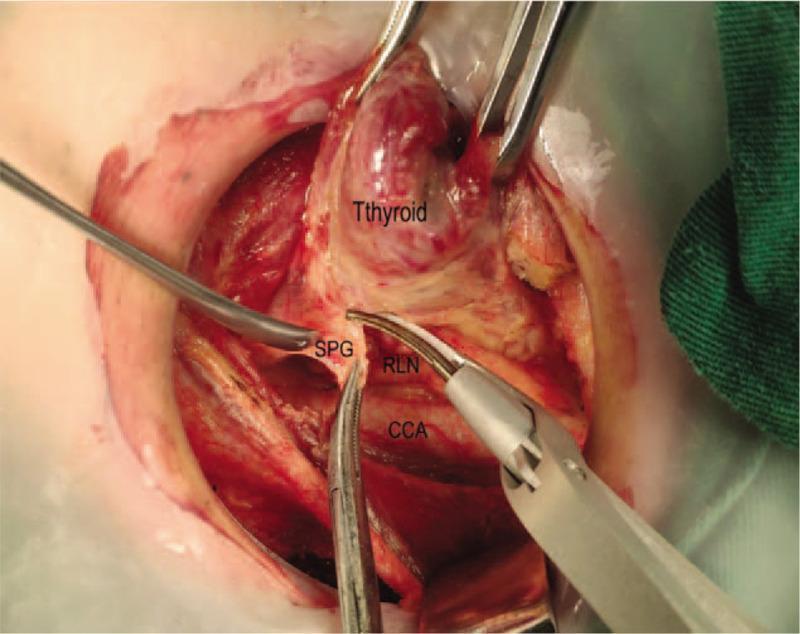
This clinical photograph shows an intraoperative view of a thyroidectomy, specifically highlighting the dissection and preservation of critical neurovascular and glandular structures. The thyroid gland is superiorly retracted. Below it, the Superior Parathyroid Gland (SPG) is identified and carefully protected. The Recurrent Laryngeal Nerve (RLN) is clearly visualized running parallel and superficial to the Common Carotid Artery (CCA). Surgical instrumentation, including dissecting forceps and a specialized thermal or ultrasonic cutting device, is being used to perform precise dissection in the tracheoesophageal groove. The image demonstrates the 'upward separation' technique aimed at isolating the thyroid lobe while maintaining the integrity of the RLN and vascular supply to the parathyroid glands. This educational visual emphasizes surgical anatomy and safety protocols for residents and surgeons performing endocrine neck surgery.
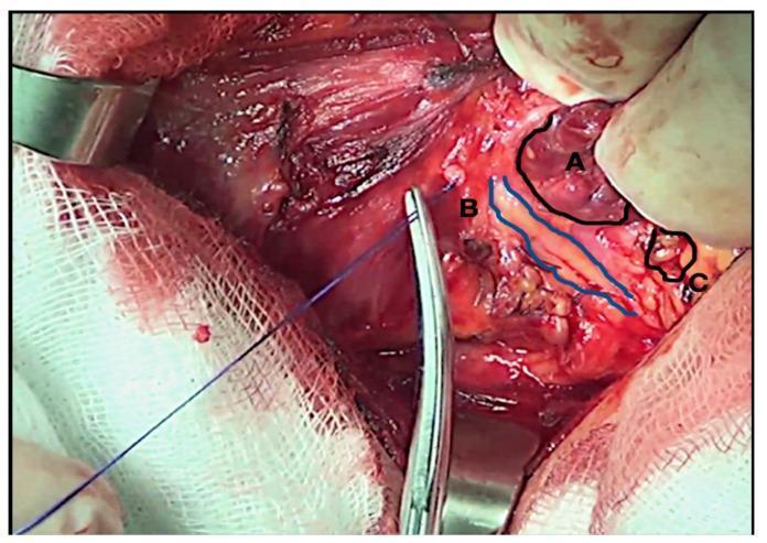
This clinical photograph displays an intraoperative view of a thyroidectomy during a recurrent laryngeal nerve (RLN) dissection, captured under 2.5x optical magnification. The surgical field highlights critical anatomical structures labeled as follows: (A) the reddish-brown, lobulated thyroid gland parenchyma; (B) the right-sided recurrent laryngeal nerve, identified as a slender, yellowish-white string-like structure outlined in blue; and (C) the inferior parathyroid gland, appearing as a small, beige-colored nodule in close proximity to the thyroid and nerve. Surgical instruments including a metallic hemostat and a blue suture thread are visible, facilitating tissue retraction and identification. White surgical gauze and metallic retractors define the periphery of the operative site. This image serves as an educational tool for endocrine surgery, illustrating the precise identification and preservation of the RLN and parathyroid glands to prevent postoperative complications such as vocal cord paralysis and hypocalcemia.
thyroid cancer papillary follicular medullary anaplastic classification
This line graph illustrates the epidemiological trends in the distribution of thyroid cancer cases by histological type from 1980 to 2012. The data, expressed as a percentage of all thyroid cancer cases, shows a prominent increase in papillary carcinoma, which rose from approximately 60% in 1980 to over 90% by 2012. Conversely, other histological subtypes show a steady decline in relative proportion over the same period. Follicular carcinoma, which initially accounted for nearly 25% of cases, decreased significantly. Medullary and anaplastic carcinomas, along with a category labeled as 'others,' remained at low percentages and trended downward toward the end of the study period. This visualization emphasizes the shifting landscape of thyroid pathology, highlighting the increasing dominance of papillary carcinoma in clinical diagnosis and incidence relative to non-papillary types. The graph serves as an educational resource for oncology and endocrinology, illustrating longitudinal changes in cancer subtypes within a specific population context.
This line graph illustrates a joinpoint analysis of time trends for the age-standardized incidence of non-papillary thyroid cancer (including follicular, medullary, and anaplastic histological types) in Israel from 1980 to 2012. The data is stratified by gender within the Jewish population. The y-axis represents the rate per 100,000, while the x-axis denotes the calendar years. The visual demonstrates a statistically significant downward trend in the incidence for both Jewish women and Jewish men over the 32-year period. Jewish women (indicated by the upper green line) started with a higher incidence rate in 1980 compared to Jewish men (indicated by the lower blue line). Both groups show fluctuations year-over-year but exhibit a clear overall decline, with incidence rates narrowing and approaching lower, more similar values by 2012. This epidemiological data highlights shifting patterns in thyroid cancer pathology, contrasting the decreasing incidence of non-papillary types with the concurrent rise of papillary carcinoma observed in the same period.
The Thyroid Gland - A Complete Surgical Perspective
1. HISTORICAL BACKGROUND
2. EMBRYOLOGY & DEVELOPMENTAL ANATOMY
- The thyroid originates as an endodermal proliferation in the floor of the primitive pharynx at the foramen cecum (base of tongue), at the junction of the anterior two-thirds and posterior third.
- It descends in the midline anterior to the hyoid bone and larynx, reaching its final pretracheal position by week 7 of gestation.
- The descent pathway is marked by the thyroglossal duct, which normally obliterates. Failure leads to a thyroglossal duct cyst (the most common congenital neck mass) - treated by Sistrunk's procedure (excision of the cyst + mid-portion of hyoid bone).
- The C-cells (parafollicular cells) that produce calcitonin migrate from the ultimobranchial bodies (4th/5th pharyngeal pouches).
| Anomaly | Description |
|---|---|
| Thyroglossal duct cyst | Most common; midline, moves with tongue protrusion |
| Ectopic thyroid | Lingual thyroid; only thyroid tissue in 70% |
| Pyramidal lobe | Present in 50% of individuals, remnant of thyroglossal duct |
| Aplasia/Hypoplasia | Rare; causes congenital hypothyroidism |
3. SURGICAL ANATOMY
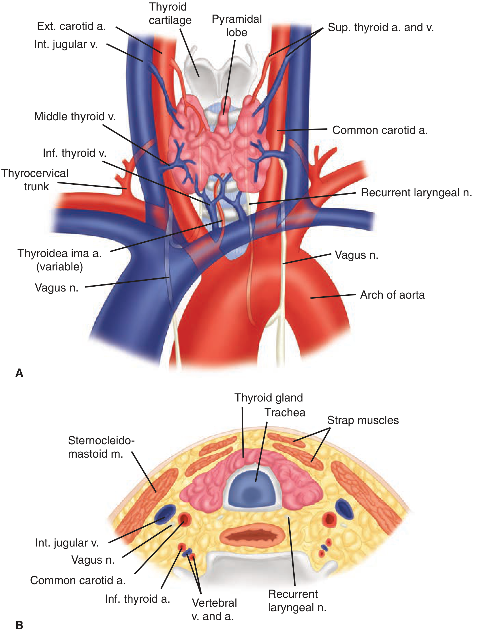
3a. Position & Relations
- Anterior: strap muscles, sternocleidomastoid
- Posterior: parathyroid glands, recurrent laryngeal nerve, tracheoesophageal groove
- Lateral: carotid sheath (common carotid artery, internal jugular vein, vagus nerve)
3b. Lobes and Isthmus
- Two lateral lobes connected by the isthmus at the level of the 2nd-3rd tracheal rings
- A pyramidal lobe arises from the isthmus in ~50% (more commonly on the left); it must be excised in total thyroidectomy
- The Tubercle of Zuckerkandl is a posterior projection of the thyroid lobe near the cricoid - a key surgical landmark for identifying the RLN
3c. Blood Supply
- Superior thyroid arteries: First branches of the external carotid arteries; divide into anterior and posterior branches at the thyroid apex. The external branch of the superior laryngeal nerve (EBSLN) runs close to these vessels - injury causes loss of cricothyroid muscle function (high-note voice changes).
- Inferior thyroid arteries: Arise from the thyrocervical trunk (from subclavian). Travel posterior to the carotid sheath and enter the thyroid lobes at their midpoint. They cross the RLN - this is the most dangerous area in thyroid surgery.
- Thyroidea ima artery: Present in 1-4% of individuals; arises directly from the aorta or innominate artery.
- Superior thyroid veins → internal jugular vein (with superior thyroid artery)
- Middle thyroid veins → internal jugular vein (least consistent; must be ligated early during mobilization)
- Inferior thyroid veins → form a plexus → brachiocephalic veins
3d. Lymphatic Drainage
- Central compartment (Level VI): Delphian nodes (precricoid), pretracheal, paratracheal nodes
- Lateral compartment (Levels II-V): Deep cervical chain
- Superior mediastinum (Level VII)
3e. Recurrent Laryngeal Nerve (RLN) - Surgical Importance
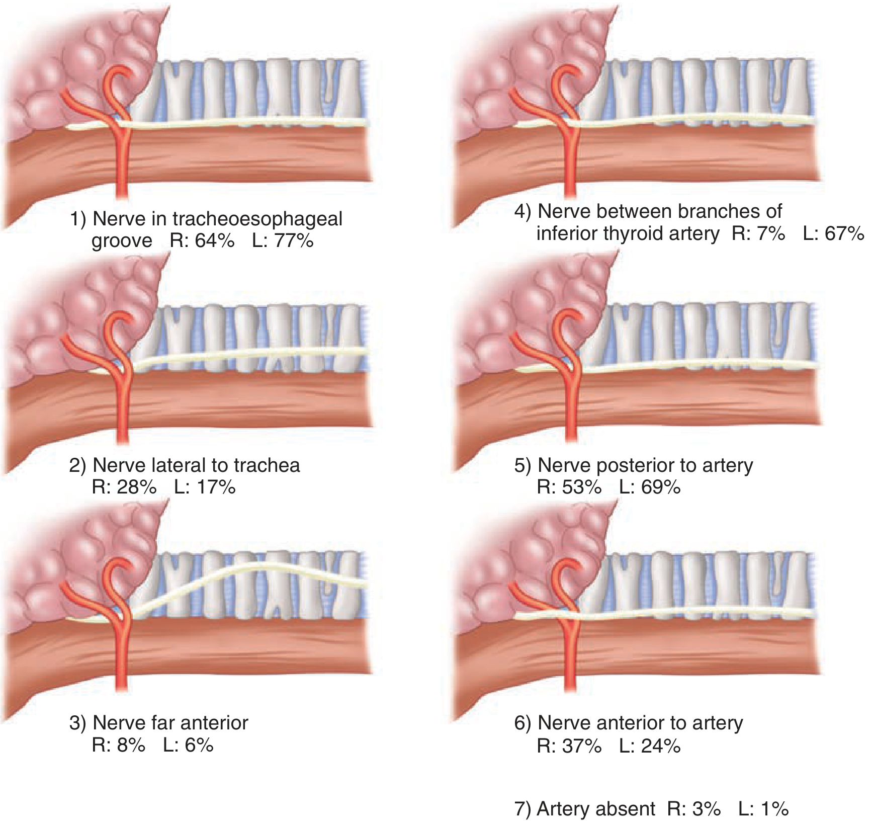
- Ligament of Berry (where nerve is tightly adherent to thyroid)
- Inferior thyroid artery crossing
- The nerve may branch before entering the larynx - a thin branch should prompt search for additional branches
4. HISTOLOGY & PHYSIOLOGY
Histology
- Follicular cells (principal cells): Form spherical follicles containing colloid (thyroglobulin). Produce T3 and T4.
- C-cells (parafollicular cells): Located between follicles; produce calcitonin. Origin from neural crest via ultimobranchial body.
- Follicles are lined by flat-to-cuboidal epithelium (inactive) or columnar epithelium (active).
Physiology - Hormone Synthesis
- Iodide trapping: Active transport into follicular cells (NIS - sodium-iodide symporter)
- Oxidation to iodine: By thyroid peroxidase (TPO)
- Organification: Iodination of tyrosine residues on thyroglobulin → MIT, DIT
- Coupling: MIT + DIT → T3; DIT + DIT → T4 (stored as colloid)
- Secretion: TSH stimulates endocytosis of colloid, proteolysis of thyroglobulin, release of T3 and T4
5. EVALUATION OF PATIENTS WITH THYROID DISEASE
Clinical Assessment
- History: Symptoms of hyper/hypothyroidism, family history (MEN syndromes, familial MTC), prior neck radiation, rate of growth
- Physical exam: Size, consistency, nodularity, fixation, cervical lymphadenopathy, vocal cord function (laryngoscopy if voice change)
Laboratory Tests
| Test | Purpose |
|---|---|
| TSH | Best screening test; suppressed in hyperthyroidism, elevated in hypothyroidism |
| Free T4 | Directly measures unbound active hormone |
| T3 | Useful in T3 toxicosis |
| Anti-TPO, Anti-Tg antibodies | Hashimoto's thyroiditis |
| TSH receptor antibodies (TRAb) | Graves' disease |
| Calcitonin | Medullary thyroid cancer screening |
| CEA | Medullary thyroid cancer |
Imaging
- Thyroid ultrasound: First-line; evaluates size, number, echogenicity, calcifications, vascularity, lymph nodes. TIRADS classification guides FNA decisions.
- Radionuclide scan (Tc-99m pertechnetate or I-123): Evaluates function - "hot" (hyperfunctioning, rarely malignant) vs "cold" (non-functioning, 5-10% malignant risk) nodules
- CT/MRI: For substernal goiter, tracheal deviation, retropharyngeal extension, and staging of malignancy
- PET scan: For recurrent/metastatic disease, especially radioiodine-refractory tumors
Fine Needle Aspiration Biopsy (FNAB)
| Bethesda | Category | Malignancy Risk | Management |
|---|---|---|---|
| I | Non-diagnostic | 5-10% | Repeat FNA |
| II | Benign | 0-3% | Follow-up |
| III | AUS/FLUS | 6-18% | Repeat FNA or molecular testing |
| IV | Follicular neoplasm | 10-40% | Lobectomy |
| V | Suspicious for malignancy | 45-75% | Lobectomy/thyroidectomy |
| VI | Malignant | 94-96% | Thyroidectomy |
6. BENIGN THYROID DISORDERS
Hyperthyroidism (Thyrotoxicosis)
- Most common cause; autoimmune with stimulatory TSH-receptor antibodies (TSI/TRAb)
- Triad: hyperthyroidism + diffuse goiter + exophthalmos (Graves' ophthalmopathy)
- Surgical indication: Failed ATD therapy, large goiter, ophthalmopathy, pregnancy contraindication to RAI, patient preference
- Pre-op preparation: Antithyroid drugs (PTU or methimazole) to achieve euthyroid state + Lugol's iodine for 10 days before surgery (reduces vascularity - "Plummer's iodine")
- Surgery: Total or near-total thyroidectomy
- Multiple autonomous nodules, usually in elderly patients with long-standing simple goiter
- RAI or surgery (subtotal/total thyroidectomy)
- Single hyperfunctioning follicular adenoma
- Surgery (lobectomy) or RAI
Hypothyroidism
- Primary (Hashimoto's) or secondary (pituitary failure)
- Usually managed medically with levothyroxine
- Hashimoto's thyroiditis: Most common cause of hypothyroidism in iodine-sufficient areas; autoimmune, elevated anti-TPO and anti-Tg antibodies. Surgery for: suspicion of malignancy, symptomatic goiter, or thyroid lymphoma (80x increased risk vs general population)
Goiter
- Simple/Nontoxic goiter: Due to iodine deficiency or goitrogens
- Substernal goiter: >50% below thoracic inlet; causes compression (dysphagia, dyspnea, SVC syndrome); surgery via cervical approach in >95% - rarely requires sternotomy
Thyroiditis
| Type | Features | Surgery? |
|---|---|---|
| Hashimoto's | Autoimmune, hypothyroidism | Only for suspicion of malignancy |
| De Quervain's (subacute granulomatous) | Viral, painful, transient hyperthyroidism then hypothyroidism | No |
| Riedel's (fibrous) | "Woody" hard thyroid, invasive fibrosis, IgG4-related | Wedge excision of isthmus to decompress trachea |
7. THYROID NODULES & MALIGNANCY
Solitary Thyroid Nodule (STN)
- Prevalence: palpable in 4-7%; ultrasound-detectable in 19-67%
- Most (95%) are benign; risk factors for malignancy: prior head/neck radiation, family history, male sex, extremes of age, rapid growth, hard consistency, fixation, associated lymphadenopathy
- Workup: TSH → if low, radionuclide scan; if normal/elevated, ultrasound + FNAB
Thyroid Cancers - Classification
| Type | Frequency | Origin | Features |
|---|---|---|---|
| Papillary | 80-85% | Follicular cells | Psammoma bodies, nuclear grooves, orphan Annie nuclei; lymphatic spread; best prognosis |
| Follicular | 10-15% | Follicular cells | Vascular + capsular invasion; hematogenous spread (bone, lung); FNA cannot distinguish from adenoma |
| Medullary | 5-8% | C-cells | Calcitonin secretion; RET proto-oncogene; MEN2A/2B; amyloid stroma |
| Anaplastic | 1-2% | Follicular cells | Most aggressive; often in elderly women; presents with rapid painful neck mass; median survival < 6 months |
| Lymphoma | <1% | B-cells | Associated with Hashimoto's; MALT or diffuse large B-cell; chemotherapy ± radiation, not surgery |
Papillary Thyroid Cancer (PTC) - Surgical Detail
- Histological hallmarks: "Orphan Annie eye" nuclei (empty-looking, ground-glass nuclei), nuclear grooves, nuclear pseudoinclusions, psammoma bodies (concentrically calcified)
- Multifocal in up to 80%, bilateral in 30%
- Excellent prognosis: 10-year survival >95% for low-risk disease
- Low risk: Intrathyroidal, no vascular invasion, no RAI-avid metastases
- Intermediate risk: Minor ETE, vascular invasion, RAI-avid neck metastases, aggressive histology
- High risk: Gross ETE, incomplete resection, distant metastases, very high Tg
- Tumors <1 cm (microPTC): Active surveillance OR thyroid lobectomy are reasonable
- Tumors 1-4 cm without high-risk features: Lobectomy may be sufficient
- Tumors >4 cm, bilateral disease, extrathyroidal extension, LN metastases: Total thyroidectomy + central neck dissection (Level VI)
- Lateral neck dissection (Levels II-V) for biopsy-proven lateral LN metastases
Follicular Thyroid Cancer (FTC) - Surgical Detail
- FNAB cannot distinguish follicular adenoma from carcinoma (requires capsular/vascular invasion on histology)
- Bethesda IV → diagnostic lobectomy → if malignant, completion thyroidectomy based on risk
- Minimally invasive FTC (capsular invasion only, no vascular invasion): Lobectomy may be curative
- Widely invasive FTC: Total thyroidectomy + consideration of RAI
Medullary Thyroid Cancer (MTC) - Surgical Detail
- All patients should have RET mutation testing (25-30% are hereditary)
- Screen for pheochromocytoma BEFORE thyroid surgery in MEN2A/2B (avoid hypertensive crisis)
- Clinically evident MTC: Total thyroidectomy + bilateral central neck dissection (central LN metastases in >70% of palpable tumors)
- Role of prophylactic lateral neck dissection is controversial; ATA recommends based on calcitonin levels (>200 pg/mL → consider contralateral lateral dissection if ipsilateral LN+ on ultrasound)
- Hereditary MTC - Prophylactic thyroidectomy: Timing based on specific RET mutation:
- Codon 918 (MEN2B): Ideally within first 6 months of life
- Codon 634 (MEN2A): By age 5 years
- Other codons: When calcitonin rises above normal or by age 5-10
Anaplastic Thyroid Cancer (ATC)
- All ATC is classified as Stage IV at diagnosis
- Rarely resectable; surgery limited to tracheostomy for airway protection or palliative debulking
- Combined modality: external-beam radiation + chemotherapy (doxorubicin as radiosensitizer)
- Novel therapies: Dabrafenib + trametinib (for BRAF V600E-mutated ATC) has shown response rates; FDA-approved
8. THYROID SURGERY - OPERATIVE TECHNIQUE
Pre-operative Assessment
- Vocal cord assessment by laryngoscopy: mandatory for any altered phonation, prior neck/upper chest surgery, known posterior extension of thyroid cancer
- Achieve euthyroid state before elective surgery
- Screen for pheochromocytoma in suspected MEN2
Patient Positioning
- Supine with a sandbag between the scapulae
- Head on donut cushion, neck extended for maximal exposure
Incision
- Kocher transverse collar incision: 3-5 cm, placed in or parallel to a natural skin crease, 1 cm below the cricoid cartilage
- Longer incisions needed for large goiters
Steps of Thyroidectomy
- Skin incision → subcutaneous tissue + platysma incised sharply
- Subplatysmal flaps raised: Superiorly to thyroid cartilage, inferiorly to suprasternal notch
- Strap muscles divided in the midline; sternohyoid separated from sternothyroid by blunt dissection
- Identify internal jugular vein and ansa cervicalis nerve laterally
- Sternothyroid muscle dissected off thyroid → exposes middle thyroid veins
- Middle thyroid veins ligated and divided (allows medial mobilization of lobe)
- Superior pole mobilization: Retract thyroid inferiorly and medially; skeletonize superior pole vessels close to thyroid to avoid injury to the EBSLN
- Identify and protect RLN: Most consistently found at the level of the cricoid cartilage; the ATA strongly recommends visual identification in all cases
- Lower pole mobilization: Inferior thyroid vessels dissected and divided close to the thyroid surface (extracapsular dissection) to preserve parathyroid blood supply
- Parathyroid gland identification: Usually within 1 cm of the crossing of inferior thyroid artery and RLN
- Ligament of Berry division: Most dangerous step - RLN passes through this structure. Avoid electrocautery near the nerve
- Isthmus and pyramidal lobe division: Pyramidal lobe dissected cephalad to thyroid cartilage notch or higher
- Hemostasis and closure
Types of Thyroid Surgery
| Procedure | Extent | Indications |
|---|---|---|
| Thyroid lobectomy | One lobe ± isthmus | Unilateral nodule, follicular neoplasm, PTC <1-4 cm (low risk), toxic adenoma |
| Near-total thyroidectomy | >95% of thyroid removed | Benign bilateral disease |
| Total thyroidectomy | Entire gland | Thyroid cancer, Graves' disease, large MNG, completion after lobectomy |
| Subtotal thyroidectomy | Bilateral, leaving remnants | Historical; less used today |
Minimally Invasive Approaches
- Video-assisted thyroidectomy (MIVAT): Small central incision with endoscopic assistance
- Robotic thyroidectomy: Via transaxillary, transoral, or retroauricular approach - no neck incision; technically demanding
- Transoral endoscopic thyroidectomy (TOETVA): No external scar
9. COMPLICATIONS OF THYROID SURGERY
9a. Recurrent Laryngeal Nerve Injury
- Most feared complication
- Temporary palsy: 3-8%; usually resolves within weeks to months
- Permanent palsy: <1% in experienced hands; up to 5% in reoperative surgery
- Unilateral injury → hoarseness, weak voice, aspiration
- Bilateral injury → bilateral vocal cord paralysis → airway obstruction → immediate tracheostomy
- Prevention: Visual identification in all cases; careful dissection near ligament of Berry; avoid electrocautery near nerve
- Intraoperative nerve monitoring (IONM) reduces risk of injury
9b. Hypoparathyroidism/Hypocalcemia
- Most common complication after total thyroidectomy
- Temporary: 20-30%; resolves within weeks to months
- Permanent: 1-3%; defined as lasting >6-12 months
- Symptoms: Chvostek's sign, Trousseau's sign, perioral numbness, tetany, seizures
- Prevention: Preserve parathyroid blood supply; identify all 4 glands; autotransplant devascularized glands (sternocleidomastoid for non-MEN patients; forearm for MEN2A patients)
- Treatment: Oral calcium + calcitriol (1,25-dihydroxyvitamin D); IV calcium gluconate for severe tetany
9c. Thyroid Storm (Thyrotoxic Crisis)
- Life-threatening exacerbation of hyperthyroidism - precipitated by surgery on inadequately prepared patients
- Presents with: hyperthermia, tachycardia, agitation, vomiting, diarrhea, confusion → coma
- Treatment: PTU/methimazole + Lugol's iodine + beta-blockers + hydrocortisone + cooling + supportive care
9d. Hemorrhage/Hematoma
- Incidence: 0.3-1%
- Post-thyroidectomy neck hematoma is a surgical emergency - can cause tracheal compression and airway loss
- Immediate bedside wound opening may be required before formal re-exploration
9e. Superior Laryngeal Nerve (External Branch) Injury
- Causes loss of cricothyroid muscle function
- Inability to tense vocal cords → loss of high-pitched voice; asymmetric vocal cord position
- More subtle than RLN injury; especially problematic in singers/voice professionals
9f. Wound Complications
- Infection (<1%), seroma, keloid formation, wound dehiscence
- Chyle leak (if lymphatics injured in neck dissection)
9g. Tracheomalacia
- Rare; occurs with long-standing large goiter causing tracheal cartilage softening
- Risk of post-extubation tracheal collapse after goiter removal
- Prevention: Pre-operative assessment; tracheostomy if at risk
10. POST-OPERATIVE MANAGEMENT
After Total Thyroidectomy for Cancer
- TSH suppression: Levothyroxine titrated to suppress TSH (goal TSH 0.1-0.5 for low risk; <0.1 for high risk)
- Radioactive iodine (RAI, I-131): For remnant ablation and treatment of metastases in intermediate- and high-risk DTC
- Requires TSH stimulation: Thyroid hormone withdrawal (TSH >30 mIU/L) OR recombinant TSH (rhTSH, Thyrogen)
- Low-iodine diet for 2 weeks before RAI
- Serum thyroglobulin (Tg): Tumor marker for differentiated thyroid cancer surveillance; should be undetectable after total thyroidectomy + RAI
- Neck ultrasound: Every 6-12 months for first few years
- Calcitonin and CEA: Surveillance markers for MTC
Targeted Therapy for Advanced Thyroid Cancer
| Drug | Target | Indication |
|---|---|---|
| Sorafenib | RET, VEGFR, RAF | RAI-refractory differentiated thyroid cancer |
| Lenvatinib | RET, VEGFR, FGFR, PDGFR | RAI-refractory DTC (PFS benefit +15.7 months vs placebo) |
| Vandetanib | RET, VEGFR, EGFR | Progressive MTC |
| Cabozantinib | RET, VEGFR, MET | Progressive MTC |
| Dabrafenib + Trametinib | BRAF + MEK | BRAF V600E-mutated ATC |
| Selpercatinib | RET | RET-mutated thyroid cancer |
11. NECK DISSECTION IN THYROID CANCER
Levels of the Neck
| Level | Anatomical Location |
|---|---|
| I | Submental + submandibular |
| II | Upper jugular |
| III | Middle jugular |
| IV | Lower jugular |
| V | Posterior triangle |
| VI | Central (pretracheal, paratracheal, prelaryngeal = Delphian) |
| VII | Superior mediastinum |
Indications
- Central neck dissection (Level VI): Prophylactic in T3/T4 PTC, clinically N0 high-risk tumors; therapeutic for biopsy-proven central LN metastases; mandatory in MTC
- Lateral neck dissection (Levels II-V): Only for biopsy-proven lateral LN metastases (therapeutic); NOT prophylactic in PTC
- Modified radical neck dissection: Preserves sternocleidomastoid, internal jugular vein, and cranial nerve XI (spinal accessory)
- Radical neck dissection: Removes all of the above (reserved for gross invasion)
12. REOPERATIVE THYROID SURGERY
- Scar tissue and fibrosis obscuring normal planes
- Altered anatomy
- Identify RLN early - IONM is particularly valuable
- Identify and protect parathyroid glands
- Consider lateral approach to avoid central scarring
- Intraoperative PTH monitoring helps confirm parathyroid function
- "4 S's" of thyroid cancer indications for total thyroidectomy: Size (>4 cm), Spread (bilateral/ETE), Syndromes (MEN), Suspicious nodes
- Parathyroid position: Superior parathyroids are dorsal to RLN; inferior parathyroids are ventral to RLN
- "Extracapsular dissection" of inferior thyroid vessels = close to thyroid surface = preserves parathyroid blood supply
- Non-recurrent RLN: Right side 0.5-1% (aberrant subclavian); Left side <0.1% (situs inversus)