
Tracheobronchial tree with anaesthesia implication 20 mark answer
tracheobronchial tree anatomy diagram trachea bronchi
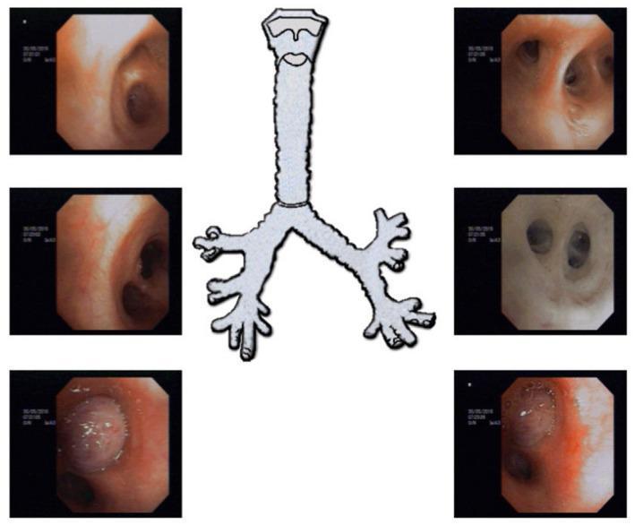
This composite diagnostic image features fiberoptic bronchoscopy stills and a central anatomical diagram of the tracheobronchial tree. The central illustration depicts the trachea and its major bifurcations into the primary and secondary bronchi. Surrounding this are endoscopic views showing various segments of the bronchial anatomy. A significant finding is demonstrated in the lower-left and lower-right endoscopic frames, which reveal a well-defined, rounded, fleshy lesion with a shiny, smooth surface located within the left upper bronchus. This mass causes complete mechanical obstruction of the bronchial lumen, preventing further advancement of the bronchoscope. The surrounding mucosa appears erythematous. This presentation is characteristic of an endobronchial carcinoid tumor, a slow-growing neuroendocrine neoplasm. The image serves as a clinical teaching tool for identifying intraluminal obstructions and understanding the diagnostic utility of bronchoscopy in pulmonary oncology and the assessment of central airway obstruction.
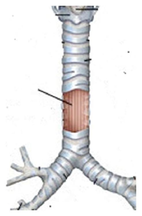
This medical anatomical diagram illustrates the tracheobronchial tree, specifically highlighting a localized pathology. The central feature is the trachea, composed of stacked horizontal cartilaginous rings, which bifurcates inferiorly at the carina into the left and right main bronchi. In the distal third of the trachea, there is a visible longitudinal defect or tear situated on the right posterolateral aspect. This area is characterized by an absence of the normal cartilaginous ring structure, revealing underlying vertical striations consistent with the membranous portion of the tracheal wall or an associated injury such as a tracheal tear or diverticulum. A black indicator line points directly to the site of the lesion. The diagram serves as an educational tool for identifying the location and appearance of tracheobronchial injuries, specifically at the right membranocartilaginous junction. The level of detail is appropriate for intermediate medical education, focusing on thoracic surgery and emergency medicine clinical scenarios.
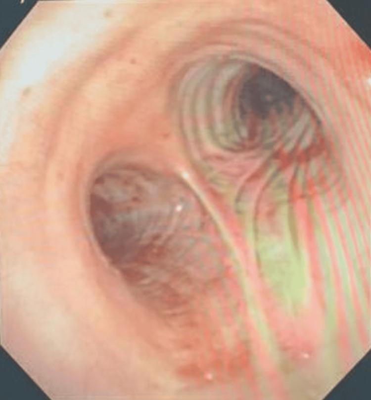
This clinical diagnostic image is a flexible bronchoscopy frame showing the internal anatomy of the tracheobronchial tree. The view centers on the primary carina, the cartilaginous ridge that marks the bifurcation of the trachea into the left and right mainstem bronchi. The bronchial mucosa appears healthy with a characteristic pinkish-red hue and visible vascularization. The cartilaginous rings are discernible as subtle horizontal indentations along the airway walls. Both the right and left main bronchi appear patent, and the airway lumens are clear of active bleeding, endobronchial lesions, or obstructive masses. In the clinical context of managing massive hemoptysis, this image demonstrates successful clearance of previous thrombi and confirms the absence of ongoing hemorrhage. This visual is significant for pulmonary and critical care medicine in assessing airway health, patency, and post-procedural outcomes following bronchoscopic intervention.
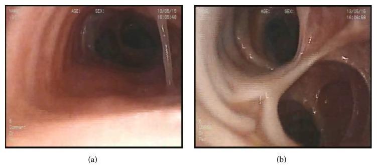
This diagnostic clinical image consists of two frames (a and b) from a flexible bronchoscopy procedure, illustrating the internal anatomy of the tracheobronchial tree in a patient with Mounier-Kuhn syndrome (tracheobronchomegaly). Frame (a) demonstrates a significantly dilated trachea with a smooth but widened mucosal surface; a thin medical instrument or suction catheter is visible on the right side of the lumen. Frame (b) provides a distal view at the level of the carina, showing the bifurcation into the right and left main bronchi. The bronchi appear pathologically enlarged with an irregular, corrugated mucosal texture and several small outpocketings or diverticula, which are characteristic of weakened connective tissue in the airway walls. The educational focus of these images is to demonstrate the macroscopic endoscopic findings of tracheobronchomegaly, specifically the excessive diameter of the conducting airways and the presence of mucosal protrusions/diverticula between the tracheal rings.
Tracheobronchial Tree with Anaesthesia Implications
1. Introduction
2. Anatomy of the Trachea
- The trachea begins at the lower border of the cricoid cartilage (C6 vertebral level) and extends to the carina (T4-T5, sternal angle of Louis).
- Average length: 10-13 cm in adults; shorter in females, children, and neonates.
- Composed of 16-20 C-shaped (horseshoe) cartilaginous rings that form the anterior and lateral walls; the posterior wall is the membranous (pars membranacea) wall, which lies in direct contact with the anterior oesophagus.
- The narrowest part of the adult trachea is at the cricoid cartilage (average diameter 17 mm in men, 13 mm in women).
- The lumen narrows slightly as it progresses toward the carina.
3. The Carina and Mainstem Bronchi
- Located at T4-T5, corresponding to the sternal angle (angle of Louis).
- The angle of tracheal bifurcation is normally 55-65° (wider in women and children).
- A widened carinal angle (>70°) is a radiological sign of left atrial enlargement - relevant when assessing pre-op chest X-rays.
- More vertical (25° from the tracheal axis), shorter (~2.5 cm in adults), and wider.
- Distance from carina to right upper lobe (RUL) bronchus take-off: 1.5-2.0 cm in men, ~1.5 cm in women. One in 250 individuals may have an anomalous RUL bronchus arising above the carina (tracheal bronchus).
- After the RUL take-off, the RMB continues as the bronchus intermedius, which then divides into the right middle lobe bronchus and right lower lobe bronchus.
- More horizontal (45° from tracheal axis), longer (average 5.0 cm in men, 4.5 cm in women), and narrower.
- Passes under the aortic arch and in front of the descending aorta.
- Divides into left upper lobe bronchus and left lower lobe bronchus.
4. Generations and Zones of the Tracheobronchial Tree
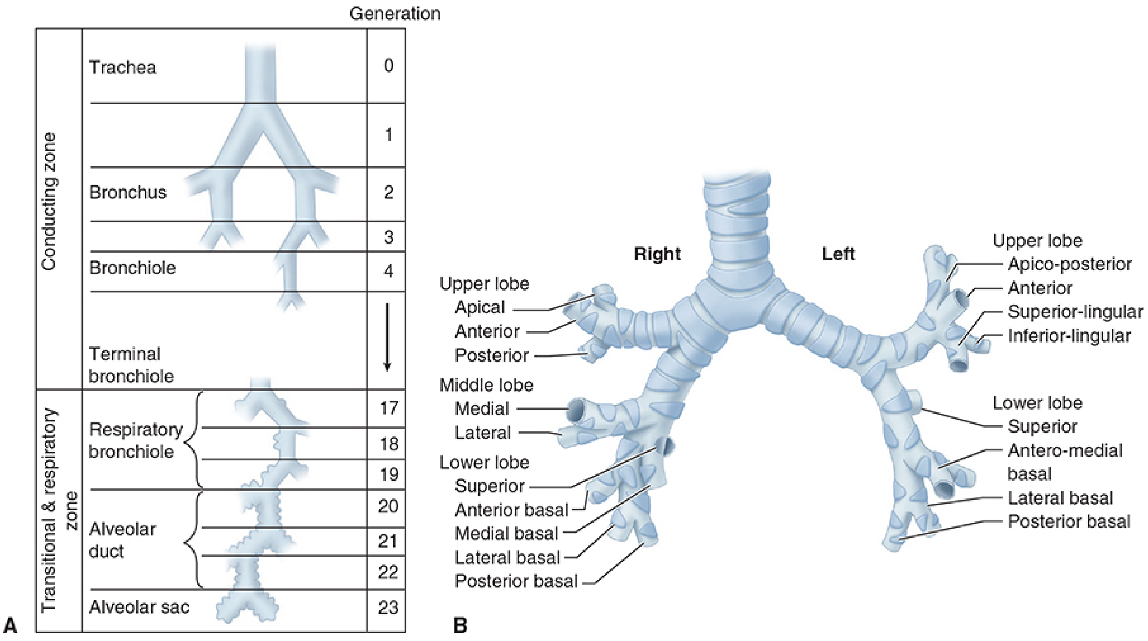
| Zone | Generations | Airways | Function |
|---|---|---|---|
| Conducting zone | 0-16 | Trachea, bronchi, bronchioles, terminal bronchioles | Conduction only - anatomical dead space |
| Transitional zone | 17-19 | Respiratory bronchioles | Conduction + some gas exchange |
| Respiratory zone | 20-23 | Alveolar ducts, alveolar sacs | Gas exchange |
- Total anatomical dead space in the conducting zone: ~150 mL in a 70 kg adult.
- Estimated alveoli: 300-500 million, providing a surface area of 50-100 m² for gas exchange.
5. Histology and Mucosal Lining - Anaesthetic Relevance
- Pseudostratified ciliated columnar epithelium (trachea and larger bronchi) - contains goblet cells secreting mucus.
- Ciliated cuboidal epithelium (bronchioles).
- Flat alveolar epithelium - Type I (gas exchange) and Type II (surfactant production) pneumocytes.
- Cilia beat in a coordinated fashion ~1000-1500 times/minute, propelling the mucus escalator toward the larynx.
- Anaesthetic agents (particularly volatile agents) depress ciliary function and inhibit mucociliary transport in a concentration- and duration-dependent manner.
- Dry, cold anaesthetic gases further impair mucociliary function.
- Clinical implication: Humidification of inspired gases and early extubation reduce the risk of post-operative sputum retention, atelectasis, and chest infections.
- Present in the trachea and bronchi down to bronchioles (generation ~11).
- Beyond this level (membranous bronchioles and smaller), patency depends entirely on radial traction from the elastic recoil of surrounding alveolar tissue.
- Airway diameter thus becomes dependent on total lung volume - explains why airway closure occurs at low lung volumes (closing capacity).
6. Key Anaesthesia Implications
6.1 Endotracheal Intubation and Tube Positioning
- Standard adult ETT is passed 2-3 cm beyond the vocal cords to sit the tip 5-7 cm above the carina (roughly at the midtracheal level).
- In adults, the mid-tracheal position corresponds to ~20-23 cm at the teeth in women and 21-25 cm in men (a rough rule: 3× the internal ETT diameter + distance to lips in cm).
- Endobronchial intubation - the commonest complication of intubation - almost always occurs into the right mainstem bronchus (RMB) because of its more vertical alignment. This results in left lung collapse and right lung hyperinflation.
- Signs: decreased breath sounds on left, rising peak airway pressure, decreased SpO2, asymmetric chest rise.
- Prevention: Confirm bilateral air entry after intubation; confirm depth with chest X-ray (ETT tip should be 3-5 cm above the carina).
6.2 Foreign Body Aspiration
- Because the RMB is more vertical, wider, and has higher airflow, inhaled foreign bodies preferentially lodge in the right bronchial tree (specifically right lower lobe or right mainstem bronchus) in adults and older children.
- In infants and young children, the right-left asymmetry is less pronounced.
- Clinical implication: During bronchoscopic retrieval of foreign bodies, the anaesthesiologist must maintain ventilation through the bronchoscope and be prepared for sudden complete airway obstruction.
6.3 One-Lung Ventilation (OLV) and Double-Lumen Tubes (DLT)
- Preferred for most elective thoracic procedures because the left bronchus is longer (4.5-5.0 cm), allowing a wider margin of safety for cuff placement without obstructing the LUL bronchus.
- After passage through the cords, the tube is rotated 90° counterclockwise (to the left) and advanced to the 27-29 cm mark at the teeth.
- The RMB is shorter (1.5-2.0 cm from carina to RUL take-off), so the right-sided DLT requires a slotted bronchial cuff to keep the RUL bronchus open.
- Indicated when left-sided DLT is contraindicated:
- Distorted left bronchial anatomy (external compression by tumor, descending aortic aneurysm)
- Left-sided pneumonectomy or sleeve resection
- Left lung transplantation
- Left-sided tracheobronchial disruption
- Auscultation alone is unreliable - fiberoptic bronchoscopy (FOB) is the gold standard.
- Through the tracheal lumen: the endobronchial portion should be seen entering the left bronchus with no bronchial cuff herniation above the carina.
- Through the bronchial lumen: the carina of the left upper and lower lobe bronchi should be visible.
- After repositioning (e.g., lateral decubitus), DLT position must be rechecked as movement can shift the tube.
6.4 Bronchial Blockers
- An alternative to DLT for OLV, especially in difficult airways.
- The blocker is placed under bronchoscopic guidance into the desired bronchus.
- Knowledge of bronchial anatomy - segmental bronchi, the bronchus intermedius, and the RUL take-off distance from the carina - is essential to position the blocker correctly.
6.5 Airway Resistance and Dead Space
- The conducting zone (generations 0-16) constitutes the anatomical dead space (~150 mL).
- The trachea and large bronchi contribute ~70% of total airway resistance at rest.
- Anaesthetic circuit dead space (mask, connectors) adds to anatomical dead space and is clinically significant in small children.
- Low-volume strategies (tidal volume 6-8 mL/kg IBW) during mechanical ventilation prevent dynamic hyperinflation and barotrauma.
6.6 Bronchospasm and Bronchodilation
- The tracheobronchial tree is richly innervated by the autonomic nervous system:
- Parasympathetic (vagal): bronchoconstriction, mucus secretion.
- Sympathetic (beta-2 adrenoceptors): bronchodilation.
- Airway instrumentation (laryngoscopy, intubation, suctioning) stimulates subglottic cough receptors (especially at the carina - the most sensitive reflex zone), triggering laryngospasm or bronchospasm.
- Preventing reflex bronchoconstriction: adequate depth of anaesthesia, IV lignocaine 1.5 mg/kg prior to intubation, use of propofol (blunts airway reflexes), volatile agents (bronchodilators - especially sevoflurane), and preoperative bronchodilators in asthmatics.
- The carina is particularly cough-sensitive; touching it during suctioning triggers powerful bronchospasm - suction catheters should not be passed beyond the distal trachea.
6.7 Tracheobronchial Secretion Clearance
- General anaesthesia impairs ciliary function, reduces functional residual capacity (FRC), and promotes secretion pooling.
- Endotracheal intubation bypasses upper airway humidification (nasal/pharyngeal mucosa adds ~44 mg H₂O/L of inspired air).
- Implications:
- Use of heat-moisture exchangers (HME) or active humidifiers on the breathing circuit.
- Regular suctioning of the airway but cautiously (avoiding deep tracheal suctioning to the carina).
- Chest physiotherapy and early mobilisation post-extubation.
6.8 Tracheobronchial Injury
- Iatrogenic tracheobronchial injury can occur with:
- Overinflation of ETT or DLT cuffs (ischaemic necrosis of tracheal mucosa).
- High-pressure mechanical ventilation (barotrauma) - particularly affects the membranous posterior tracheal wall.
- Traumatic intubation with a stylet.
- Management: Immediate bronchoscopy, conservative management for partial tears, surgical repair for complete disruption.
6.9 Anatomical Variants and Paediatric Considerations
- Tracheal bronchus: RUL bronchus arising from the trachea above the carina (1 in 250). A right-sided DLT in such a patient will obstruct the RUL. Preoperative CT or bronchoscopy is essential.
- Bridging bronchus and other rare variants can make OLV challenging.
- Neonates and infants:
- Trachea is short (~4 cm in neonates), so the margin between tracheal and endobronchial intubation is very narrow.
- To avoid endobronchial intubation in infants, the ETT tip should pass only 1-2 cm beyond the glottis.
- Neck flexion advances the ETT (bends it deeper); neck extension withdraws it - remember "flexion-in, extension-out."
6.10 Laser Surgery and Tracheobronchial Lesions
- Laser bronchoscopy (Nd:YAG via flexible fibers, CO2 via rigid bronchoscope) treats tracheobronchial lesions (tumors, stenoses, granulomas).
- Anaesthetic risks: airway fire (especially in high FiO2 environments), airway perforation, post-procedure oedema.
- Nd:YAG laser (1064 nm) penetrates tissue deeply via flexible fibers and can be used alongside flexible fiberoptic bronchoscopes.
- Anaesthetic management involves FiO2 <40% when possible, use of laser-safe ETTs (metallic/spiral-wrapped), and having the surgeon coordinate with the anaesthesiologist before each laser burst.
7. Summary Table: Key Anatomical Facts and Their Anaesthetic Implications
| Anatomical Feature | Key Measurement | Anaesthetic Implication |
|---|---|---|
| Tracheal length (adult) | 10-13 cm | ETT should sit 5-7 cm above carina; adjust for short necks |
| Cricoid diameter | 17 mm (M), 13 mm (F) | Narrowest adult airway; guides ETT size selection; site of cricoid pressure |
| Carina level | T4-T5 / sternal angle | Chest X-ray landmark for ETT tip positioning |
| RMB angle from trachea | ~25° (more vertical) | Endobronchial intubation almost always goes right; foreign bodies lodge right |
| LMB angle from trachea | ~45° (more oblique) | Left DLT placed safely; longer length gives margin of safety |
| Carina to RUL take-off | 1.5-2.0 cm | Right DLT placement requires slotted cuff to ventilate RUL |
| LMB length | 4.5-5.0 cm | Wider margin for left DLT placement |
| Anatomical dead space | ~150 mL | Ventilator settings; significant in paediatrics |
| 23 airway generations | 0-23 | Conducting zone (0-16) = dead space; gas exchange from gen 17 |
8. Conclusion
- Morgan and Mikhail's Clinical Anesthesiology, 7e, Chapter 23 (Tracheobronchial Tree section) - Morgan & Mikhail, pp. 911-914
- Miller's Anesthesia, 10e, Chapter 49 (Double-lumen tubes, right-sided DLT, positioning) - Miller, pp. 7151-7152
- Miller's Anesthesia, 10e, Chapter 66 (Laser ENT and tracheobronchial lesions)