
Cardiac patient posted fo non cardiac surgery mangement anesthesia residency for 5 marks
Cardiac Patient Posted for Non-Cardiac Surgery - Anesthetic Management
Overview
1. PREOPERATIVE EVALUATION
Step-wise ACC/AHA Algorithmic Approach
- Is surgery emergent? - If yes, proceed with risk stratification and postoperative surveillance; no time for testing.
- Active cardiac conditions? - Must be treated FIRST before elective surgery:
- Unstable angina / recent MI (within 60 days)
- Decompensated heart failure (NYHA Class IV)
- Significant arrhythmias (high-degree AV block, symptomatic VT, AF with rapid rate)
- Severe valvular disease (severe AS: AVA <1 cm², severe symptomatic MR)
- Surgical risk category:
- Low risk (<1% MACE): Superficial, endoscopy, cataract
- Intermediate risk (1-5%): Abdominal, orthopedic, head & neck
- High risk (>5%): Aortic/major vascular, peripheral vascular
- Functional capacity (METs):
- ≥4 METs (climb stairs, walk uphill) = adequate; proceed without further testing
- <4 METs = poor functional capacity; consider further testing
- RCRI (Revised Cardiac Risk Index) - 1 point each for:
- High-risk surgery
- History of IHD
- History of heart failure
- History of cerebrovascular disease
- Diabetes on insulin
- Serum creatinine >2 mg/dL
- Score 0-1 = low risk; ≥3 = high risk (~11% MACE)
- ACS NSQIP - alternative risk calculator for perioperative MACE.
Biomarkers
- BNP / NT-proBNP: Prognostic value for perioperative cardiac risk; ACC/AHA does not yet recommend routine use for decision-making
- Cardiac troponin: Measure only when signs/symptoms of myocardial ischemia; routine screening of asymptomatic patients not recommended
- (Fuster and Hurst's The Heart, 15th Ed.)
Investigations
- ECG (baseline for patients with known CVD or risk factors)
- Echo - for HF assessment, valvular disease
- Stress testing - only if it will change management AND functional capacity is unknown/poor
Anticoagulant/Antiplatelet Therapy
- Aspirin: Continue unless bleeding risk outweighs benefit; generally continue for cardiac stents
- Dual antiplatelet (DAPT):
- Bare metal stent (BMS): Wait at least 30 days before elective surgery
- Drug-eluting stent (DES): Wait at least 6-12 months before elective surgery
- Warfarin: Bridge therapy with LMWH in high-risk patients (mechanical valves, AF with high CHADS₂)
- DOACs: Hold 24-48 hours pre-op (renal function dependent)
2. INTRAOPERATIVE ANESTHETIC MANAGEMENT
Choice of Anesthetic Technique
General Anesthesia (GA)
- Intravenous induction agents:
- Propofol - vasodilation and myocardial depression; reduce dose in cardiac patients
- Etomidate - most hemodynamically stable induction agent; drug of choice in compromised cardiac function (does not depress myocardium significantly)
- Ketamine - sympathomimetic (raises HR, BP); useful in low-output states but avoid in tachycardia/ischemia
- Inhalational agents:
- Volatile agents (sevoflurane, desflurane, isoflurane) - all cause dose-dependent myocardial depression and vasodilation
- Ischemic preconditioning effect: volatile agents (especially sevoflurane, isoflurane) confer myocardial protection via preconditioning
- Desflurane may cause tachycardia with rapid increases - avoid in ischemic heart disease
- Opioids - fentanyl, remifentanil: minimal hemodynamic effects; useful adjuncts to reduce volatile agent requirements
- Neuromuscular blockade - prefer agents with minimal cardiovascular side effects; rocuronium (preferred); vecuronium (minimal cardiac effects); succinylcholine causes transient bradycardia
Neuraxial and Regional Anesthesia
- Spinal anesthesia: Profound sympathetic block → hypotension; use cautiously in cardiac patients; treat hypotension promptly (phenylephrine preferred over ephedrine for IHD)
- Epidural anesthesia: Slower onset of sympathetic block; allows titration; used for thoracic epidural analgesia (TEA) which has cardioprotective effect
- Thoracic epidural: reduces cardiac preload and afterload; useful in IHD patients; reduces perioperative MI incidence
- Neuraxial + anticoagulation: Strict ASRA guidelines must be followed (e.g., hold LMWH 12h before neuraxial block)
- Regional/nerve blocks: Excellent for peripheral procedures; avoids systemic effects of GA
| Condition | HR | Preload | Afterload | Contractility |
|---|---|---|---|---|
| IHD | Low-normal (60-80) | Normal | Normal | Normal |
| HF (systolic) | Normal | Optimize (euvolemia) | Reduce | Augment |
| AS | Normal-slow | Maintain (no preload drop) | Maintain | Normal |
| MR/AR | Slightly high | Normal-low | Reduce | Normal |
| HCM | Low | High | High | Avoid inotropes |
3. INTRAOPERATIVE MONITORING
Non-invasive (Standard)
- 5-lead ECG (leads II and V5 - maximum sensitivity for ischemia detection)
- Pulse oximetry
- Non-invasive BP (every 1-5 min)
- Capnography (ETCO₂)
- Temperature monitoring
Invasive Monitoring (for high-risk patients)
- Arterial line (A-line): Beat-to-beat BP; waveform analysis; blood gas sampling; for all high-risk cardiac patients
- Central venous catheter (CVC): CVP monitoring; vasoactive drug infusion
- Pulmonary artery catheter (PAC): For severe HF, pulmonary hypertension, complex procedures; allows measurement of PCWP, CO, SVR; controversial routine use
- Transesophageal Echocardiography (TEE): Real-time cardiac function; wall motion abnormality detection (ischemia); valvular function; volume status; highly sensitive for ischemia; increasingly standard for high-risk cardiac patients
- Transthoracic Echocardiography (TTE): Preoperative baseline; less useful intraoperatively
- Cardiac output / Goal-Directed Therapy (GDT): FloTrac, LiDCO, PICCO; optimize fluid management and cardiac output in real-time
- (Fuster and Hurst's The Heart, 15th Ed.; Miller's Anesthesia, 10th Ed.)
4. INTRAOPERATIVE GOALS
- Avoid tachycardia (increases O₂ demand, reduces diastolic filling)
- Avoid hypotension (MAP >65 mmHg; coronary perfusion pressure depends on diastolic BP)
- Avoid hypertension (increases afterload and O₂ demand)
- Maintain normovolemia (euvolemia; avoid fluid overload in HF)
- Maintain normothermia (hypothermia → coagulopathy, arrhythmias)
- Adequate analgesia (pain → catecholamine surge → tachycardia, hypertension)
- Optimize oxygen delivery; maintain Hb >8-10 g/dL in ischemic heart disease
5. POSTOPERATIVE MANAGEMENT
Cardiac Complications
- Myocardial Infarction (MI): Most common cause of perioperative cardiac death; peaks on POD 3; typically pain-free (silent); diagnosed by troponin rise
- Heart Failure: Fluid shifts, fluid overload, myocardial dysfunction
- Arrhythmias: Common; manage with rate control, cardioversion as needed
- Deep Vein Thrombosis/PE: Anticoagulation strategy important
Enhanced Recovery After Surgery (ERAS)
- Multimodal analgesia to reduce opioid requirements
- Early mobilization
- Optimal fluid management
- Nausea/vomiting prophylaxis
- (Fuster and Hurst's The Heart, 15th Ed.)
Medications to Continue Perioperatively
- Beta-blockers: Continue; abrupt withdrawal increases risk of perioperative MI; if starting new beta-blocker, begin ≥1 week before surgery (not on day of surgery)
- Statins: Continue perioperatively (plaque stabilization effect)
- ACE inhibitors / ARBs: Consider holding on morning of surgery (risk of refractory hypotension with induction); restart as soon as hemodynamically stable
- Nitrates: Continue
- Antiarrhythmics: Continue
Summary Table: Key Points for 5-Mark Answer
| Domain | Key Points |
|---|---|
| Pre-op assessment | ACC/AHA stepwise algorithm, RCRI, NSQIP, functional capacity (METs), active cardiac conditions |
| Risk stratification | Low/intermediate/high surgical risk; stent timing (BMS 30d, DES 6-12 months) |
| Anesthesia choice | No technique proven superior; etomidate for hemodynamic stability; TEA cardioprotective |
| Hemodynamic goals | Disease-specific (IHD: avoid tachycardia; AS: avoid preload drop; MR: reduce afterload) |
| Monitoring | A-line, TEE for high-risk; 5-lead ECG, GDT |
| Drugs | Continue beta-blockers/statins; hold ACE-I on morning of surgery; DAPT timing critical |
| Post-op | Troponin monitoring, pain control, ERAS protocols |
- Fuster and Hurst's The Heart, 15th Edition - Chapter 73: Perioperative Anesthetic Management of Patients with CVD
- Miller's Anesthesia, 10th Edition - Chapter 52: Anesthesia and the Cardiac Patient for Noncardiac Surgery
- ACC/AHA 2014 Perioperative Guidelines for Noncardiac Surgery
Cardiac risk index
Cardiac Risk Index
1. GOLDMAN'S CARDIAC RISK INDEX (Original, 1977)
Goldman's 9 Factors and Points:
| Risk Factor | Points |
|---|---|
| S3 gallop or JVD (signs of heart failure) | 11 |
| MI within the preceding 6 months | 10 |
| >5 PVCs/min documented pre-op | 7 |
| Rhythm other than sinus, or PACs on pre-op ECG | 7 |
| Age >70 years | 5 |
| Emergency surgery | 4 |
| Intraperitoneal, intrathoracic, or aortic surgery | 3 |
| Significant aortic stenosis | 3 |
| Poor general medical status (PO₂ <60, PCO₂ >50, K⁺ <3, HCO₃ <20, BUN >50, Cr >3, SGOT raised, signs of chronic liver disease, bedridden) | 3 |
| Total | 53 |
Goldman Classes and Cardiac Complication Risk:
| Class | Points | Life-threatening complications | Cardiac deaths |
|---|---|---|---|
| I | 0-5 | 0.7% | 0.2% |
| II | 6-12 | 5% | 2% |
| III | 13-25 | 11% | 2% |
| IV | >26 | 22% | 56% |
- (Miller's Anesthesia, 10th Ed.)
2. DETSKY MODIFIED CARDIAC RISK INDEX
- Modification of Goldman's CRI
- Adds unstable angina and pulmonary edema as separate weighted items
- Uses a nomogram to calculate pretest probability of complications based on surgery type
- Was advocated as starting point for risk stratification in American College of Physicians guidelines
- (Miller's Anesthesia, 10th Ed.)
3. LEE'S REVISED CARDIAC RISK INDEX (RCRI) - Current Standard
RCRI - 6 Predictors:
| # | Risk Factor | Points |
|---|---|---|
| 1 | High-risk surgery - intraperitoneal, intrathoracic, or suprainguinal vascular procedure | 1 |
| 2 | Ischemic heart disease - history of MI, positive stress test, use of nitrates, current chest pain from IHD, ECG with Q waves | 1 |
| 3 | History of congestive heart failure - pulmonary edema, bilateral rales, S3 gallop, paroxysmal nocturnal dyspnea, CXR showing redistribution | 1 |
| 4 | History of cerebrovascular disease - stroke or TIA | 1 |
| 5 | Diabetes mellitus requiring insulin | 1 |
| 6 | Creatinine >2.0 mg/dL (>176 μmol/L) - preoperative renal insufficiency | 1 |
RCRI Score and Risk of Major Cardiac Events (MACE):
| RCRI Score | Risk of MACE |
|---|---|
| 0 | 1.6% |
| 1 | 4.0% |
| 2 | 7.9% |
| ≥3 | 12.9% |
- (Miller's Anesthesia, 10th Ed.; Barash Clinical Anesthesia, 9th Ed.)
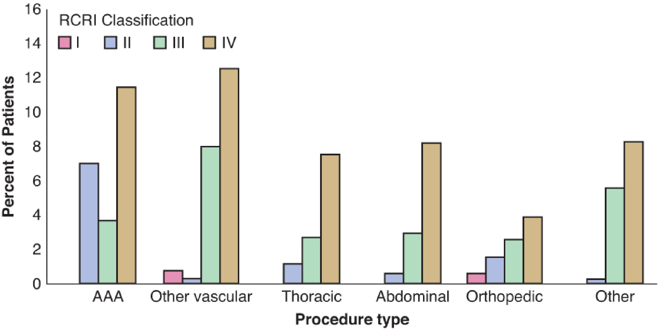
RCRI Risk by Procedure Type:
4. ACS NSQIP / MICA (Newer Tools)
| Tool | Description |
|---|---|
| ACS NSQIP Surgical Risk Calculator | Web-based; uses current CPT codes + 21 patient variables; predicts multiple outcomes including MACE; better discriminative ability than RCRI but more complex |
| MICA (Myocardial Infarction and Cardiac Arrest) | Derived from NSQIP database; specifically predicts MI and cardiac arrest |
5. Comparison: Goldman vs RCRI
| Feature | Goldman CRI (1977) | RCRI (1999) |
|---|---|---|
| Study population | 1001 patients | 4315 patients |
| Risk factors | 9, unequal weighting | 6, equal weighting (1 point each) |
| Max score | 53 | 6 |
| Classes | I-IV | Score 0-≥3 |
| Current use | Historical; largely replaced | Current clinical standard |
| Validated in vascular surgery | Controversial | Better validated |
| ACC/AHA recommendation | Not current | Recommended |
6. Clinical Application
- Emergency surgery? → Proceed, manage perioperatively
- Active cardiac conditions? → Stabilize first
- Low-risk surgery? → Proceed
- Good functional capacity (≥4 METs)? → Proceed
- RCRI ≥2 + poor functional capacity → Consider further testing (stress imaging) only if result will change management
| ASA Grade | Description | 30-day Mortality |
|---|---|---|
| I | Healthy | 0.1% |
| II | Mild systemic disease, no functional limitation | 0.7% |
| III | Severe systemic disease, definite functional limitation | 3.5% |
| IV | Severe systemic disease, constant threat to life | 18.3% |
| V | Moribund, unlikely to survive 24h | 93.3% |
| E | Emergency | - |
- Miller's Anesthesia, 10th Ed. - Chapter 27 & 28
- Barash Clinical Anesthesia, 9th Ed. - Chapter 23
- Bailey and Love's Short Practice of Surgery, 28th Ed. - Chapter 21
- Lee TH et al., Circulation 1999;100:1043-1049
- Goldman L et al., NEJM 1977;297:845-850
Periop mi pathophysiology flowchart
Detailed medical flowchart titled "Perioperative Myocardial Infarction (POMI) - Pathophysiology" showing two parallel pathways: LEFT PATHWAY - TYPE 1 (Plaque Rupture/Thrombosis): Surgical Stress → Sympathetic Activation → Catecholamine Surge → Hemodynamic Instability (tachycardia, hypertension) → Increased Shear Stress on Coronary Plaque → Vulnerable Atherosclerotic Plaque Rupture → Exposure of Subendothelial Collagen → Platelet Activation and Aggregation → Thrombus Formation → Complete Coronary Occlusion → STEMI RIGHT PATHWAY - TYPE 2 (Supply-Demand Mismatch): Surgical Stress → Multiple triggers: Tachycardia (reduced diastolic filling time), Hypotension (reduced coronary perfusion pressure), Anemia (reduced O2 carrying capacity), Hypothermia, Hypoxia → Decreased Myocardial O2 Supply → Combined with: Increased O2 Demand (pain, hypertension, tachycardia) → Supply-Demand Imbalance → Subendocardial Ischemia → Partial Occlusion / Severe Stenosis → NSTEMI CENTER/BOTTOM - COMMON FINAL PATHWAY: Both pathways converge to: Myocardial Cell Death → Troponin Release (peaks POD 1-3) → Myocardial Stunning / Hibernation → LV Dysfunction → Heart Failure / Arrhythmias / Death CONTRIBUTING FACTORS BOX on the side: - Pro-inflammatory state (IL-6, CRP rise) - Hypercoagulable state (↑fibrinogen, ↓fibrinolysis) - Endothelial dysfunction - Coronary artery spasm - Pre-existing CAD / vulnerable plaques Use blue for Type 1 pathway, orange for Type 2, red for the converging final pathway, clean medical diagram style with clear arrows and labels
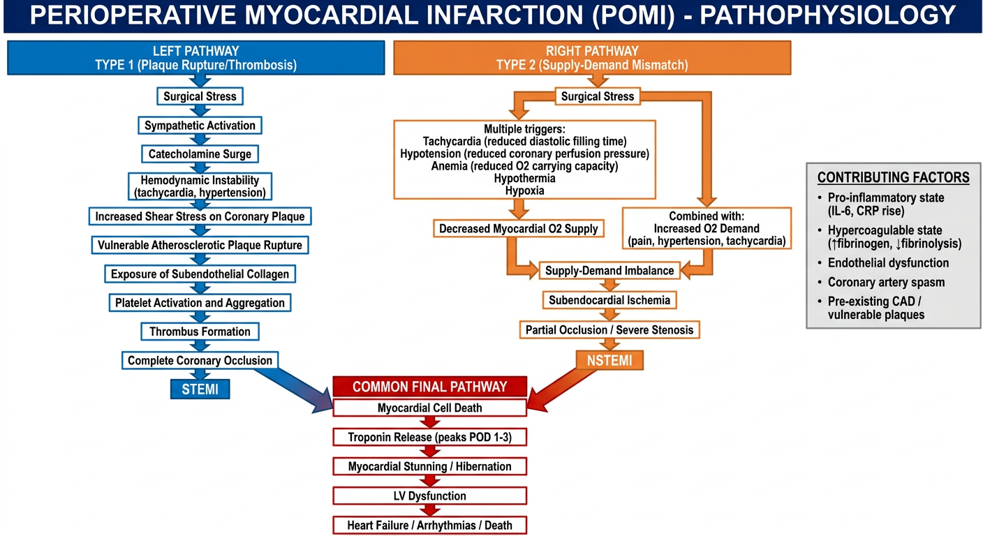
Perioperative MI (POMI) - Pathophysiology
Classification of AMI Types (Universal Definition)
| Type | Mechanism | Common Perioperative Scenario |
|---|---|---|
| Type 1 | Spontaneous - atherosclerotic plaque rupture with thrombosis | Major vascular, cardiac surgery |
| Type 2 | Demand ischemia - O₂ supply/demand imbalance | Any surgery; most common periop type |
| Type 3 | Sudden cardiac death with ischemia evidence | Unwitnessed arrest |
| Type 4 | PCI-related | Post-PCI period |
| Type 5 | CABG-related | Post-bypass |
KEY PATHOPHYSIOLOGICAL MECHANISMS IN DETAIL
1. SURGICAL STRESS RESPONSE (The Trigger)
Surgery / Tissue Injury
        ↓
Hypothalamic-Pituitary-Adrenal (HPA) Axis Activation
        +
Sympatho-adrenal Activation
        ↓
↑ Catecholamines (adrenaline, noradrenaline)
↑ Cortisol, ↑ Glucagon
        ↓
Tachycardia + Hypertension + Vasoconstriction
2. PRO-INFLAMMATORY / PRO-COAGULANT STATE (Unique to Surgery)
- ↑ IL-6, IL-1, TNF-α → C-reactive protein surge
- ↑ Fibrinogen, ↑ Factor VIII, ↑ vWF
- ↓ Fibrinolysis (↑ PAI-1)
- Platelet activation enhanced by surgical trauma and catecholamines
- Endothelial dysfunction from ischemia-reperfusion and inflammation
3. TYPE 1 PATHWAY - Plaque Rupture
Pre-existing Vulnerable Atherosclerotic Plaque
(lipid-rich core, thin fibrous cap)
        ↓
Catecholamine surge → ↑ Heart rate + ↑ Blood pressure
        ↓
↑ Shear stress on coronary vessel wall
        +
Inflammatory mediators (MMP activation → cap thinning)
        ↓
PLAQUE RUPTURE / EROSION
        ↓
Subendothelial collagen + lipid core exposed
        ↓
Platelet adhesion → Activation → Aggregation (GP IIb/IIIa)
        ↓
Fibrin deposition (↑ coagulation factors perioperatively)
        ↓
THROMBUS FORMATION
        ↓
Complete coronary occlusion
        ↓
STEMI (transmural infarct)
4. TYPE 2 PATHWAY - Supply-Demand Mismatch
DECREASED O₂ SUPPLY:
- Tachycardia → ↓ diastolic filling time → ↓ coronary flow
- Hypotension → ↓ coronary perfusion pressure (CPP = DBP - LVEDP)
- Anemia → ↓ O₂ carrying capacity
- Hypoxia / Respiratory depression
- Coronary artery spasm (cold, pain, catecholamines)
- Hypovolemia

        +

INCREASED O₂ DEMAND:
- Tachycardia (most important - doubles O₂ demand)
- Hypertension → ↑ wall stress / afterload
- Pain → catecholamine surge
- Shivering / Hypothermia → ↑ metabolic demand
- Anaemia compensation (↑ CO)

        ↓

O₂ Supply < O₂ Demand

        ↓

Subendocardial ischemia first
(subendocardium most vulnerable - highest wall stress, lowest perfusion)

        ↓

ST depression, T-wave changes, troponin rise

        ↓

NSTEMI (non-transmural, demand-type)
5. COMMON FINAL PATHWAY
Myocardial cell death (irreversible ischemia >20 min)
        ↓
Troponin I/T release (peaks POD 1-3)
        ↓
┌─────────────────────────────────────────┐
│                                         │
Myocardial Stunning          Myocardial Hibernation
(reversible dysfunction       (chronic ischemia,
 after brief ischemia)        viable but non-contractile)
│                                         │
└─────────────────────────────────────────┘
        ↓
LV systolic dysfunction → ↓ EF
        ↓
┌───────────────────────────────────────────────────────┐
│               COMPLICATIONS                           │
│  Ventricular arrhythmias (VF - most common early death│
│  Acute heart failure / cardiogenic shock              │
│  Mechanical complications (papillary muscle rupture,  │
│  free wall rupture, VSD)                              │
│  Reinfarction                                         │
└───────────────────────────────────────────────────────┘
TIMING OF PERIOPERATIVE MI
| Time | Mechanism | Notes |
|---|---|---|
| Intraoperative | Acute ischemia from hemodynamic instability | Less common |
| POD 1-3 (most common) | Hypercoagulable state peaks; pain; catecholamine surge post-op | Most periop MIs occur here |
| POD 3-5 | Inflammatory peak, fluid shifts | Delayed presentation |
CLINICAL FEATURES (Often Silent Perioperatively)
- 60-90% are SILENT in the perioperative period - pain masked by residual analgesia, sedation, general anesthesia
- Diagnosis relies on: troponin rise + ECG changes ± symptoms
- May present as: unexplained hypotension, new arrhythmia, ST changes, heart failure, cardiac arrest
- MINS (Myocardial Injury after Non-cardiac Surgery) = troponin elevation without symptoms - still associated with 30-day mortality
RISK FACTORS AMPLIFYING PERIOP MI RISK
| Factor | Effect |
|---|---|
| Pre-existing CAD | Vulnerable plaques already present |
| Diabetes | Silent ischemia, autonomic neuropathy |
| Hypertension + LVH | Increased O₂ demand, diastolic dysfunction |
| Anemia | ↓ O₂ delivery |
| Renal failure | Uremic platelet dysfunction + vascular disease |
| High-risk surgery | Major vascular, intraperitoneal, intrathoracic |
| Emergency surgery | No time for optimization |
- Morgan & Mikhail's Clinical Anesthesiology, 7th Ed. - Chapter on Acute MI Pathophysiology
- Fuster and Hurst's The Heart, 15th Ed. - Chapter 73
- Miller's Anesthesia, 10th Ed. - Chapter 27/28