
Cataract ophthalmology 3rd year mbbs
cataract lens opacity types senile nuclear cortical
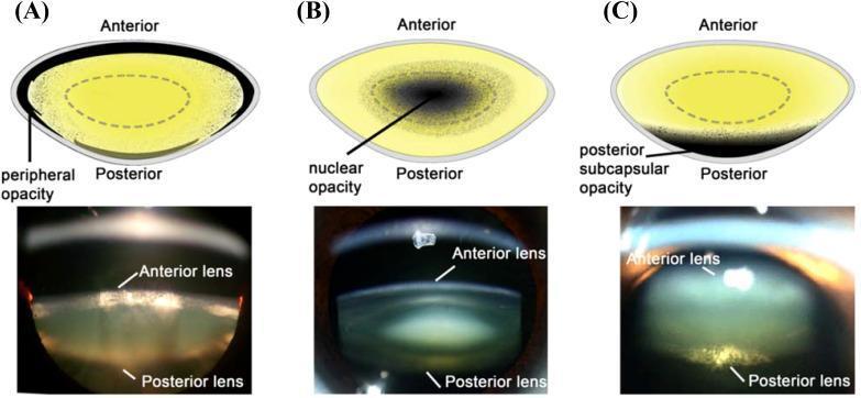
Educational comparison of cataract types featuring schematic diagrams (top row) and Scheimpflug slit-lamp photographic images (bottom row) classified into three categories: (A) Cortical cataract, (B) Nuclear cataract, and (C) Posterior subcapsular cataract. In the cortical cataract section, the diagram highlights 'peripheral opacity' along the outer lens cortex, mirrored in the slit-lamp image by granular light-scattering in the peripheral layers. The nuclear cataract section demonstrates 'nuclear opacity' concentrated in the central core of the crystalline lens, appearing as a dense, dark focal point in both the illustration and the photographic cross-section. The posterior subcapsular cataract section depicts 'posterior subcapsular opacity' localized just beneath the posterior lens capsule, shown as a distinct white plaque-like reflection at the back of the lens in the clinical image. Each image is annotated with 'Anterior' and 'Posterior' landmarks to orient the observer to the lens anatomy and the depth of the opacification. This visual material is used to teach diagnostic ophthalmology and the pathophysiology of age-related and metabolic lens changes.
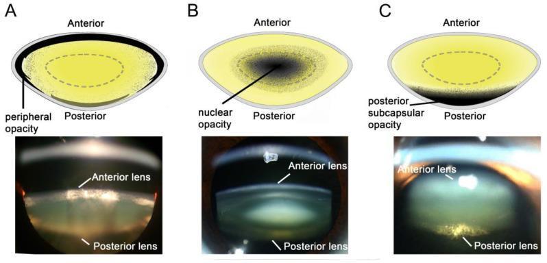
Educational comparison of the three primary types of human cataracts, presented via schematic diagrams (top) and corresponding Scheimpflug slit-lamp photographic images (bottom). The visual is organized into three panels: (A) Cortical cataract, showing characteristic wedge-shaped or peripheral opacities that originate at the lens equator and extend toward the center, often associated with diabetic osmotic stress. (B) Nuclear cataract, demonstrating central densification and opacification within the lens nucleus, typically linked to age-related oxidative protein damage. (C) Posterior subcapsular cataract (PSC), illustrating a localized plaque-like opacity at the posterior pole of the lens just beneath the capsule, frequently associated with corticosteroid use or diabetes. Each schematic identifies the anterior and posterior orientations and highlights the specific zone of lens clouding. The clinical photographs provide a cross-sectional view of the lens, with labels indicating the anterior and posterior lens surfaces, allowing for visualization of the depth and distribution of light scattering within the crystalline lens structure.
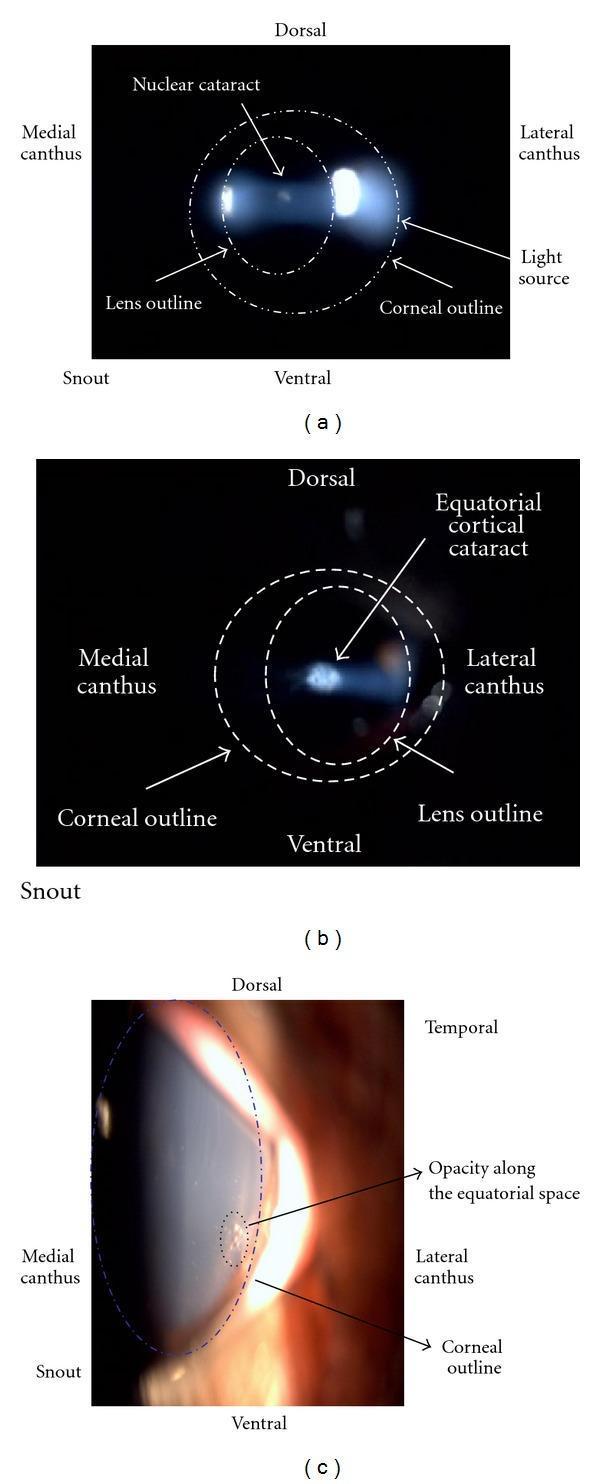
A composite of three ophthalmic images (a, b, and c) illustrating different types and modalities for visualizing lens opacities (cataracts). Image (a) is a slit lamp photograph of an eye showing a nuclear cataract, characterized by a central, concentrated opacity located within the lens outline along the light axis. Image (b) shows an equatorial cortical cataract via slit lamp examination, presenting as a less defined opacity toward the periphery of the lens. Image (c) provides an external ocular photograph of the same eye in (b), highlighting a focal opacity along the equatorial space near the lateral canthus. The images include anatomical annotations (dorsal, ventral, medial canthus, lateral canthus, and snout) to provide orientation within a veterinary or animal research model context. Dotted outlines distinguish the corneal and lens boundaries. This comparison demonstrates the morphological differences between nuclear and cortical lens changes and the varying visualization achieved through different clinical imaging techniques.
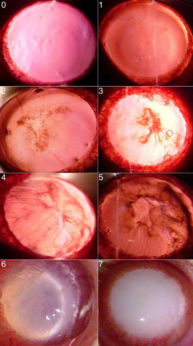
This composite of eight slit-lamp biomicroscopy clinical photographs illustrates the progression and grading of nuclear cataracts using the Oxford clinical grading system, ranging from Grade 0 to Grade 7. The images demonstrate the transformation of the crystalline lens from total transparency to complete opacification. Grade 0 shows a clear lens with a red reflex. Early stages (Grades 1–2) exhibit subtle nuclear density and visible wide sutures. Intermediate stages (Grades 3–5) show increasing nuclear densification and the formation of distinct cortical or nuclear clefts. Advanced stages (Grades 6–7) feature dense radial opacities and progress to a mature cataract characterized by total whole-lens opacification, which completely obscures internal structures and the posterior red reflex. This visual series is an educational tool for ophthalmology and optometry to standardize the assessment of lenticular opacity and cataract severity in clinical practice.
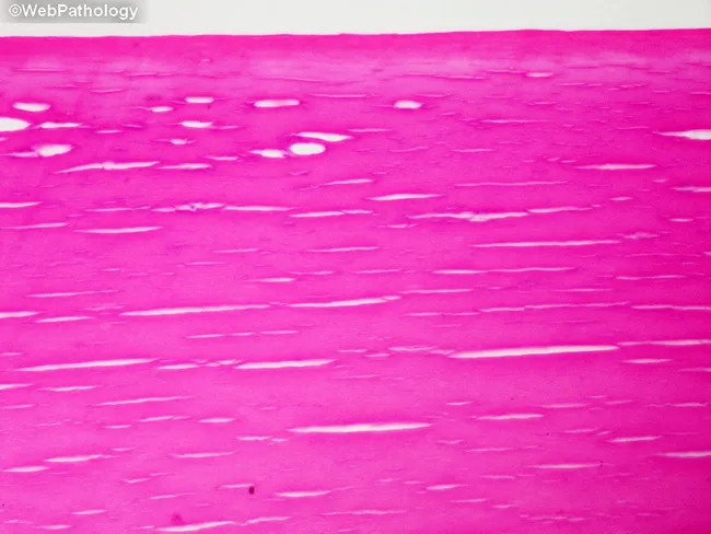
Light microscopy of an H&E-stained crystalline lens section depicting nuclear sclerosis in cataract. The image focuses on the lens nucleus, with a longitudinal sectional plane through the central optical portion. The nucleus shows markedly increased eosinophilia, reflecting dense, protein-rich crystallin accumulation, and overall homogenization of the normally granular lamellar architecture. Normal, alternating lens fiber lamellae are partially obscured by progressive crowding and coalescence of advanced nuclear cataract material. The tissue appears more compact and optically dense, with reduced delimitation between fibers and diminished cytoplasmic clear spaces. The staining accentuates the eosinophilic, pale pink matrix consistent with protein aggregation and cross-linking typical of senile nuclear cataract. Clinically, this pattern corresponds to opacity of the central visual axis, decreased light transmission, and a tendency toward a myopic shift. The histologic features align with chronic degenerative changes in aging lens, including protein denaturation, crystallin aggregation, and lens hardening (sclerosis). Diagnostic significance: confirms nuclear sclerosis type cataract pathogenesis; aids educational correlation between microscopic appearance and opacification. Potential clinical uses: histopathology teaching slide, correlation with slit-lamp findings, research into age-related lens protein chemistry, and evaluation of cataract progression in experimental models. Also note the absence of inflammatory infiltrates.
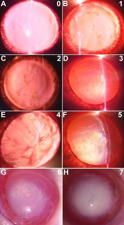
A series of eight clinical slit-lamp photographs (labeled A-H and graded 0-7) demonstrating the progression of cataract formation in a diabetic model, categorized according to the Oxford grading system. The images illustrate the transition from a clear lens (Grade 0) to total lenticular opacity (Grade 7). Key pathological landmarks include widening of the lens sutures (Grade 1), the emergence of radiating opacities from the sutures (Grade 2), and the development of a dense nucleus (Grade 3). More advanced stages demonstrate the formation of cortical clefts (Grades 4-5) and dense radial opacification (Grade 6). The final stage (Grade 7) depicts a mature, opaque nuclear cataract involving the entire lens volume. This comparison chart is used in ophthalmology to study the pathophysiology of diabetic cataracts and to evaluate the efficacy of therapeutic interventions on lens clarity. The visual sequence emphasizes diagnostic features such as nuclear density, suture morphology, and the structural integrity of the lens fibers.
phacoemulsification cataract surgery IOL intraocular lens implant
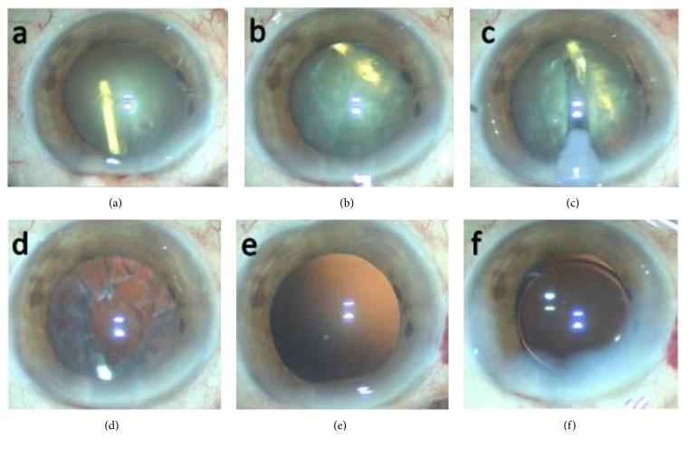
A series of six clinical intraoperative photographs (a–f) documenting a phacoemulsification cataract surgery complicated by an accidental intralenticular Ozurdex (dexamethasone) implant. (a) Displays a completed anterior continuous curvilinear capsulorrhexis (CCC). (b) Shows the intralenticular dexamethasone implant, visible as a rod-shaped semi-opaque structure, rotating within the opacified crystalline lens nucleus. (c) Illustrates the phacoemulsification probe creating a central groove in the lens nucleus to facilitate fragmentation. (d) Shows the eye after removal of the nucleus and implant, with remaining cortical remnants visible against the red reflex. (e) Demonstrates an intact and clear posterior capsule following complete irrigation and aspiration of cortical material. (f) Final stage showing the successful implantation of a three-piece hydrophobic acrylic intraocular lens (IOL) within the capsular bag. This series highlights the surgical management of lens trauma and iatrogenic implant displacement during cataract extraction.
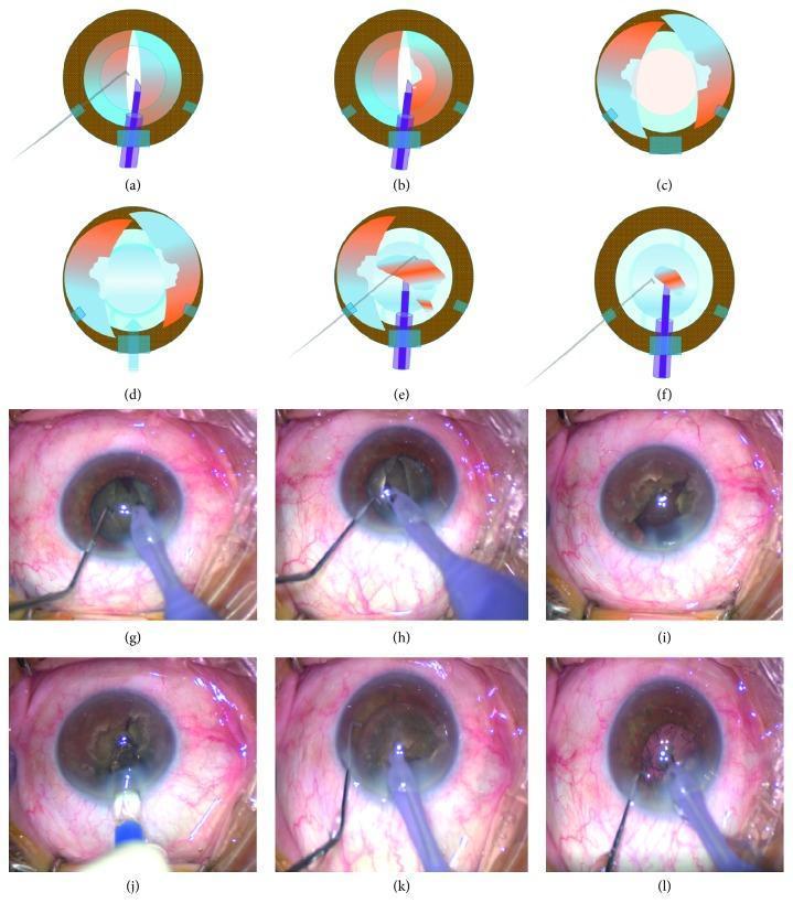
A multi-panel series of anatomical diagrams (a-f) and corresponding intraoperative clinical photographs (g-l) illustrating the 'crack, reduce, and implant' phacoemulsification technique for cataract surgery. The sequence demonstrates the mechanical management of a dense lens nucleus within the anterior segment of the eye. Panels a and g show the initial vertical groove formation and cracking of the nucleus into two halves. Panels b and h depict the 'shaving' or hollowing out of the core of each nuclear half to reduce mass while maintaining the capsular bag integrity. Panels c and i illustrate the prolapse of these nuclear halves toward the iris plane assisted by ophthalmic viscosurgical devices (OVD). Panels d and j show the early implantation of a foldable intraocular lens (IOL) into the capsular bag while nuclear fragments are still present, serving to protect the posterior capsule. The final panels (e, f, k, l) demonstrate the subsequent chopping of the remaining nuclear material into small fragments for emulsification and aspiration far from the corneal endothelium. The series highlights surgical strategies for protecting intraocular structures during the management of high-grade cataracts.
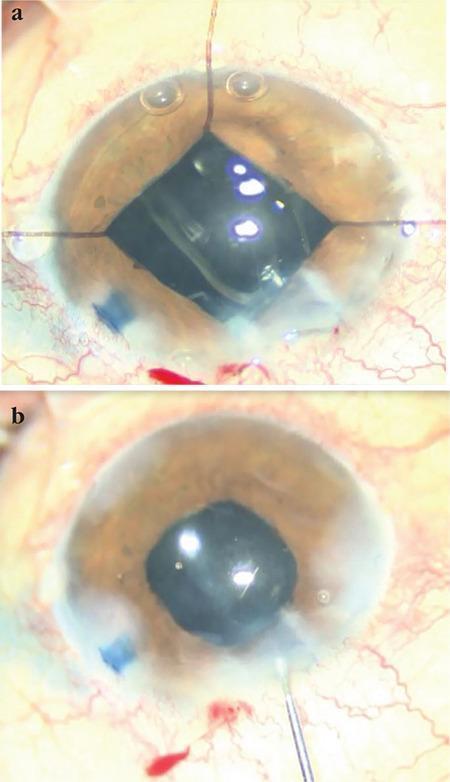
Two-panel clinical photograph capturing stages of a phacoemulsification cataract surgery with intraocular lens (IOL) implantation. Panel (a) illustrates an intraoperative view where four iris retractors are deployed to maintain a mechanical pupillary dilation, creating a characteristic square-shaped pupillary aperture. This technique is typically used in cases of small pupil syndrome or posterior synechiae. Within the pupillary opening, the surgical field is visible with multiple light reflections and evidence of Trypan blue staining of the capsule. Panel (b) shows the postoperative or end-of-procedure state with the iris retractors removed and the pupil returning to a more natural, circular morphology. A clear, foldable IOL is seen centrally positioned within the capsular bag in the posterior chamber. A surgical instrument remains visible at the limbal incision site in the lower right quadrant. The surrounding sclera shows expected postoperative conjunctival injection. The images demonstrate ophthalmic surgical management of complicated cataracts using iris expansion devices.
Cataract - Ophthalmology (3rd Year MBBS)
1. Definition
2. Anatomy of the Lens (Quick Review)
- Zonules (suspensory ligaments of Zinn) hold the lens in position
- Capsule - acellular membrane enclosing all lens material
- Anterior epithelium - cuboid cells beneath the anterior capsule; the germinative zone at the equator continuously divides and differentiates into lens fibers
- Lens substance - central nucleus surrounded by cortex
- Lens is avascular; depends entirely on aqueous humour for nutrition
3. Types of Cataract
A. By Age of Onset
| Type | Age |
|---|---|
| Congenital | Present at birth / develops in infancy |
| Juvenile | < 10 years |
| Presenile | 40-50 years |
| Senile / Age-related | > 50 years (most common) |
B. Age-Related (Senile) Cataract - Most Important for Exams
i. Subcapsular Cataract
- Anterior subcapsular: directly under the lens capsule; associated with fibrous metaplasia of the lens epithelium
- Posterior subcapsular (PSC): lies just in front of the posterior capsule; granular/plaque-like on oblique slit lamp; black and vacuolated on retroillumination
- Vacuoles are swollen lens epithelial cells (Bladder cells / Wedl cells)
- Located at the nodal point of the eye - profound effect on vision
- Causes glare (e.g., from oncoming headlights); symptoms worsen with miosis (near work, bright sunlight)
- Associated with: steroids (topical/systemic), diabetes, trauma, irradiation
ii. Nuclear Sclerotic Cataract
- Exaggeration of normal ageing changes in the nucleus
- Characterized by yellowish hue due to deposition of urochrome pigment
- Best assessed with an oblique slit lamp beam
- Causes myopic shift ("second sight of the aged" - elderly patient can read without glasses again) due to increased refractive index of the nucleus
- Advanced: nucleus appears brown (brunescent) or, rarely, black
- Retroillumination shows a good red reflex but subtle nucleus-cortex distinction
iii. Cortical Cataract
- Opacities start as clefts and vacuoles between lens fibers due to cortical hydration
- Evolve into cuneiform (wedge-shaped) or radial spoke-like opacities
- Often first appear in the inferonasal quadrant
- Causes glare (like PSC)
- Associated strongly with diabetes (osmotic mechanism via sorbitol)
iv. Christmas Tree Cataract (Rare)
- Polychromatic needle-like formations in the deep cortex and nucleus
- Associated with myotonic dystrophy
Types of Cataract - Slit Lamp Appearance
4. Stages of Maturity (Senile Cataract)
| Stage | Features |
|---|---|
| Immature | Lens partially opaque; iris shadow present on oblique illumination |
| Mature | Lens completely opaque; no iris shadow; no red reflex |
| Hypermature | Shrunken, wrinkled anterior capsule due to leakage of water out of the lens |
| Morgagnian | Hypermature cataract where the cortex is completely liquefied and the nucleus sinks inferiorly |
Iris shadow test: In immature cataract, the iris casts a shadow on the lens on oblique illumination. In mature cataract, no shadow (lens fully opaque). The shadow disappears as the cataract matures.
5. Cataract in Systemic Disease
Diabetes Mellitus
- Hyperglycaemia → high glucose in aqueous → diffuses into lens → metabolized to sorbitol → accumulates → osmotic overhydration
- Classic diabetic cataract (rare, young patients): snowflake cortical opacities - can mature within days or resolve spontaneously
- More commonly: accelerated age-related nuclear sclerosis
- Fluctuating refraction with blood glucose: hyperglycaemia → myopia; hypoglycaemia → hypermetropia
Myotonic Dystrophy
- ~90% develop fine iridescent cortical opacities in the 3rd decade (may resemble Christmas tree cataract)
- Evolve into visually disabling wedge-shaped / star-shaped subcapsular opacities by the 5th decade
Neurofibromatosis Type 2 (NF2)
- Cataract in >60% of patients; posterior subcapsular or capsular opacities; develop in early adulthood
Atopic Dermatitis
- Anterior or posterior subcapsular "shield-like" cataract; may occur in young adults
6. Secondary (Complicated) Cataract
| Cause | Feature |
|---|---|
| Chronic anterior uveitis (most common cause) | PSC earliest; progresses to anterior + posterior opacities; worsened by posterior synechiae |
| Acute angle-closure glaucoma | Glaukomflecken - small grey-white anterior subcapsular opacities; pathognomonic of previous acute congestive angle closure; focal infarcts of lens epithelium |
| High myopia | PSC + early nuclear sclerosis |
| Retinitis pigmentosa | PSC |
| Intraocular tumours | Local toxic effects |
7. Traumatic Cataract
- Blunt trauma: "rosette-shaped" or stellate posterior subcapsular opacity
- Penetrating trauma: rapid opacification after capsular rupture
- Electrical / lightning: posterior subcapsular or anterior subcapsular; bilateral
- Radiation (ionizing): posterior subcapsular; latent period of months to years
- Infrared (glass-blower's cataract): exfoliation of anterior capsule; "true exfoliation"
8. Congenital Cataract
- 1/3 associated with systemic disease
- 1/3 inherited (most commonly autosomal dominant)
- 1/3 unknown cause
Associated Metabolic Disorders
| Disorder | Cataract Type |
|---|---|
| Galactosaemia (AR; GALT deficiency) | "Oil droplet" opacity (first weeks of life); exclusion of galactose may reverse changes |
| Lowe syndrome (oculo-cerebro-renal; X-linked recessive) | Universal cataract; microphakia; associated congenital glaucoma in ~50% |
| Mannosidosis (AR; α-mannosidase deficiency) | Spoke-like opacities in posterior lens cortex |
| Hypo/pseudohypoparathyroidism | Lamellar or zonular cataract |
Associated Intrauterine Infections (TORCH)
| Infection | Feature |
|---|---|
| Rubella (most important) | Pearly nuclear or diffuse unilateral/bilateral cataract (~15%); part of the congenital rubella syndrome (cataract + cardiac defects + deafness) |
| Toxoplasmosis | Cataract + chorioretinitis + microphthalmos + optic atrophy |
| CMV | Cataract + chorioretinitis + microphthalmos |
| Varicella zoster | Cataract + chorioretinitis |
Management of Congenital Cataract
- Urgent surgery to prevent amblyopia (most important complication)
- Visually significant cataracts should be operated on within weeks of diagnosis
- Optical rehabilitation post-surgery (contact lens or glasses) + occlusion therapy for amblyopia
- IOL implantation - generally deferred in infants; preferred after age 2
9. Complications of Untreated Mature/Hypermature Cataract
| Complication | Mechanism |
|---|---|
| Phacolytic glaucoma | Lens proteins leak through intact capsule → macrophages engulf → block trabecular meshwork → raised IOP |
| Phacomorphic glaucoma | Swollen (intumescent) lens → pupillary block → angle closure glaucoma |
| Phacoanaphylactic uveitis (phacoantigenic uveitis) | Immune reaction to leaked lens proteins |
| Subluxation / dislocation | Weakening of zonules in hypermature stage |
10. Clinical Examination & Investigations
Visual Acuity
- Snellen chart
- PL (perception of light) and PR (projection of rays) - used when media too opaque
Slit Lamp Examination
- Essential to classify type of cataract
- Oblique illumination for nuclear sclerosis
- Retroillumination for PSC and cortical spokes
Key Tests Before Surgery
| Test | Purpose |
|---|---|
| Keratometry / IOL Master | Calculate IOL power (biometry) |
| A-scan ultrasonography | Axial length measurement |
| B-scan ultrasonography | Exclude retinal detachment (when fundus not visible) |
| Specular microscopy | Corneal endothelial cell count |
| PL and PR | Test retinal function before surgery |
| Contrast sensitivity | Functional visual assessment |
Important: Cataract never produces an afferent pupillary defect (APD). If APD is present, it implies substantial posterior pole/optic nerve pathology.
11. Management / Surgery
Indications for Surgery
- Visual impairment affecting patient's quality of life (most common indication)
- Complications (phacolytic glaucoma, phacomorphic glaucoma)
- Therapeutic (e.g., fundus visualization for diabetic retinopathy management)
- Cosmetic (mature white cataract)
Types of Surgery
A. Phacoemulsification (PHACO) - Gold Standard
- Most widely used technique worldwide
- Uses ultrasonic vibrations (28,000-40,000 Hz) to emulsify the nucleus
- Small incision (~2.2-2.8 mm), self-sealing
- Steps:
- Clear corneal or scleral tunnel incision
- Continuous curvilinear capsulorhexis (CCC) - circular opening in anterior capsule
- Hydrodissection - fluid wave separates cortex from capsule
- Emulsification of nucleus (divide-and-conquer or chop techniques)
- Irrigation/aspiration (I/A) of cortex
- IOL implantation in capsular bag
- Trypan blue dye stains the anterior capsule (useful when red reflex is poor in dense cataracts)
- OVD (ophthalmic viscosurgical device / viscoelastic) used throughout to protect corneal endothelium
B. Extracapsular Cataract Extraction (ECCE)
- Larger incision (~10-12 mm)
- Nucleus expressed whole through anterior capsule opening
- Posterior capsule left intact
- Indicated for: very dense/hard (brunescent/black) nuclear cataracts where phaco is risky, limited resources
C. Intracapsular Cataract Extraction (ICCE)
- Entire lens removed including capsule
- Now largely obsolete
- Complication: aphakia, vitreous prolapse, retinal detachment
12. Intraocular Lens (IOL)
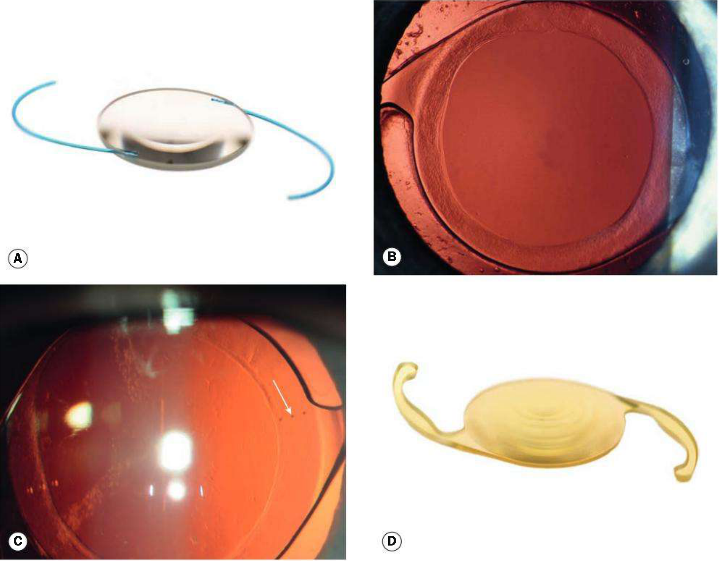
- Implanted in the capsular bag (in-the-bag position - ideal)
- Material: most commonly foldable hydrophobic or hydrophilic acrylic
- Square/sharp optic edge - reduces posterior capsule opacification (PCO)
- Toric IOL - corrects pre-existing corneal astigmatism
- Multifocal/bifocal IOL - provides near and distance vision
- Blue light filter IOLs - protect retina from high-energy visible light
- IOL power calculation: uses keratometry (corneal curvature) + axial length (A-scan) with formulae (SRK/T, Haigis, Hoffer Q, Barrett)
13. Postoperative Complications of Cataract Surgery
| Complication | Notes |
|---|---|
| Posterior Capsule Opacification (PCO) | Most common late complication; "after-cataract"; Elschnig's pearls (proliferating lens epithelial cells); treated with Nd:YAG laser capsulotomy |
| Endophthalmitis | Most dreaded; usually within 1 week; organisms: Staph. epidermidis (most common), Staph. aureus; treat with intravitreal antibiotics |
| Cystoid Macular Oedema (CMO / Irvine-Gass syndrome) | Most common cause of reduced VA post-PHACO; peaks 4-6 weeks postoperatively; treated with NSAIDs/steroids |
| Corneal decompensation / bullous keratopathy | Endothelial damage during surgery |
| Raised IOP | Retained viscoelastic; steroid-induced |
| Retinal detachment | Especially in high myopes; incidence increased post-ICCE |
| Wound leak | Early complication |
| Dropped nucleus | Vitreous prolapse during phaco; fragment falls to vitreous |
14. Anaesthesia for Cataract Surgery
- Topical anaesthesia (proxymetacaine/lidocaine drops) - most common
- Peribulbar block - injection around the globe; akinesia + analgesia
- Retrobulbar block - injection behind globe; risk of optic nerve damage, retrobulbar haemorrhage
- Sub-Tenon's block - cannula passed behind Tenon's capsule
- General anaesthesia - children, very anxious patients, unable to cooperate
15. High-Yield Summary Table for MCQs
| Feature | Key Point |
|---|---|
| Commonest cataract worldwide | Age-related / senile |
| Commonest type of age-related | Nuclear sclerotic |
| Most visually significant type | Posterior subcapsular (PSC) |
| PSC associated with | Steroids, diabetes, trauma, radiation, uveitis |
| Nuclear cataract causes | Myopic shift ("second sight") |
| Cortical cataract - location first | Inferonasal quadrant |
| Vacuoles/Bladder cells | PSC (Wedl cells) |
| Morgagnian cataract | Nucleus sinks in liquefied cortex |
| Glaukomflecken | Pathognomonic of previous acute angle-closure glaucoma |
| Congenital rubella cataract | Pearly nuclear opacity; part of TORCH |
| Snowflake cataract | Classic diabetic cataract (young) |
| Oil droplet cataract | Galactosaemia |
| No APD in cataract | If APD present - look for posterior segment pathology |
| Gold standard surgery | Phacoemulsification + IOL implantation |
| Most common late complication of surgery | PCO - treated by Nd:YAG laser |
| Most dreaded complication | Endophthalmitis |
| Most common cause of reduced VA post-surgery | Cystoid macular oedema (Irvine-Gass) |
| Glass-blower's cataract | True exfoliation of anterior capsule (infrared radiation) |
| IOL power formula | SRK/T (most widely used for axial length) |