
laproscopic instruments with images
Reading File
Now let me search the medical library and web simultaneously for laparoscopic instruments with images.
Finding Sources
Searching the Web
laparoscopic instruments names and images types
Reading File
Reading File
Finding Sources
Finding Sources
Loading Image
Loading Image
Reading File
Loading Image
Loading Image
Finding Sources
Searching the Web
laparoscopic instruments Veress needle trocar laparoscope energy devices images diagram
Loading Image
Loading Image
Reading Web Page
https://doultanigroup.com/blog/laparoscopic-instruments-guid…
Searching the Web
harmonic scalpel LigaSure stapler laparoscopic energy device images surgery
Finding Sources
Loading Image
Loading Image
Now I have enough material for a comprehensive, image-rich guide. Let me compile the full answer.
Laparoscopic Instruments - Complete Guide with Images
Laparoscopic (minimally invasive) surgery uses a set of specialized instruments inserted through small abdominal incisions (ports). They fall into several broad categories:
1. Access & Insufflation Instruments
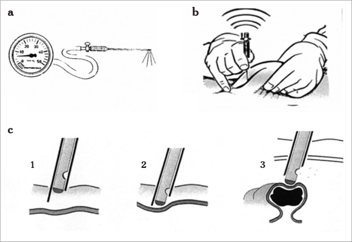
Veress Needle
A spring-loaded, blunt-tipped needle used to create pneumoperitoneum by insufflating CO₂ gas before trocar insertion. The spring mechanism retracts the sharp outer needle once it penetrates the peritoneum, protecting viscera.
- Confirmation of correct placement: hanging drop test, free saline flow, pressure reading < 10 mmHg
- Target intra-abdominal pressure: 12-15 mmHg
The diagram below shows the Veress needle, its pressure gauge connection, and the three stages of peritoneal entry:
Trocars / Cannulas
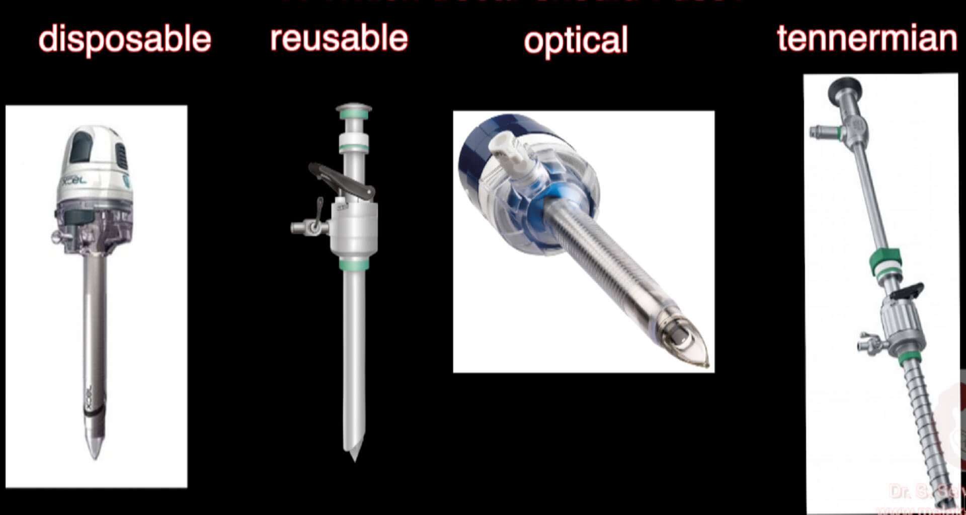
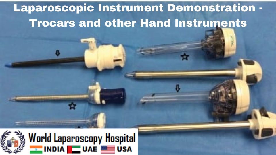
Trocars create the ports through which all laparoscopic instruments pass. The term now refers to both the initial entry device and the hollow cannula left in place.
Types:
| Type | Description |
|---|---|
| Disposable (bladed) | Sharp tip, single-use |
| Reusable (pyramidal/conical) | Metal, sterilizable |
| Optical (visualizing) | Clear plastic tip allows camera-guided entry |
| Hasson (open/blunt) | Used for open-entry technique; safer in re-operated abdomens |
| Tennermian / Threaded | Screw-thread design anchors to abdominal wall |
Common sizes: 5 mm, 10/12 mm (for specimen extraction, staplers, clip appliers)
Key trocar placement rules (Bailey & Love's, p. 124):
- All secondary trocars inserted under direct vision
- Always inserted perpendicular to the abdominal wall
- Non-midline trocars placed lateral to rectus sheath (mid-clavicular line) to avoid epigastric vessels
- Port sites > 5 mm require fascial closure to prevent hernia
2. Optical Instruments
Laparoscope (Telescope)
A rigid rod-lens system with a camera head attached. The image is displayed on a monitor.
| Specification | Options |
|---|---|
| Diameter | 5 mm or 10 mm (10 mm gives brighter image) |
| Viewing angle | 0° (straight) or 30° (most common - allows angled viewing) |
| Light source | Xenon or LED via fibre-optic cable |
The 30° scope is the standard choice for most procedures as it allows the surgeon to look "around corners" by rotating the scope.
Camera System
- HD or 4K chip-on-tip camera head attaches to the laparoscope eyepiece
- Image transmitted to a monitor (typically 26-32 inch, positioned directly in the surgeon's line of sight)
- 3D laparoscopes are now available to improve depth perception
3. Grasping & Dissecting Instruments
All hand instruments share a common design: long shaft (33 cm or 43 cm), handle with ring grip or pistol grip, and a jaw tip. They are 5 mm or 10 mm in diameter.
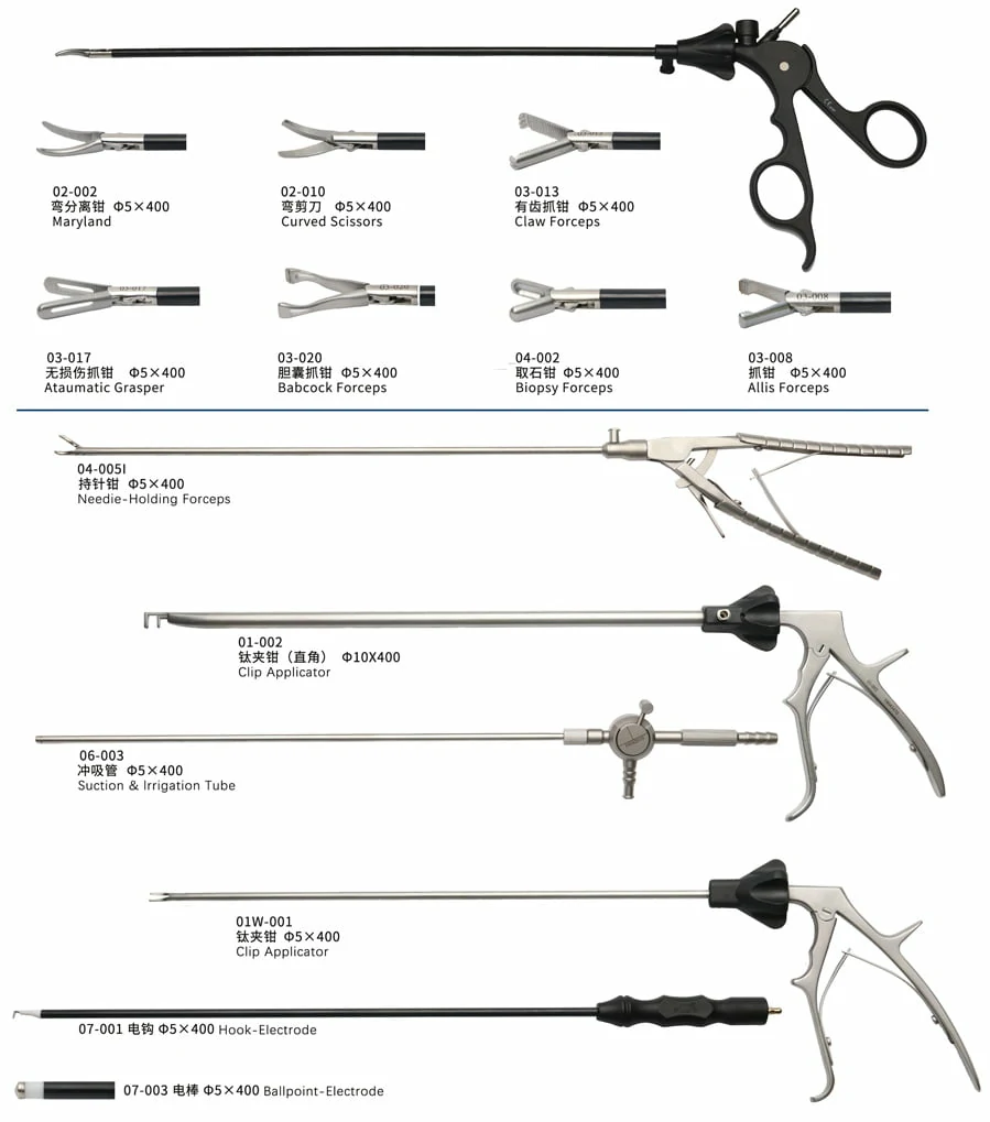
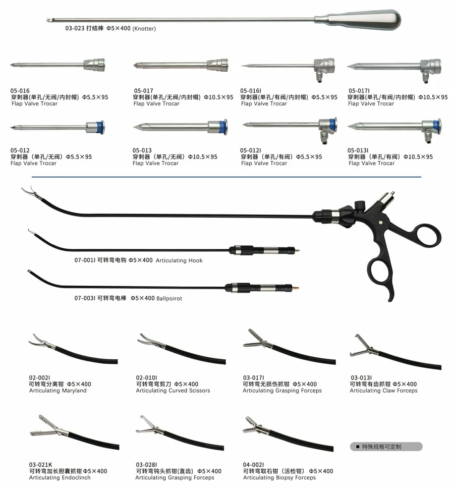
Key Graspers:
| Instrument | Jaw Type | Primary Use |
|---|---|---|
| Babcock forceps | Atraumatic, broad | Bowel, fallopian tube, appendix - gentle tissue handling |
| Atraumatic grasper | Smooth jaws | General tissue retraction without serosal injury |
| Claw / Toothed grasper | Rat-tooth / claw jaws | Firm grip on gallbladder, peritoneum |
| Allis forceps | Interlocking serrated | Grasping tougher tissues |
| Maryland dissector | Curved angled jaw | Dissection around vessels and ducts |
| Bowel grasper | Large flat atraumatic | Large bowel mobilization |
| Fundus grasper | Fixed flat jaw | Uterine fundus manipulation |
| Biopsy forceps | Cup jaw | Taking tissue specimens |
Scissors:
| Type | Use |
|---|---|
| Hook scissors (curved) | Most versatile; can also cauterize with monopolar current |
| Straight scissors | Cutting sutures, fine dissection |
| Metzenbaum scissors | Blunt dissection around structures |
4. Energy Devices
Energy devices perform cutting AND hemostasis simultaneously - replacing the need for separate clipping and cutting.
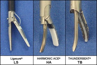
Monopolar Electrosurgery
- Hook electrode: Most common; used for dissection and cutting
- Ball electrode: Coagulation of oozing surfaces
- L-shaped (spatula) electrode: Blunt dissection
- Current passes through patient to a return pad; risk of capacitive coupling and inadvertent burns
Bipolar Electrosurgery
- Current flows only between the two jaws - no current through patient's body
- Safer than monopolar near bowel/vessels
- Bipolar Maryland forceps: Vessel sealing up to 3 mm
Advanced Bipolar / Vessel Sealers
LigaSure (Medtronic/Valleylab):
- Combines pressure + bipolar thermal energy
- Seals vessels up to 7 mm diameter reliably
- Fuses collagen and elastin in vessel wall
- Minimal lateral thermal spread (~1.5 mm)
- Source: Maingot's Abdominal Operations
Ultrasonic Devices

Harmonic Scalpel (Ethicon/Johnson & Johnson):
- Uses ultrasonic vibrations at 55,500 Hz to simultaneously cut and coagulate
- Blade vibrates at 80 µm amplitude; generates heat by friction (50-100°C)
- No electrical current through patient
- Cuts and seals vessels up to 5 mm
- Advantage: less smoke, less lateral heat than electrocautery
Thunderbeat (Olympus):
- Combines bipolar energy + ultrasonic energy in a single instrument
- Fastest vessel sealing among the three
- Seals vessels up to 7 mm
5. Clip Appliers & Stapling Devices
| Instrument | Use |
|---|---|
| Automatic clip applier (titanium/absorbable) | Clip vessels and ducts (cystic duct in cholecystectomy, vas deferens, appendiceal base) |
| Laparoscopic linear stapler (Endo-GIA) | Simultaneous staple + cut; used for bowel resection, vascular pedicles, lung resection. Fires 2-3 rows of staples on each side |
| Circular stapler (EEA/CDH) | Colorectal anastomosis - introduced transanally or through a port |
6. Suturing Instruments
(Fischer's Mastery of Surgery, p. 570-571)
| Instrument | Description |
|---|---|
| Laparoscopic needle holder | Straight or curved head; straight or ergonomic curved handle - reduces wrist angulation |
| Knot pusher | Allows extracorporeal knot tying - knot tied outside body, pushed down the suture |
| Endo Stitch (Medtronic) | Self-loading needle device; passes needle back and forth between jaws for reliable bites |
| Loop ligature (Endoloop) | Pre-tied loop for ligating vessels/ducts with one hand |
| Articulating needle holders | Allow extra degrees of freedom; useful in single-port (SILS) surgery |
7. Suction/Irrigation
- Suction-irrigation cannula: 5 mm shaft with dual lumen; washes operative field and aspirates blood/fluid
- CO₂ insufflated cavity means suction must be used judiciously to maintain pneumoperitoneum
8. Specimen Retrieval
| Device | Use |
|---|---|
| Endobag (specimen retrieval bag) | Placed around specimen laparoscopically; bag closed and extracted through an enlarged port site - prevents port site tumor implantation in oncological cases |
| Morcellator | Mechanical fragmentation of large specimens (fibroid tissue) - use now restricted due to concerns about spreading occult malignancy |
Summary: Commonly Used Laparoscopic Instrument Set
Based on Maingot's Abdominal Operations (Table 37-4):
- Video camera unit
- Light source (Xenon/LED)
- CO₂ insufflator
- 30° laparoscope (5 or 10 mm)
- Suction/irrigator
- Cannulas (Hasson and 10/12 mm + 5 mm)
- Scissors with cautery attachment
- Babcock graspers
- Intracorporeal vascular ligation device
- Clip applier
- LigaSure or Harmonic scalpel
- Linear stapler (for bowel/vascular procedures)
- Circular stapler (for pelvic/colorectal cases)
- Needle holder + knot pusher (for suturing)
Instrument Sizes at a Glance
| Size | Instruments |
|---|---|
| 3.5-5 mm | Graspers, scissors, dissectors, hook electrode, clip applier (small) |
| 10-12 mm | Laparoscope (10 mm), clip applier (large), linear stapler, specimen retrieval bag |
| 15-30 mm | Enlarged extraction port |
| Length | Standard 33 cm; bariatric/long instruments 38-43 cm for obese patients |
Sources: Maingot's Abdominal Operations | Fischer's Mastery of Surgery (8th ed., p. 570) | Bailey & Love's Short Practice of Surgery (28th ed., p. 124) | Campbell-Walsh Urology
This is a shared conversation. Sign in to Orris to start your own chat.