
Wallerian degeneration of axons flowchart
Reading File
Wallerian degeneration is the process by which the distal segment of a severed axon degenerates after injury. Let me pull the detailed content from the medical library.
Searching sources...
Finding Sources
Finding Sources
Reading File
Reading File
Reading File
Loading Image
Reading File
I now have all the content needed to build a comprehensive flowchart answer. Here it is:
Wallerian Degeneration of Axons - Flowchart
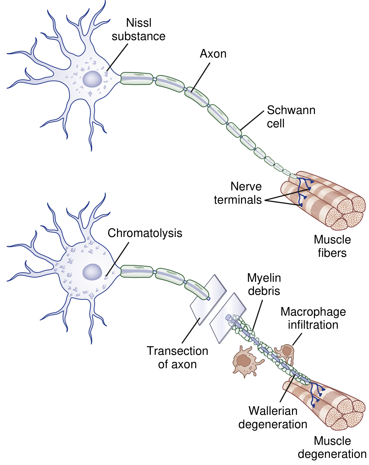
Fig. 64.4 - Bradley and Daroff's Neurology in Clinical Practice
Step-by-Step Flowchart
AXON INJURY (Grade II - V nerve lesion)
         |
         v
AXONAL TRANSECTION / DISRUPTION
  - Interruption of axoplasmic transport
  - Retrograde + anterograde flow blocked
         |
         |————————————————————————————————|
         v                                v
  DISTAL SEGMENT CHANGES          PROXIMAL SEGMENT CHANGES
  ("Dying forward")                (Retrograde changes)
         |                                |
         v                                v
Rapid Ca²⁺ & Na⁺ influx          Axon breaks down back
through disrupted plasma          to 1st node of Ranvier
membrane                                  |
         |                                v
         v                        CHROMATOLYSIS of cell body:
Activation of apoptosis-like       - Breakup & dispersion of
cascade (calpains, proteases)        rough ER (Nissl substance)
         |                           - Eccentric nucleus
         v                           - Switch: axon maintenance
Recruitment of LEUKOCYTES             → protein synthesis
+ Cytokine signaling cascade           for regeneration
         |
         v
 By Day 3:
Schwann cells retract from
nodes of Ranvier
         |
         v
Schwann cells & macrophages
activated → begin digesting MYELIN
         |
         v
Myelin breaks down into blocks/ovoids
= "Digestion chambers of Cajal"
         |
         v
Macrophages convert myelin fragments
→ neutral fats + cholesterol esters
→ carried to bloodstream
         |
         v
Entire distal axon degenerates
≈ 1 week
         |
         v
TARGET ORGAN (muscle) denervated
→ muscle degeneration / atrophy
After Degeneration: Regeneration Phase
WALLERIAN DEGENERATION COMPLETE
         |
         v
Schwann cells dedifferentiate
+ upregulate adhesion molecules
+ neurotrophins (laminin, cadherins, NCAM)
         |
         v
Bands of Büngner form
(Schwann cell cords aligned along
original basal lamina tubes)
         |
         v
AXON SPROUT grows from proximal stump
(Growth cone = filopodia + lamellipodia)
         |
         v
Growth cone guided distally
by semaphorins, ephrins, netrins
through Bands of Büngner
         |
         v
Axon reaches target muscle
→ Remyelination
→ Reinnervation
(Rate: ~1 mm/day)
Timeline Summary
| Time | Event |
|---|---|
| Immediately | Ca²⁺/Na⁺ influx; anterograde/retrograde transport fails |
| Hours | Leukocyte recruitment; cytokine signaling |
| Day 3 | Schwann cells retract from nodes of Ranvier |
| Days 3-7 | Myelin digestion by Schwann cells + macrophages |
| ~1 week | Full distal axon degeneration complete |
| Days 4+ | Collateral sprouting from surviving axons begins |
| 3-6 months | Clinical recovery via collateral sprouting |
| 6-24 months | Main recovery via proximal-to-distal axon regeneration |
Key Cellular Players
| Cell | Role |
|---|---|
| Schwann cells | Retract from nodes; digest myelin; dedifferentiate; form Bands of Büngner; remyelinate regenerating axon |
| Macrophages | Infiltrate injury site; phagocytose myelin ovoids + axonal debris; clear path for regrowth |
| Neuronal cell body | Undergoes chromatolysis; shifts gene expression toward regeneration |
| Growth cone | Tip of regenerating axon; navigates via chemotrophic signals |
Key distinction to remember: Wallerian degeneration is "dying forward" (distal to injury, outward) - contrasted with "dying-back" axonal degeneration in metabolic/toxic neuropathies where degeneration proceeds from distal to proximal. - Adams and Victor's Principles of Neurology, 12th Ed.
Sources: Bradley and Daroff's Neurology in Clinical Practice, Adams and Victor's Principles of Neurology 12th Ed., Neuroscience: Exploring the Brain 5th Ed.
This is a shared conversation. Sign in to Orris to start your own chat.