
Amelogenesis
Reading File
Amelogenesis is the process of enamel formation by ameloblasts during tooth development. Let me search the medical library for comprehensive coverage.
Searching sources...
Finding Sources
Finding Sources
The histology textbook has the most detailed coverage. Let me read the enamel/amelogenesis section from it, plus the embryology content.
Reading File
Reading File
I have detailed content. Let me read the core amelogenesis stages section more completely, and also get the images.
Reading File
Reading File
Let me get the rest of the amelogenesis section (Tomes process, maturation stage, and protein content) plus a key diagram image.
Reading File
Loading Image
Excellent - I have the diagram and histomicrograph. Let me fetch one more image showing the Tomes process/amelogenesis schematic and read the rest of the maturation stage.
Reading File
Loading Image
I now have rich, comprehensive content to write a detailed answer on amelogenesis. Let me also grab the maturation-stage details I truncated earlier.
Reading File
I now have all the content needed for a thorough, well-illustrated answer. Here is the full response:
Amelogenesis
Amelogenesis is the process of dental enamel formation by a matrix-mediated biomineralization mechanism. It is carried out exclusively by ameloblasts - polarized columnar epithelial cells derived from the inner enamel epithelium of the enamel organ. Mature enamel is the hardest substance in the human body (96-97% mineral by weight), and once formed, ameloblasts degenerate permanently - meaning enamel cannot be regenerated after eruption.
1. Origin of Ameloblasts - Tooth Development Context
Tooth development passes through three stages before amelogenesis begins:
| Stage | Key Events |
|---|---|
| Bud stage | Oral epithelium invaginates into mesenchyme |
| Cap stage | Inner enamel epithelium differentiates; tall columnar cells appear |
| Bell stage | Enamel organ fully differentiates into four layers; dental lamina degenerates |
The enamel organ in the bell stage contains four distinct layers:
- Outer enamel epithelium - convex surface layer
- Inner enamel epithelium - concave surface; cells differentiate into ameloblasts
- Stratum intermedium - immediately internal to inner enamel epithelium; contains alkaline phosphatase, active in calcification
- Stellate reticulum - loosely arranged stellate cells occupying the inner portion
The neural crest-derived preodontoblasts adjacent to the inner enamel epithelium become columnar odontoblasts and form dentin first. Only after this inductive signal do the inner enamel epithelial cells differentiate into ameloblasts. The dental lamina degenerates just before dentinogenesis and amelogenesis begin, detaching the developing tooth from the oral epithelium.
- Histology: A Text and Atlas with Correlated Cell and Molecular Biology, p. 1444
2. Stages of Amelogenesis
Stage 1 - Matrix Production (Secretory Stage)
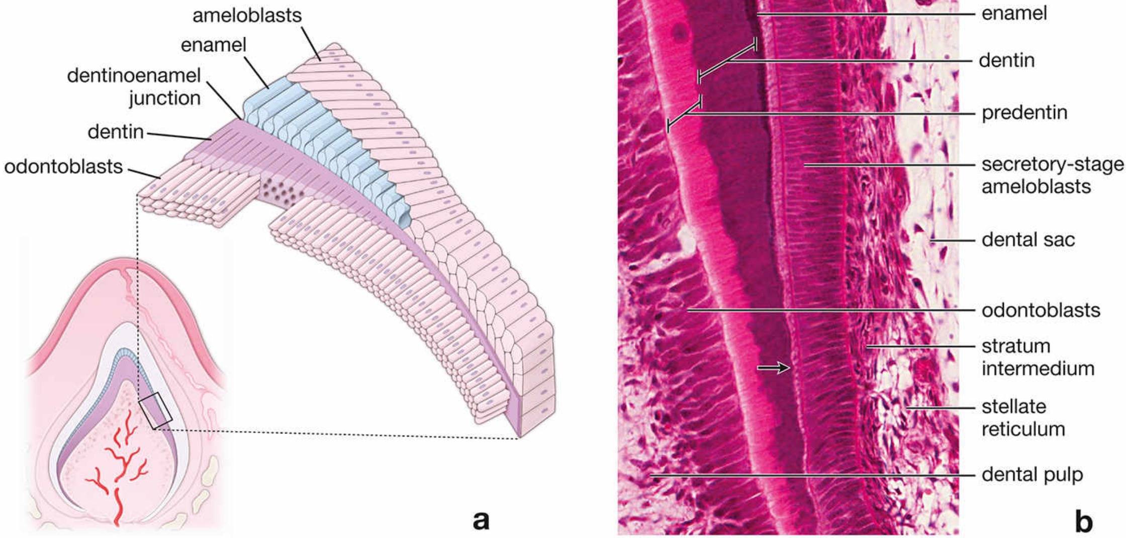
Diagram (a) and H&E photomicrograph (b) of early amelogenesis. Secretory-stage ameloblasts deposit enamel matrix directly onto previously formed dentin. The stratum intermedium, stellate reticulum, and dental pulp are visible (H&E x240). - Histology: A Text and Atlas, Fig. 16.13
- Dentin is deposited first by odontoblasts; only then does enamel matrix deposition begin on the dentin surface.
- Secretory-stage ameloblasts are tall, polarized columnar cells organized like osteoblasts - using rER, Golgi apparatus, and secretory granules to produce a proteinaceous organic matrix.
- They lay down enamel matrix continuously until the full thickness of future enamel is achieved.
- As enamel is deposited, ameloblasts migrate away from the dentinoenamel junction (DEJ), leaving enamel rods behind in their wake.
The Tomes Process
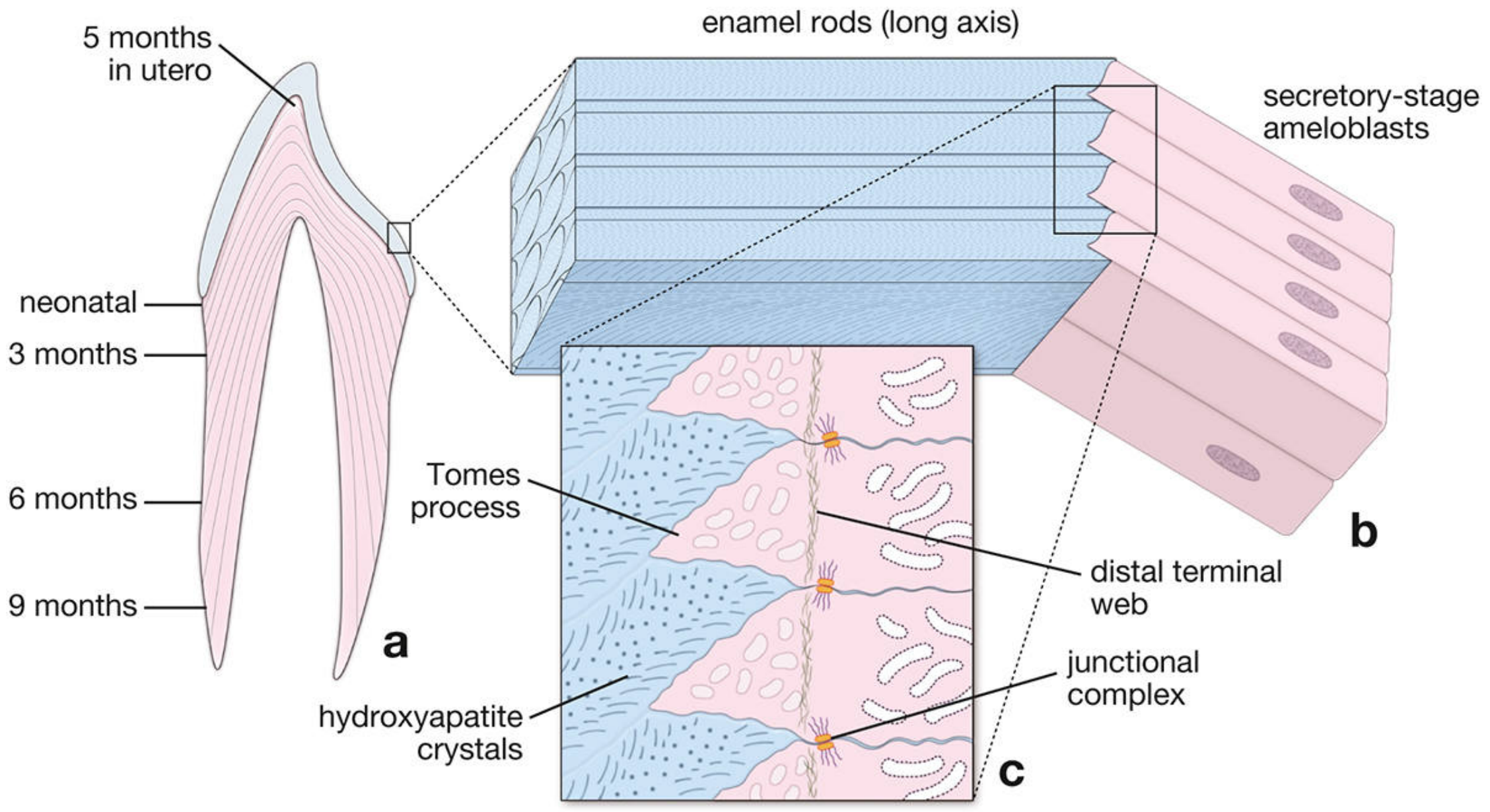
Schematic of amelogenesis (a) showing enamel rod contour lines reflecting incremental dentin development from 5 months in utero through postnatal months; (b) enamel rods in long axis; (c) close-up of Tomes processes, hydroxyapatite crystals, distal terminal web, and junctional complexes. - Histology: A Text and Atlas, Fig. 16.14
- At the apical pole of each secretory-stage ameloblast is the Tomes process, which is surrounded by developing enamel.
- A proximal terminal web (actin-rich band at the base of the cell) and a distal terminal web (at the base of the Tomes process) are the two eosinophilic staining regions visible in H&E.
- Junctional complexes at both the apical and basal poles maintain cell orientation and integrity as ameloblasts migrate.
- The direction of the completed enamel rod permanently records the migratory path taken by the ameloblast - this is why enamel rods run in decussating curves (Hunter-Schreger bands) in mature enamel.
- Histology: A Text and Atlas with Correlated Cell and Molecular Biology, p. 1447
Stage 2 - Matrix Maturation
Once full enamel thickness is reached, the ameloblasts transform into maturation-stage ameloblasts - now functioning primarily as a transporting epithelium rather than a secretory one.
Key features:
- The stratum intermedium disappears at this stage - the outer layers of the enamel organ collapse and merge together.
- A papillary layer forms adjacent to the basal pole of maturation-stage ameloblasts, with rich capillary supply to facilitate transport.
- Maturation-stage ameloblasts undergo modulation - a cyclic alternation between two morphological states:
| Morphology | % of cells | Function |
|---|---|---|
| Striated (ruffled) border | ~70% | Secretes bicarbonate ions (HCO3-); contains plasma membrane Ca2+-ATPases (PMCA) that pump Ca2+ into maturing enamel |
| Smooth-ended border | ~30% | Secretes proteolytic enzymes to degrade and reabsorb the organic matrix |
- This cyclic modulation corresponds to the cyclic influx of calcium into the enamel.
- The net result: organic matrix (amelogenins, ameloblastins) is removed and enamel becomes heavily mineralized with hydroxyapatite crystals.
- Histology: A Text and Atlas with Correlated Cell and Molecular Biology, p. 1450
3. Enamel Matrix Proteins
Four principal proteins are secreted into the extracellular matrix during amelogenesis:
| Protein | Role |
|---|---|
| Amelogenins (encoded by AMELX) | Most abundant (~90%). Establish and maintain spacing between enamel rods in early enamel development. Removed during maturation. |
| Ameloblastins | Signaling proteins secreted from early secretory to late maturation stage. Guide enamel mineralization; control elongation of enamel crystals; form junctional complexes between individual enamel crystals. Removed during maturation. |
| Enamelins (encoded by ENAM) | Distributed throughout the enamel layer; undergo proteolytic cleavage as enamel matures. Low-molecular-weight cleavage products are retained on enamel crystal surfaces in mature enamel. |
| Tuftelins | Earliest-detected proteins at the DEJ. Acidic and insoluble; aid in nucleation of enamel crystals. Found in enamel tufts (hypomineralized zones at the DEJ). |
After maturation, only enamelins and tuftelins remain in mature enamel. Ameloblasts degenerate after enamel is fully formed, around the time of tooth eruption through the gum.
- Histology: A Text and Atlas with Correlated Cell and Molecular Biology, p. 1451
4. Summary of Key Steps
Oral epithelium → Bud → Cap → Bell stage
↓
Inner enamel epithelium → Ameloblasts
↓
Odontoblasts lay down dentin FIRST
↓
SECRETORY STAGE: Ameloblasts produce enamel matrix (amelogenin-rich)
via Tomes process → enamel rods form as ameloblasts migrate outward
↓
MATURATION STAGE: Matrix proteins degraded + removed
Calcium pumped in via PMCA → hydroxyapatite crystal growth
↓
Mature enamel (96-97% mineral; hardest substance in body)
↓
Ameloblasts degenerate at eruption → enamel cannot regenerate
5. Clinical Correlate - Amelogenesis Imperfecta
When the genetic control of amelogenesis is disrupted, the result is amelogenesis imperfecta (AI) - a group of at least 14 clinical entities involving aberrant enamel formation in the absence of systemic disease.
- Inheritance: Multiple patterns - autosomal dominant, autosomal recessive, X-linked
- Genes involved: AMELX (amelogenin), ENAM (enamelin), MMP20 (enamelysin/matrix metalloproteinase 20)
- Incidence: 1 in 700 (Sweden) to 1 in 1200 (USA)
- Subtypes (based on phenotype):
- Hypoplastic - thin enamel; defective secretory stage
- Hypocalcified - soft enamel, normal thickness; defective mineralization
- Hypomature - enamel not fully hardened; defective maturation stage
- Enamel may be hard or soft, pitted or smooth, thin or normal in thickness.
- The Developing Human: Clinically Oriented Embryology, p. 1206
Key facts for exams:
- Ameloblasts are ectodermal in origin (inner enamel epithelium)
- Dentin precedes enamel deposition - odontoblasts must deposit dentin first to signal ameloblast differentiation
- Tomes process = apical secretory extension of the ameloblast
- Ameloblasts are lost after eruption - enamel cannot self-repair
- Mature enamel contains enamelins + tuftelins only (amelogenins/ameloblastins are removed)
- Neonatal line = the incremental line in enamel at the time of birth
This is a shared conversation. Sign in to Orris to start your own chat.