I now have all the material I need from Harrison's 22E, Goldman-Cecil, Sleisenger, Schwartz's Surgery, and the 2025 PubMed review. Let me now write the comprehensive algorithm.Here is your complete, ward-ready, exam-ready algorithm - sourced directly from Harrison's 22E (2025), Goldman-Cecil Medicine, Sleisenger & Fordtran, Schwartz's Surgery, and the 2025 Am Fam Physician review (
Nelson et al., 2025, PMID 39823630).
THE COMPLETE JAUNDICE ALGORITHM
For Ward, ER, FCPS Part 2 TOACS & MRCP
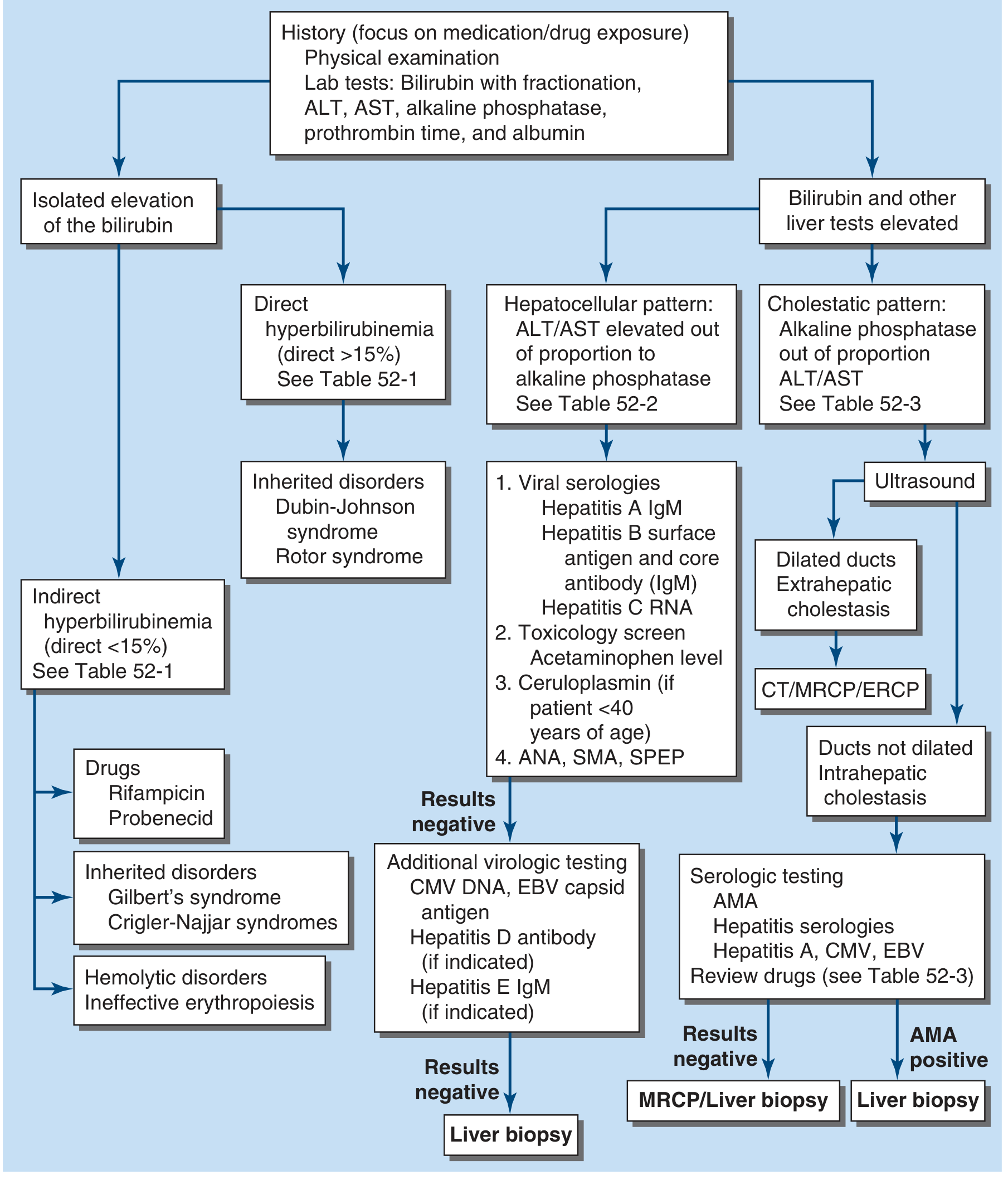
THE MASTER FLOWCHART (Harrison's 22E, Fig. 52-1)
SECTION 1: THE GOLDEN RULE - FIRST 3 QUESTIONS IN YOUR HEAD
When a jaundiced patient walks in, IMMEDIATELY ask yourself:
1. Is bilirubin elevated alone, or are other LFTs also elevated?
2. Is it conjugated (direct) or unconjugated (indirect)?
3. Is the pattern hepatocellular or cholestatic?
These three questions drive everything. Everything else is detail.
SECTION 2: THE "3 ZONES" CLASSIFICATION - PREHEPATIC / HEPATIC / POSTHEPATIC
Memory Mnemonic: "PRE-HEP-POST" or think of a factory analogy:
- PRE = Too much raw material (excess bilirubin production)
- HEP = Factory broken (liver can't process)
- POST = Pipe blocked (can't export what's made)
| Feature | Prehepatic (Pre) | Hepatic (Hep) | Posthepatic (Post) |
|---|
| Bilirubin type | Unconjugated (indirect) | Both (mixed) | Conjugated (direct) |
| Urine colour | Normal/pale | Dark | Dark (bilirubinuria) |
| Stool colour | Normal | Normal-pale | Pale/clay-coloured |
| Urobilinogen | Increased | Variable | Absent |
| Pruritus | Absent | Variable | Prominent |
| Key lab | Raised indirect bili, anaemia, reticulocytosis | Raised ALT/AST >> ALP | Raised ALP >> ALT/AST |
| Typical cause | Haemolysis, ineffective erythropoiesis | Hepatitis, cirrhosis | CBD stone, Ca head of pancreas |
SECTION 3: HISTORY TAKING - THE STRUCTURED APPROACH
The "DRUG-FAT-SIS" Mnemonic for history:
D - Drugs - ALL medications, herbal, OTC, anabolic steroids, contraceptives, anti-TB drugs (rifampicin, INH, pyrazinamide), statins, paracetamol dose/overdose
R - Risk factors for viral hepatitis - IV drug use, tattoos, sexual history, blood transfusion, healthcare worker, travel
U - Urine & stool change - Dark urine = conjugated; pale/clay stool = obstruction; ask specifically
G - Gallstones/prior surgery - Previous biliary surgery, RUQ pain attacks, prior cholecystectomy
F - Fever + Pain - Fever + RUQ pain + jaundice = Charcot's triad = CHOLANGITIS (emergency)
A - Alcohol - Quantify units/week; duration; AST:ALT ratio >2:1 = alcoholic hepatitis
T - Travel/tainted food - Travel to endemic area = Hep A/E; raw shellfish; Leptospirosis (Weil's disease)
S - Symptoms suggesting malignancy - Painless jaundice + weight loss + anorexia = Red flag for Ca head of pancreas/cholangiocarcinoma
I - Itching (pruritus) - Suggests cholestasis; bile salts deposited in skin
S - Systemic/family - Family history of jaundice/liver disease (Wilson's, haemochromatosis, Gilbert's); autoimmune history
Additional HIGH-YIELD History Points:
| Symptom cluster | Think of |
|---|
| Jaundice after fasting/stress/illness, mild, no other symptoms | Gilbert's syndrome (most common benign cause) |
| Young patient, neuropsychiatric + jaundice + Kayser-Fleischer rings | Wilson's disease |
| Jaundice + arthralgia/myalgia BEFORE jaundice | Viral hepatitis (prodrome) |
| Jaundice + sudden severe RUQ pain + rigor + fever | Choledocholithiasis + cholangitis |
| Painless progressive jaundice + weight loss + new-onset diabetes | Carcinoma head of pancreas |
| Jaundice + fatigue + pruritus in middle-aged woman | PBC (Primary Biliary Cholangitis) |
| Jaundice + inflammatory bowel disease | PSC (Primary Sclerosing Cholangitis) |
| Jaundice in a returning traveller with fever, rigors | Malaria, Hep A/E, Leptospirosis |
| Jaundice + pallor + splenomegaly, NO hepatomegaly | Haemolytic anaemia |
| Jaundice + pancytopenia | Think bone marrow failure, lymphoma infiltration, leukaemia |
| Jaundice + right heart failure symptoms, JVD, pulsatile liver | Congestive hepatopathy (Budd-Chiari or CCF) |
| Post-operative jaundice | Ischaemic hepatitis, haemolysis, drug reaction, cholestasis |
SECTION 4: GENERAL PHYSICAL EXAMINATION - THE STRUCTURED HEAD-TO-TOE APPROACH
STEP 1: GENERAL INSPECTION (look before you touch)
Assess from the foot of the bed:
- Nutritional state - Temporal wasting, muscle wasting = chronic disease (cirrhosis, malignancy)
- Depth of jaundice - Mild yellowing (sclera only) vs deep orange-green (chronic obstruction)
- Consciousness - Encephalopathy? (flapping hands, confusion)
- Build - Cachexia = malignancy, chronic liver disease
STEP 2: SKIN SIGNS - "Spider Farmer Has Dupuytren And Leuconychia, He Bruises Easily"
| Sign | Meaning |
|---|
| Spider naevi (>5, upper body) | Chronic liver disease (CLD) |
| Palmar erythema | CLD (hyperestrogenism) |
| Dupuytren's contracture | Alcoholic liver disease |
| Leuconychia (white nails) | Hypoalbuminaemia |
| Terry's nails (white proximal, pink distal) | Cirrhosis |
| Bruising / purpura | Coagulopathy |
| Scratch marks (excoriations) | Cholestasis - pruritus |
| Xanthelasma / xanthomata | PBC, chronic cholestasis |
| Gynaecomastia | CLD (hyperestrogenism) |
| Testicular atrophy | CLD (alcohol) |
| Parotid enlargement | Alcoholic liver disease |
| Caput medusae | Portal hypertension |
| Kayser-Fleischer rings (slit lamp) | Wilson's disease |
STEP 3: EYES & FACE
- Scleral icterus - Best place to detect early jaundice (bilirubin >2.5-3 mg/dL). Look in good daylight, pull down lower lid, compare to healthy person's sclera.
- Anaemia - Conjunctival pallor (haemolysis, bone marrow failure, chronic disease)
- Kayser-Fleischer rings - Brown-gold rings at corneal periphery (Wilson's disease - use slit lamp to confirm)
- Xanthelasma - PBC
STEP 4: HANDS
- Flapping tremor (asterixis) - hepatic encephalopathy
- Leuconychia - hypoalbuminaemia
- Palmar erythema - CLD
- Clubbing - CLD, IBD (PSC), hepatopulmonary syndrome
- Dupuytren's - alcoholic liver disease
- Bruising - coagulopathy
STEP 5: LYMPH NODES - KEY ONES TO FEEL
| Node | Clinical Significance |
|---|
| Virchow's node (left supraclavicular) | Intra-abdominal malignancy (Ca stomach, pancreas, colon) |
| Sister Mary Joseph's nodule (periumbilical) | Intra-abdominal malignancy with peritoneal spread |
| Generalised lymphadenopathy | Lymphoma, leukaemia, EBV, CMV |
STEP 6: ABDOMINAL EXAMINATION - THE CORE EXAM
Inspection first:
- Distension = ascites
- Caput medusae = portal hypertension
- Surgical scars = prior biliary surgery
Palpation:
| Finding | Clinical Significance |
|---|
| Hepatomegaly - smooth, tender | Viral/alcoholic hepatitis, CCF, early cirrhosis |
| Hepatomegaly - hard, nodular | Malignancy (HCC, metastases), cirrhosis (later) |
| Hepatomegaly - pulsatile | Tricuspid regurgitation / right heart failure |
| Splenomegaly + jaundice, NO hepatomegaly | Haemolysis, portal hypertension |
| Hepatosplenomegaly | Portal hypertension, lymphoma, haematological malignancy |
| Murphy's sign positive | Acute cholecystitis |
| Palpable non-tender gallbladder + jaundice | Courvoisier's sign = malignant obstruction (Ca head of pancreas) |
| Hard, irregular mass RUQ | Malignancy |
| Ascites | Cirrhosis, malignancy, CCF |
Courvoisier's Law (HIGH-YIELD EXAM POINT):
"A palpable, non-tender gallbladder in a jaundiced patient suggests malignant obstruction of the common bile duct, NOT gallstones" - because gallstones cause chronic fibrosis and a shrunken non-distensible gallbladder.
Percussion:
- Dullness in flanks + shifting dullness = ascites
- Liver dullness span: Normal 8-12 cm in mid-clavicular line. Dullness reduced = small/shrunken liver (late cirrhosis, acute liver failure)
STEP 7: CARDIOVASCULAR
- JVD + peripheral oedema + jaundice = Congestive hepatopathy (CCF causing hepatic congestion)
- Right pleural effusion without obvious ascites = advanced cirrhosis (hepatic hydrothorax)
STEP 8: NEUROLOGICAL
- Asterixis (flapping tremor) = hepatic encephalopathy
- Confusion, slurring, behavioural change = encephalopathy
- Kayser-Fleischer rings + neuropsychiatric features = Wilson's disease
SECTION 5: THE INVESTIGATION ALGORITHM - STEP BY STEP
FIRST-LINE INVESTIGATIONS (Order immediately on any jaundiced patient):
Blood tests:
- Total bilirubin + fractionation (direct vs indirect)
- ALT, AST, ALP, GGT
- Albumin, Prothrombin time / INR
- Full blood count (CBC)
- Blood urea, creatinine, electrolytes
Urine:
- Urine bilirubin (dipstick) - present = conjugated hyperbilirubinemia (hepatic/posthepatic)
- Urobilinogen - increased in haemolysis/hepatitis, absent in complete obstruction
INTERPRETING THE PATTERN - THE "ENZYME COMPASS":
HEPATOCELLULAR INJURY             CHOLESTATIC PATTERN
ALT/AST very high (>500 IU)   vs.  ALP very high + ALT/AST mildly raised
Bilirubin raised (mixed)            Bilirubin raised (conjugated)
PT prolonged (doesn't correct       PT may correct with Vit K
with Vit K in severe disease)       (bile needed for Vit K absorption)
Specific patterns:
| Pattern | Think |
|---|
| ALT/AST >1000 IU | Acute viral hepatitis, drug-induced (paracetamol), ischaemic hepatitis |
| AST:ALT ratio >2:1 | Alcoholic liver disease |
| ALP very high, ALT/AST mildly elevated | Cholestasis, infiltrative disease, bone disease |
| GGT elevated WITH ALP | Liver source confirmed (not bone); also elevated in alcohol |
| Isolated unconjugated hyperbilirubinemia, normal rest | Gilbert's syndrome |
| Prolonged PT + low albumin | Advanced/chronic liver disease |
| PT prolonged + corrects with Vit K | Obstructive jaundice (malabsorption of fat-soluble vitamins) |
SECOND-LINE INVESTIGATIONS (based on pattern):
If hepatocellular pattern:
- Hepatitis A IgM, Hepatitis B sAg + core Ab (IgM), Hepatitis C RNA
- Toxicology screen, paracetamol level
- ANA, ASMA, anti-LKM1 = Autoimmune hepatitis
- Ceruloplasmin + 24hr urine copper = Wilson's (in patients <40)
- Serum ferritin + transferrin saturation = Haemochromatosis
- CMV DNA, EBV capsid antigen, Hepatitis D Ab, Hepatitis E IgM
If cholestatic pattern:
- USS abdomen FIRST (cheap, accessible, non-invasive) - look for dilated biliary ducts
- If ducts dilated (extrahepatic cholestasis): CT abdomen / MRCP / ERCP
- If ducts NOT dilated (intrahepatic cholestasis): AMA (antimitochondrial Ab) for PBC, hepatitis serologies, drug review
- AMA positive = PBC - proceed to liver biopsy
- AMA negative, no cause found = MRCP or liver biopsy
If isolated unconjugated hyperbilirubinemia:
- Blood film, reticulocyte count, LDH, haptoglobin, Coombs test = haemolysis screen
- No haemolysis + otherwise well = Gilbert's syndrome (no further workup needed)
If malignancy suspected:
- CT chest/abdomen/pelvis with contrast
- CA 19-9 (pancreatic cancer), AFP (HCC), CEA (colorectal metastases)
- ERCP/MRCP for biliary obstruction characterisation
- Biopsy/EUS for tissue diagnosis
If bone marrow failure suspected (pancytopenia + jaundice):
- Blood film (dysplastic cells, blasts?)
- Reticulocyte count
- Bone marrow aspirate + trephine biopsy
- LFTs pattern (usually hepatic infiltration pattern - raised ALP, mild bilirubin)
SECTION 6: DISEASE-SPECIFIC APPROACH - MATCHING PRESENTATION TO DIAGNOSIS
SCENARIO-BASED QUICK GUIDE FOR WARD ROUNDS:
Young patient, jaundice + fever + arthralgia 2 weeks before:
- Think viral hepatitis - send Hep serology, ALT/AST pattern
Young patient, exam stress/fasting, mild jaundice, nothing else:
- Gilbert's syndrome - fractionated bili (indirect up, direct normal), otherwise normal LFTs - REASSURE
Middle-aged alcoholic, tender hepatomegaly, fever, high AST:ALT >2:1:
- Alcoholic hepatitis - Maddrey's Discriminant Function = 4.6 x (PT patient - PT control) + bilirubin (mg/dL); if >32 = severe, consider steroids
Middle-aged woman, pruritus, fatigue, xanthelasma:
- PBC - AMA, ALP high, check cholesterol
Young patient, neuropsychiatric + Kayser-Fleischer rings:
- Wilson's disease - ceruloplasmin, 24hr urine copper, slit-lamp
Elderly patient, painless progressive jaundice + weight loss + new-onset diabetes:
- Carcinoma head of pancreas - CT triple phase, CA19-9, ERCP/MRCP
RUQ pain + fever + jaundice (Charcot's triad) + hypotension + confusion (Reynolds' pentad):
- ASCENDING CHOLANGITIS - EMERGENCY - blood cultures, IV antibiotics, urgent ERCP
Jaundice + pallor + no hepatomegaly + splenomegaly + raised reticulocytes:
- Haemolytic anaemia - blood film, Coombs test, haemoglobin electrophoresis
Jaundice + IBD (UC) + ALP raised + PSC on MRCP:
- Primary Sclerosing Cholangitis - MRCP (beading of ducts), p-ANCA, colonoscopy
Jaundice + ascites in a young patient after exertion:
- Think Budd-Chiari syndrome - Doppler USS of hepatic veins (absent flow)
SECTION 7: CLINICAL SIGNS TABLE - "WHAT SIGN POINTS TO WHAT"
| Clinical Sign | Diagnosis to Think Of |
|---|
| Kayser-Fleischer rings | Wilson's disease |
| Xanthelasma + scratch marks | PBC, chronic cholestasis |
| Spider naevi + gynaecomastia + parotid enlargement | Alcoholic cirrhosis |
| Pulsatile hepatomegaly + JVD | Tricuspid regurgitation, CCF |
| Hepatomegaly + hard/nodular | HCC, metastases, lymphoma |
| Virchow's node | GI malignancy |
| Sister Mary Joseph's nodule | Peritoneal malignancy |
| Courvoisier's sign | Malignant biliary obstruction |
| Murphy's sign | Acute cholecystitis |
| Asterixis | Hepatic encephalopathy |
| Dupuytren's + parotid + spider naevi | Alcohol-related CLD |
| Fever + rigors + RUQ pain + jaundice | Cholangitis (Charcot's triad) - EMERGENCY |
SECTION 8: VIVA HOT QUESTIONS - FOR FCPS TOACS & MRCP
Q1: What is the first investigation in a jaundiced patient?
Fractionated serum bilirubin (total + direct/indirect) PLUS LFT panel (ALT, AST, ALP, albumin, PT). Also urine for bilirubin and urobilinogen. First imaging = USS abdomen.
Q2: What is Courvoisier's law?
A palpable, non-tender gallbladder in a jaundiced patient suggests malignant obstruction of the bile duct, NOT gallstones. Gallstones cause chronic inflammation and fibrosis of the GB wall, making it unable to distend.
Q3: What is Charcot's triad and why is it an emergency?
Fever + RUQ pain + Jaundice = Ascending cholangitis. Reynolds' pentad adds hypotension + altered consciousness. It is septic shock from biliary sepsis. Requires immediate blood cultures, IV broad-spectrum antibiotics, and urgent biliary decompression (ERCP).
Q4: How do you distinguish obstructive jaundice from parenchymal liver disease on labs?
Obstructive: ALP >> ALT/AST, conjugated bilirubin, PT corrects with Vit K, dilated ducts on USS.
Parenchymal: ALT/AST >> ALP, mixed bilirubin, PT may not correct with Vit K, no duct dilation.
Q5: What is Gilbert's syndrome and when do you say "nothing more needed"?
Benign inherited condition, reduced UGT1A1 enzyme activity. Presents with mild isolated unconjugated (indirect) hyperbilirubinemia during fasting, stress, or illness. ALL other LFTs are normal. Blood film normal. No haemolysis. REASSURE - no treatment needed.
Q6: A 25-year-old with jaundice, low ceruloplasmin, Kayser-Fleischer rings - what test confirms Wilson's disease?
24-hour urine copper (>100 mcg/day is diagnostic). Liver biopsy with copper quantification (>250 mcg/g dry weight) is gold standard if uncertain.
Q7: What is the Maddrey Discriminant Function?
Used in alcoholic hepatitis: 4.6 x (PT_patient - PT_control) + serum bilirubin (mg/dL). Score >32 = severe alcoholic hepatitis = consider prednisolone 40 mg/day. Score <32 = conservative management.
Q8: What is the MELD score used for?
Model for End-stage Liver Disease. Used to prioritise liver transplant. = 3.78 x ln[bilirubin mg/dL] + 11.2 x ln[INR] + 9.57 x ln[creatinine mg/dL] + 6.43. Score >15 = consider transplant listing.
Q9: In PBC, what is the diagnostic antibody and what is the treatment?
Anti-mitochondrial antibody (AMA) in >90% cases. Treatment = Ursodeoxycholic acid (UDCA) 13-15 mg/kg/day. If inadequate response: Obeticholic acid.
Q10: When do you do liver biopsy in jaundice?
When etiology remains unclear despite full biochemical and imaging workup. Also for: autoimmune hepatitis confirmation, grading/staging of chronic hepatitis, unexplained cholestasis with non-dilated ducts and negative serology.
SECTION 9: COMMON MISTAKES & BLUNDERS TO AVOID
THE "8 DEADLY SINS" IN JAUNDICE MANAGEMENT:
1. Missing cholangitis (the most dangerous mistake)
- Mistake: Calling RUQ pain + jaundice "gallstones" and sending home or waiting
- Correct: Charcot's triad = EMERGENCY. Start IV antibiotics immediately. Arrange urgent ERCP. Do NOT delay.
2. Missing the drug history
- Mistake: Not asking about herbal medicines, supplements, OTC drugs, recent antibiotics (flucloxacillin causes cholestatic jaundice weeks later), statins, OCP
- Correct: ALWAYS ask a detailed drug history including herbals and recent changes. Drug-induced liver injury is one of the most commonly missed diagnoses.
3. Reassuring a patient with "painless jaundice + weight loss" without malignancy workup
- Mistake: Attributing painless jaundice in an elderly patient to Gilbert's or viral hepatitis
- Correct: Painless progressive jaundice + weight loss = Ca head of pancreas until proven otherwise. Order CT triple phase + CA19-9.
4. Forgetting to check PT/INR in an acutely jaundiced patient
- Mistake: Only checking bilirubin and transaminases
- Correct: PT/INR is a marker of SYNTHETIC FUNCTION. A prolonged PT that does NOT correct with Vitamin K = severe parenchymal disease / acute liver failure. This changes management urgently.
5. Giving Vitamin K without checking whether it corrects the PT
- Mistake: Just giving Vit K without checking effect
- Correct: Give IV Vit K 10 mg and recheck PT in 24 hours. Correction = obstructive; no correction = liver synthetic failure.
6. Ordering ERCP before USS in a new jaundiced patient
- Mistake: Jumping to invasive investigation
- Correct: USS abdomen is ALWAYS the first imaging modality. Cheap, non-invasive, excellent for gallstones and ductal dilation. Reserve ERCP for therapeutic intervention.
7. Not checking blood film and reticulocyte count in unconjugated hyperbilirubinemia
- Mistake: Treating unconjugated jaundice as Gilbert's without ruling out haemolysis
- Correct: ALWAYS check blood film + reticulocyte count + LDH + haptoglobin before diagnosing Gilbert's. Haemolysis must be excluded.
8. Forgetting that jaundice in cirrhosis may not be from the liver
- Mistake: Assuming new jaundice in a known cirrhotic = disease progression
- Correct: Known cirrhotic + new/worsening jaundice = rule out superimposed cause: hepatocellular carcinoma (AFP, triphasic CT), spontaneous bacterial peritonitis, portal vein thrombosis, drug hepatotoxicity, or superimposed viral hepatitis.
SECTION 10: THE EMERGENCY ER APPROACH - WITHIN THE FIRST 30 MINUTES
IMMEDIATE TRIAGE CHECKLIST:
RED FLAGS = Admit and act NOW:
ER Immediate Orders on a Jaundiced Patient:
- IV access - take bloods at the same time: FBC, LFTs, PT/INR, renal function, blood cultures (if febrile)
- Fractionated bilirubin + full LFT panel
- Urine dipstick: bilirubin + urobilinogen
- USS abdomen - same day if possible
- Fluids if dehydrated/hypotensive
- IV Vitamin K 10 mg if coagulopathic
- Lactulose + protein restriction if encephalopathy
- IV broad-spectrum antibiotics (e.g., piperacillin-tazobactam or ceftriaxone + metronidazole) if cholangitis suspected
SECTION 11: WARD MANAGEMENT ORIENTATION - THE DAILY CHECKLIST
Every day on rounds for your jaundiced patient, assess:
Grades of Hepatic Encephalopathy (West Haven):
| Grade | Features |
|---|
| 1 | Trivial confusion, altered sleep |
| 2 | Asterixis, moderate confusion |
| 3 | Marked confusion, somnolence, incoherence |
| 4 | Coma |
QUICK-REFERENCE SUMMARY CARD (Laminate this for your coat pocket)
JAUNDICE FIRST 3 QUESTIONS:
1. Isolated bili rise OR other LFTs too?
2. Conjugated (direct) or Unconjugated (indirect)?
3. Hepatocellular (ALT>ALP) or Cholestatic (ALP>ALT)?

URINE DARK + STOOLS PALE = Conjugated = Obstruction
URINE NORMAL + PALLOR + SPLENOMEGALY = Haemolysis

ALARMS:
- Fever + RUQ pain + jaundice = CHOLANGITIS (emergency)
- Painless jaundice + weight loss = MALIGNANCY
- Altered consciousness + jaundice = LIVER FAILURE

COURVOISIER'S LAW:
Palpable non-tender GB + jaundice = MALIGNANT obstruction

KEY DRUG HISTORY: Herbals, OCP, anti-TB, statins, paracetamol

INVESTIGATIONS ORDER:
1. FBC + LFTs + PT/INR
2. Fractionated bilirubin
3. USS abdomen
4. Then targeted: serology / MRCP / CT / Biopsy
Sources: Harrison's Principles of Internal Medicine 22E (2025) Ch. 52; Goldman-Cecil Medicine (2023) Ch. 133; Sleisenger & Fordtran's GI & Liver Disease, Ch. 21; Schwartz's Principles of Surgery 11th Ed., Ch. 30;
Nelson M et al. Evaluation of Jaundice in Adults. Am Fam Physician. 2025;
Pavlovic Markovic A et al. Jaundice as a Diagnostic and Therapeutic Problem. Dig Dis. 2022.