
Anuerysmal bone cyst
I'll start by reading the medical library skill, then search for aneurysmal bone cyst content in parallel.
Reading File
Now let me search the library and PubMed in parallel.
Finding Sources
Searching PubMed
"aneurysmal bone cyst"[MeSH Terms]
Excellent results. Now let me read the most comprehensive sources in parallel - Robbins Pathology, Campbell's Operative Orthopaedics, and Grainger's Radiology.
Reading File
Reading File
Reading File
Now let me read the Campbell's Operative Orthopaedics dedicated section (block10) and fetch the key images in parallel.
Reading File
Loading Image
Reading File
Now let me read the full Campbell's treatment section and the Robbins histology/clinical features section in parallel.
Reading File
Reading File
Finding Sources
Let me get the rest of the Campbell's treatment text and fetch the histology image.
Reading File
Loading Image
I now have comprehensive information from multiple authoritative textbooks. Here is the full answer:
Aneurysmal Bone Cyst (ABC)
Definition
An aneurysmal bone cyst is a locally destructive, blood-filled, reactive lesion of bone - not a true neoplasm. It consists of multiloculated, hemorrhagic cystic spaces within an expanding shell of bone.
Epidemiology
- Primarily affects patients younger than 20 years (1st-2nd decades)
- Slight female predominance
- Any bone can be involved
Common Locations
| Site | Notes |
|---|---|
| Proximal humerus | Most common long bone site |
| Distal femur | Frequent |
| Proximal tibia | Frequent |
| Spine (posterior elements) | 15-20% of cases; may extend into vertebral body |
| Flat bones | Less common |
The lesion arises eccentrically in the metaphysis and does not cross the growth plate.
Pathogenesis
Two mechanisms are recognized:
1. Primary (de novo):
Spindle-shaped cells show rearrangements of chromosome 17p13, causing fusion of USP6 gene coding sequences to regulatory elements of COL1A1 (most common) or other genes. This leads to USP6 overexpression - a deubiquitinating enzyme that upregulates NF-kB, which in turn increases matrix metalloprotease expression, causing cystic bone resorption.
2. Secondary (up to 30% of cases):
ABC-like changes arise within other bone tumors, including:
- Giant cell tumors
- Chondroblastomas
- Osteoblastomas
- Fibrous dysplasia
- Nonossifying fibromas
- Chondromyxoid fibromas
A local circulatory disturbance causing increased venous pressure and hemorrhage has also been proposed.
Morphology
Radiography
- Eccentric, expansile, lytic, metaphyseal lesion with well-defined margins
- Thin shell of overlying cortical bone (periosteum elevated but intact)
- May occasionally have a permeative appearance mimicking malignancy
CT
- Delineates complex anatomy (spine, pelvis)
- Identifies the thin cortical rim
- Shows multiloculated architecture
MRI (most informative)
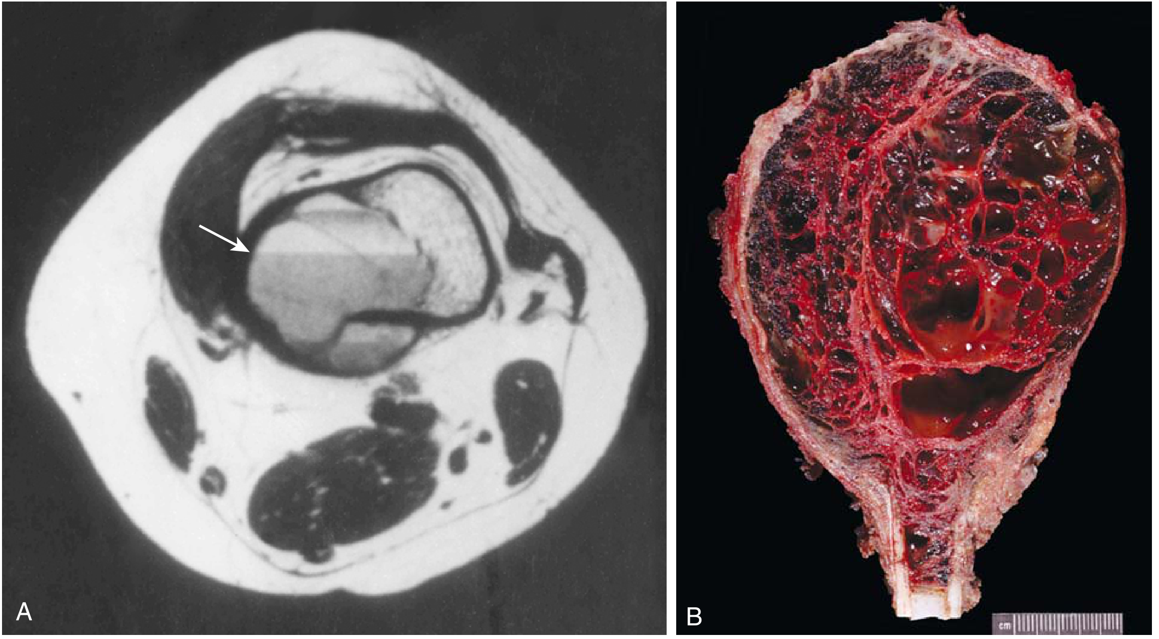
- Fluid-fluid levels - the hallmark finding (arrow in image below)
- Double-density fluid levels + intralesional septations distinguish ABC from unicameral bone cyst
- Multiloculated cavities clearly demonstrated
Bone Scan
- Diffuse or peripheral tracer uptake with central area of decreased uptake ("doughnut sign")
Gross
- Multiple blood-filled cystic spaces separated by thin tan-white septa
- Surrounded by a thin layer of bone covered by raised periosteum
Microscopy
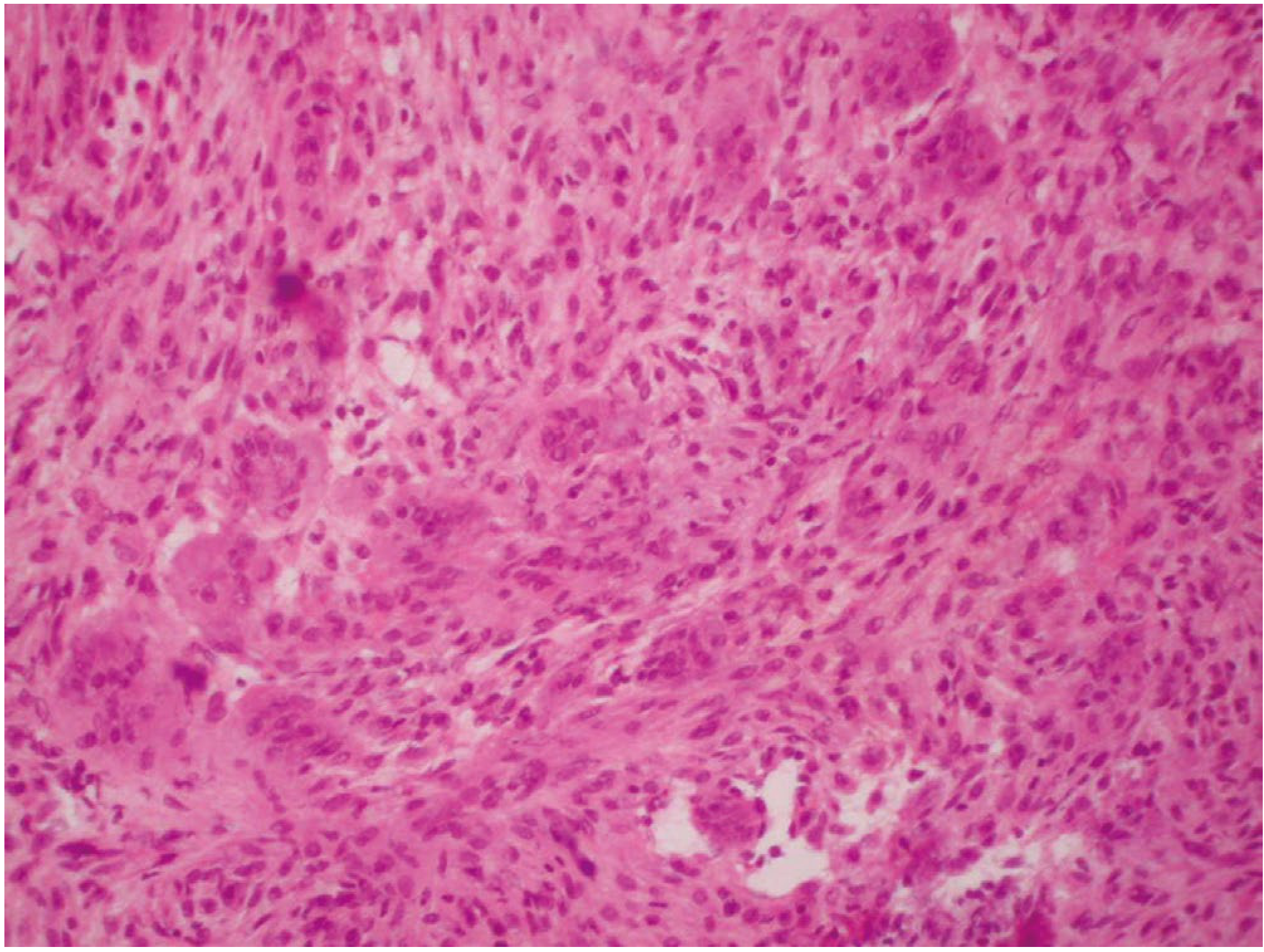
- Cystic spaces lack an endothelial lining
- Septa contain:
- Plump uniform fibroblasts and histiocytes
- Multinucleate osteoclast-like giant cells
- Hemosiderin-laden macrophages
- Reactive woven bone
- Chronic inflammatory cells
Clinical Features
- Localized pain and swelling (present for weeks to months)
- Limitation of motion
- Rapid growth can mimic a malignancy clinically
- Spinal lesions may cause neurologic deficits or radicular pain
- Although benign, ABCs are locally aggressive
Differential Diagnosis
The radiographic and clinical appearance overlaps with:
- Giant cell tumor - most important mimic (especially in adults)
- Enchondroma
- Unicameral (simple) bone cyst - distinguished by centrally located unilocular cyst with clear fluid
- Telangiectatic osteosarcoma - most dangerous mimic; biopsy mandatory
Solid Variant
A solid variant of ABC exists, frequently referred to as giant cell reparative granuloma, which lacks the classic blood-filled spaces.
Treatment
| Setting | Preferred Treatment |
|---|---|
| Standard long bone lesion | Extended curettage + bone graft substitute |
| Expendable bones | Marginal resection |
| Spine/pelvis | Preoperative arterial embolization to minimize blood loss, then curettage |
| Inaccessible lesions | Percutaneous sclerotherapy (fluoroscopic guidance) |
| High surgical risk | Denosumab (anti-RANKL monoclonal antibody) |
| Historical/rarely used | Low-dose radiation (risk of malignant transformation precludes routine use) |
Key surgical note: Tourniquet control is advised due to heavy bleeding potential.
Recurrence
- After curettage: 10-20% recurrence rate (Robbins: up to 50% in some series)
- Risk factors for recurrence:
- Age younger than 15 years
- Centrally located cysts
- Incomplete removal of cystic cavity contents
- Recurrent cysts can be retreated with the same approach
Summary Comparison: ABC vs Unicameral Bone Cyst
| Feature | ABC | Unicameral BC |
|---|---|---|
| Age | 1st-2nd decade | 1st-2nd decade |
| Sex | Slight female predominance | Male:female 2:1 |
| Location | Eccentric, metaphyseal | Central, metaphyseal |
| Common sites | Proximal humerus, distal femur, tibia, spine | Proximal humerus, proximal femur |
| MRI | Fluid-fluid levels, double-density, septa | Simple cyst, no septa |
| Fluid | Blood-filled | Straw-colored fluid |
| Presentation | Pain | Usually silent; pathologic fracture |
| Treatment | Extended curettage | Observation/aspiration/injection |
Sources:
- Robbins & Kumar Basic Pathology, p. 787 (Robbins Pathology)
- Campbell's Operative Orthopaedics 15th Ed 2026, pp. 1172, 4548-4549
- Grainger & Allison's Diagnostic Radiology, p. 1959
This is a shared conversation. Sign in to Orris to start your own chat.