I have all the data needed now. Let me update the todos and compile the comprehensive answer:Here is the complete, detailed, exam-oriented answer covering all your MUHS 2nd MBBS Pathology questions. All content is sourced from Robbins Pathologic Basis of Disease (the standard reference aligned with Harsh Mohan's textbook).
MUHS 2nd MBBS Pathology - Renal Pathology Complete Notes
TOPIC 1: RENAL CELL CARCINOMA
Definition
Renal cell carcinoma (RCC) arises from the renal tubular epithelium and is located predominantly in the cortex. It represents 80-85% of all primary malignant renal neoplasms and about 2-3% of all adult cancers.
Epidemiology
- Peak incidence: 6th-7th decade
- Men:Women = 2:1
- Risk factors: Smoking, hypertension, obesity, cadmium exposure, acquired polycystic disease (30x risk)
Classification of RCC
| Type | Frequency | Origin | Key Feature |
|---|
| Clear Cell | 65% | Proximal tubule | VHL gene mutation (chr 3p25) |
| Papillary | 10-15% | Proximal tubule | MET gene abnormality (chr 7q) |
| Chromophobe | 5% | Intercalated cells of collecting duct | Multiple chromosome losses |
Pathogenesis - Clear Cell Carcinoma (MOST IMPORTANT)
- Loss/inactivation of VHL gene on chromosome 3p25 is the molecular hallmark
- VHL protein normally degrades HIF (Hypoxia Inducible Factor)
- Without VHL → HIF stabilizes → ↑VEGF (promotes angiogenesis) → highly vascular tumor
- Sporadic tumors: monoallelic deletion of 3p + mutation of second allele
- Familial: VHL disease (autosomal dominant) - bilateral/multiple CCCs in 40-60%
Pathogenesis - Papillary RCC
- MET gene (tyrosine kinase receptor) on chr 7q - activating mutations or increased copy number
- Frequently multifocal and bilateral
GROSS MORPHOLOGY
┌─────────────────────────────────────────────────────┐
│ CLEAR CELL CARCINOMA - GROSS │
│ │
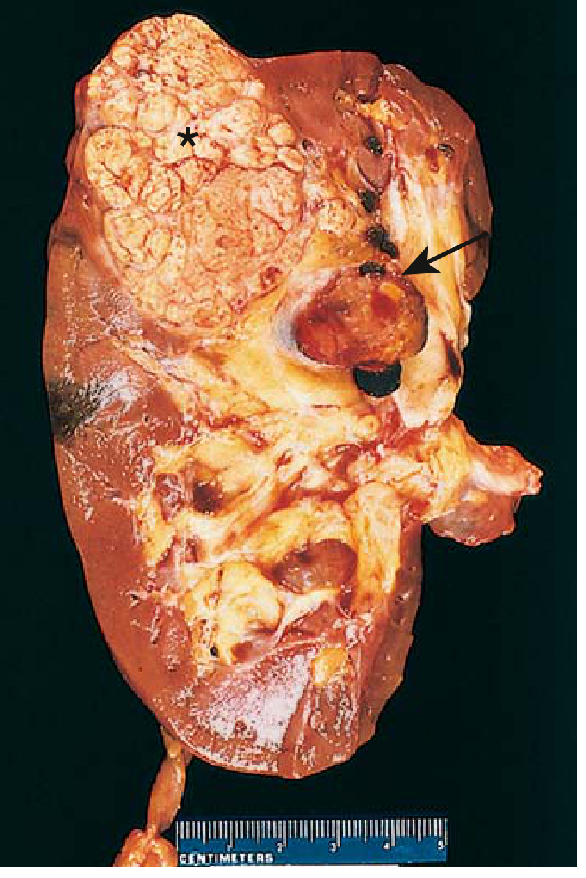
│ • Large, solitary, spherical mass (3-15 cm) │
│ • Located in CORTEX │
│ • Cut surface: YELLOW TO ORANGE (lipid-rich) │
│ • Areas of necrosis, hemorrhage, cystic softening │
│ • Often pseudocapsulated (well-circumscribed) │
│ • May grow into renal vein / inferior vena cava │
│ (tumor thrombus - CLASSICAL FEATURE) │
└─────────────────────────────────────────────────────┘
- Papillary RCC: Frequently multifocal, bilateral; often early-stage tumors
- Chromophobe RCC: Pale lightly eosinophilic cut surface
Classic Triad (only in ~10%): Hematuria + Flank pain + Palpable mass
MICROSCOPIC MORPHOLOGY
Clear Cell Carcinoma:
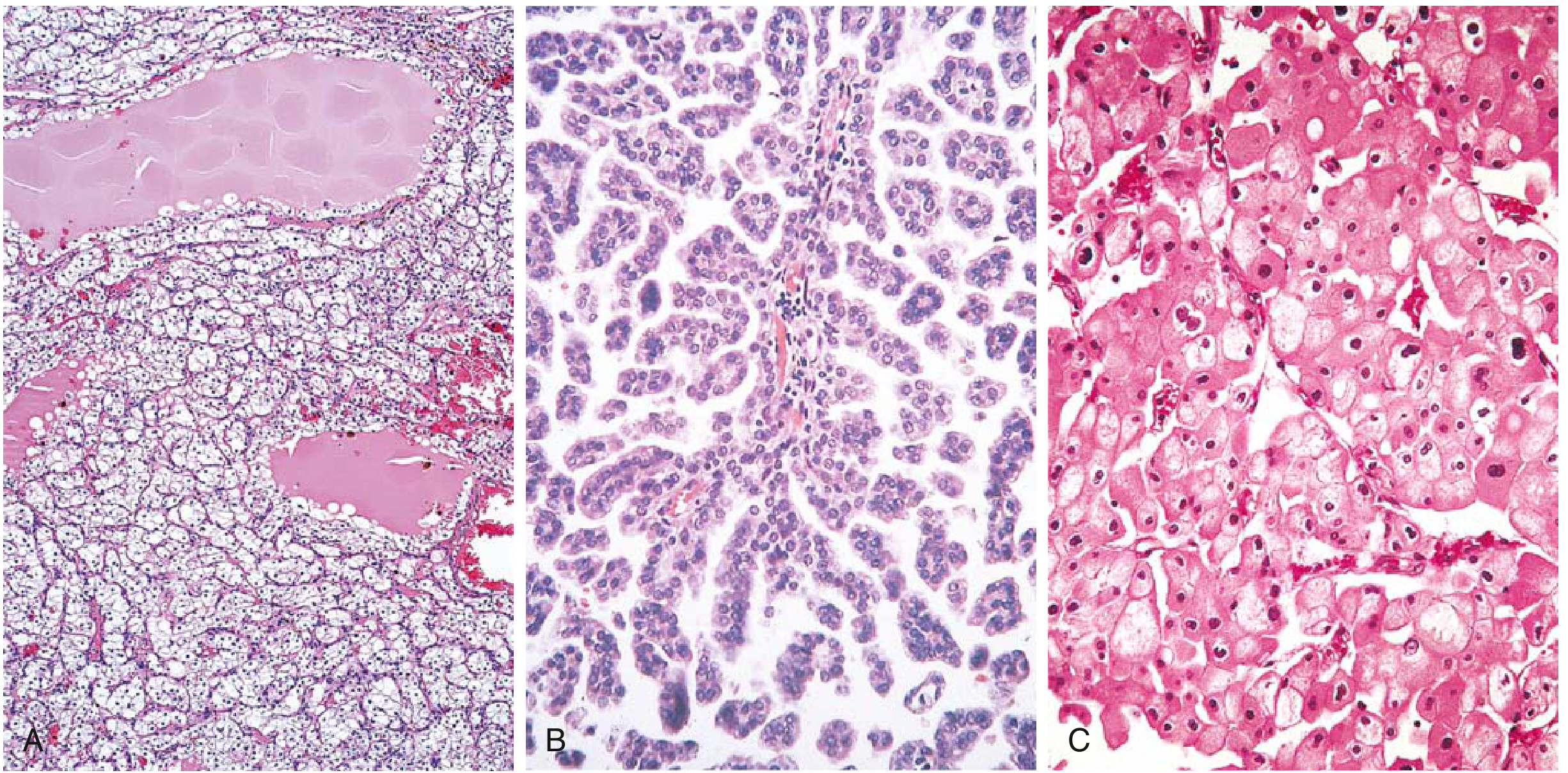
- Cells arranged in solid, tubular, or cystic patterns separated by a delicate vascular network
- Tumor cells have clear cytoplasm (dissolved lipid and glycogen in routine sections)
- Nuclei variable from small/bland to large/irregular
Papillary RCC:
- Papillary architecture with fibrovascular cores
- May be multifocal; psammoma bodies may be seen
Chromophobe RCC:
- Cells are pale (chromophobe) but NOT clear like clear cell type
- Large cells with prominent cell membranes ("plant cell" appearance)
CLINICAL FEATURES
- Hematuria (most common symptom)
- Classic triad: hematuria + flank pain + palpable mass (10%)
- Paraneoplastic syndromes (IMPORTANT for exam):
- Polycythemia (ectopic EPO)
- Hypercalcemia (PTHrP)
- Hypertension (renin)
- Cushing syndrome (ACTH)
- Feminization/masculinization (gonadotropins)
- Spread: hematogenous (lung "cannonball" mets), lymphatic; tumor thrombus in renal vein/IVC
Source: Robbins & Kumar Basic Pathology, Robbins Pathologic Basis of Disease
TOPIC 2: CLASSIFICATION OF GLOMERULAR DISEASES
A. Clinical Syndromes of Glomerular Disease
┌───────────────────────────────────────────────────────────────────────┐
│ GLOMERULAR SYNDROMES (MUHS Exam Important) │
├────────────────────┬──────────────────────────────────────────────────┤
│ NEPHRITIC │ Hematuria (RBC casts), Proteinuria (subnephrotic)│
│ SYNDROME │ Azotemia, Oliguria, Hypertension, Edema │
├────────────────────┼──────────────────────────────────────────────────┤
│ NEPHROTIC │ Massive proteinuria (>3.5g/day), Hypoalbuminemia │
│ SYNDROME │ Generalized edema, Hyperlipidemia, Lipiduria │
├────────────────────┼──────────────────────────────────────────────────┤
│ RAPIDLY │ Acute renal failure + features of nephritic │
│ PROGRESSIVE GN │ syndrome; crescents on biopsy │
├────────────────────┼──────────────────────────────────────────────────┤
│ CHRONIC GN │ Slowly progressive renal failure, proteinuria, │
│ │ hematuria, hypertension │
├────────────────────┼──────────────────────────────────────────────────┤
│ ASYMPTOMATIC │ Incidental hematuria/proteinuria without │
│ HEMATURIA/ │ systemic symptoms │
│ PROTEINURIA │ │
└────────────────────┴──────────────────────────────────────────────────┘
B. Classification by Primary Glomerular Diseases
Diseases Presenting as NEPHRITIC Syndrome:
- Acute proliferative (post-streptococcal) GN
- Rapidly progressive (crescentic) GN
- IgA nephropathy (Berger disease)
Diseases Presenting as NEPHROTIC Syndrome:
- Minimal Change Disease (MCD) - #1 in children
- Focal Segmental Glomerulosclerosis (FSGS)
- Membranous Glomerulopathy - #1 in adults
- Membranoproliferative GN (MPGN)
Diseases Presenting as BOTH:
TOPIC 2B: MEMBRANOUS GLOMERULOPATHY
Definition
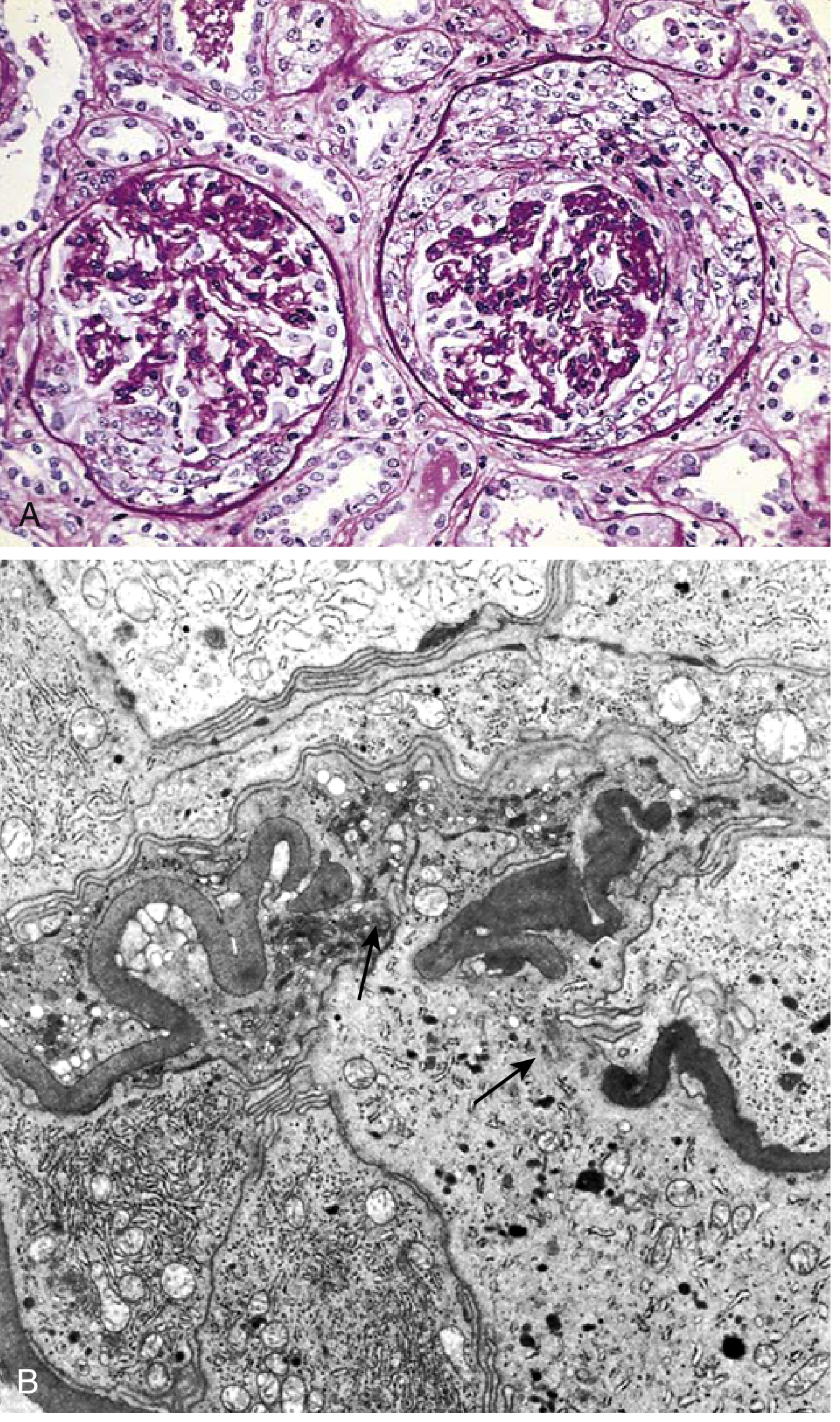
A form of chronic immune complex-mediated glomerular disease characterized by diffuse thickening of the glomerular capillary wall due to subepithelial immune complex deposits.
Causes
| Primary (Idiopathic) ~75% | Secondary ~25% |
|---|
| Anti-PLA2R antibodies (60%) | Hepatitis B, C |
| Anti-NELL1, THSD7A, EXT1/2 | Syphilis (T. pallidum) |
| Anti-NCAM1 antibodies | SLE, Rheumatoid arthritis |
| Malignancy (lung, colon) |
| Drugs (penicillamine, gold, NSAIDs, captopril) |
Pathogenesis
- Main antigen: M-type phospholipase A2 receptor (PLA2R) on podocytes
- Anti-PLA2R antibodies (mainly IgG4 subclass) bind to PLA2R on basal surface of glomerular epithelial cells
- → Complement activation → Immune aggregates shed → Subepithelial immune complex deposits
- C5b-9 membrane attack complex injures podocytes → loss of foot processes → proteinuria
- No significant neutrophil/monocyte infiltration (paucicellular injury)
PATHOGENESIS DIAGRAM:
Anti-PLA2R Ab → binds podocyte PLA2R → immune complex formed
↓
Subepithelial deposits form (IgG4 + complement)
↓
C5b-9 MAC activates → podocyte injury
↓
Foot process effacement → Massive proteinuria (NEPHROTIC SYNDROME)
↓
GBM material deposited between deposits → "SPIKE" formation
MORPHOLOGY OF MEMBRANOUS GLOMERULOPATHY
Light Microscopy:
- Early: Glomeruli appear normal or uniform, diffuse thickening of capillary wall
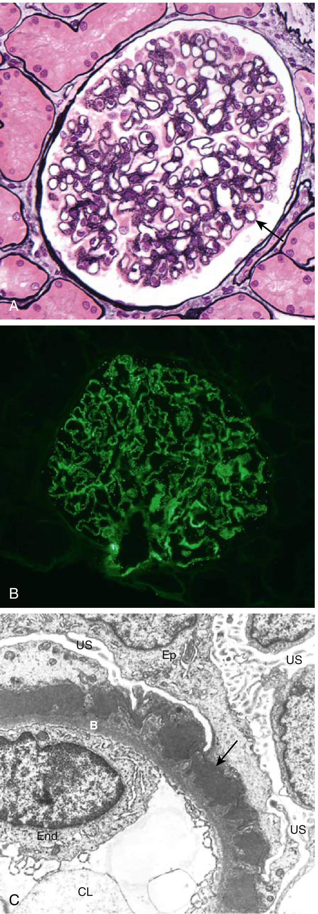
- Silver stain: Characteristic "spike and dome" pattern - spikes of GBM material project between subepithelial deposits
- Advanced: Spikes close over deposits → markedly thickened, irregular GBM
- May progress to segmental sclerosis → global sclerosis
- No cellular proliferation, no inflammatory infiltrate (paucicellular)
Electron Microscopy (MOST DIAGNOSTIC):
- Subepithelial electron-dense deposits between GBM and podocytes
- Effacement of podocyte foot processes
- GBM material laid down between deposits → spikes
- Progressive: spikes form dome-like protrusions → deposits buried inside thickened GBM
Immunofluorescence:
- Granular deposits of IgG and C3 along capillary walls (diffuse, global)
- IgG4 predominant in PLA2R-associated disease
STAGES OF MEMBRANOUS GN (Ehrenreich-Churg):
Stage I: Subepithelial deposits only - LM near normal
Stage II: GBM spikes between deposits (Silver stain)
Stage III: Deposits enclosed within thickened GBM
Stage IV: Advanced scarring, glomerulosclerosis
Clinical Features:
- Most common cause of nephrotic syndrome in adults (ages 30-50, male predominance)
- Insidious onset of nephrotic syndrome
- "Rule of thirds": 1/3 spontaneous remission, 1/3 proteinuria without progression, 1/3 progressive renal failure
- Increased risk of renal vein thrombosis (due to loss of antithrombin III)
TOPIC 3: CHRONIC PYELONEPHRITIS
Definition
Chronic tubulointerstitial inflammation and scarring involving calyces and pelvis, resulting from repeated or persistent bacterial infections. Pelvocalyceal damage is the key diagnostic feature distinguishing it from other chronic interstitial nephritides.
Types / Pathogenesis
- Obstructive Chronic Pyelonephritis: Recurrent infections due to obstruction (calculi, strictures, prostatic hypertrophy)
- Reflux Nephropathy (most common): Vesicoureteral reflux (VUR) allows infected urine to reflux into renal pelvis → intrarenal reflux → polar scars
GROSS MORPHOLOGY
┌────────────────────────────────────────────────────────────┐
│ CHRONIC PYELONEPHRITIS - GROSS FEATURES │
│ │
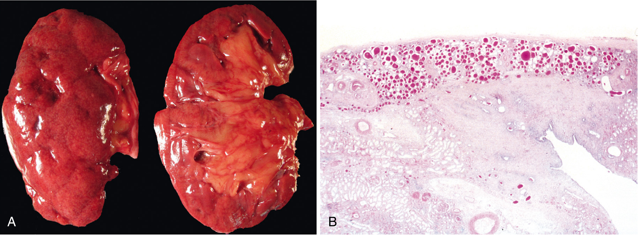
│ • Kidneys: IRREGULARLY SCARRED │
│ • Involvement: ASYMMETRIC (unlike chronic GN = symmetric) │
│ • Hallmark: COARSE, DISCRETE, CORTICOMEDULLARY SCARS │
│ overlying DILATED, BLUNTED, DEFORMED CALYCES │
│ • Flattening of papillae │
│ • Scars predominantly in UPPER AND LOWER POLES │
│ (sites of greatest VUR) │
│ • Remaining parenchyma may show compensatory hypertrophy │
└────────────────────────────────────────────────────────────┘
KEY DISTINGUISHING POINT:
| Chronic Pyelonephritis | Chronic Glomerulonephritis |
|---|
| Scarring | Coarse, irregular, asymmetric | Fine, diffuse, symmetric |
| Calyceal involvement | YES (blunted, deformed) | No |
MICROSCOPIC MORPHOLOGY
- Predominantly tubular and interstitial changes:
- Tubular atrophy in some areas
- Hypertrophy or dilation in others
- "Thyroidization" - Dilated tubules with flattened epithelium filled with proteinaceous casts resembling thyroid colloid (PATHOGNOMONIC feature)
- Interstitial fibrosis and chronic inflammatory infiltrate (lymphocytes, plasma cells)
- Fibrosis around calyceal epithelium
- Glomerular changes (secondary):
- Periglomerular fibrosis
- Fibrous obliteration
- Ischemic changes (wrinkled GBM)
- Secondary FSGS in late stages (causes proteinuria)
Special variant: Xanthogranulomatous Pyelonephritis
- Rare form of chronic pyelonephritis
- Characterized by foamy macrophages (xanthoma cells) + plasma cells + lymphocytes + PMNs + giant cells
- Associated with Proteus infections and obstruction
- Large yellowish-orange nodules - can mimic RCC grossly (important!)
CLINICAL FEATURES
- Silent onset OR recurrent acute pyelonephritis (fever, back pain, pyuria, bacteriuria)
- Loss of tubular concentrating ability → polyuria, nocturia
- Hypertension common
- Radiologic: asymmetrically contracted kidneys, coarse scars, blunted/deformed calyces
- Late: secondary FSGS → significant proteinuria → poor prognosis sign
- Bacteriuria often absent in late stages
TOPIC 4: RAPIDLY PROGRESSIVE GLOMERULONEPHRITIS (RPGN)
Definition
A clinical syndrome characterized by:
- Rapidly progressive renal failure (days to weeks)
- Features of nephritic syndrome
- Severe oliguria
- Glomerular crescents in majority of glomeruli on biopsy → also called Crescentic GN
CLASSIFICATION (3 Types - MOST IMPORTANT FOR EXAM)
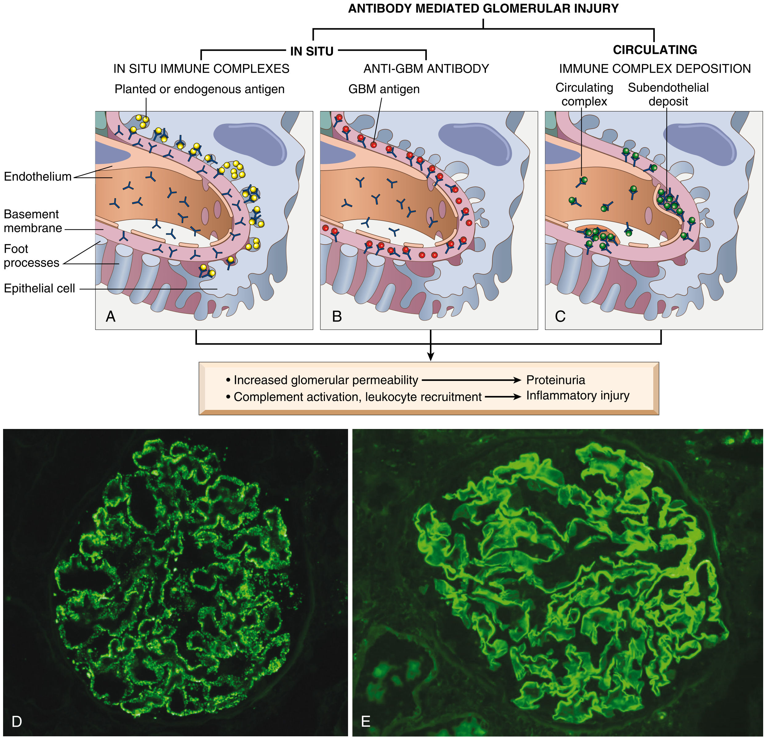
Fig: Antibody-mediated glomerular injury - (A) In situ immune complexes, (B) Anti-GBM antibody - linear pattern, (C) Circulating immune complex deposition - granular pattern on IF
╔══════════════╦════════════════════════╦═══════════════════════╗
║ TYPE ║ MECHANISM ║ IF PATTERN ║
╠══════════════╬════════════════════════╬═══════════════════════╣
║ Type I ║ Anti-GBM antibody ║ LINEAR IgG + C3 ║
║ (Anti-GBM) ║ Autoantibodies vs α3 ║ along GBM ║
║ ~20% ║ chain of collagen IV ║ ║
║ ║ ± Goodpasture syndrome ║ ║
╠══════════════╬════════════════════════╬═══════════════════════╣
║ Type II ║ Immune complex ║ GRANULAR IgG + C3 ║
║ (Immune ║ deposition (post-GN, ║ (mesangium + GBM) ║
║ complex) ║ SLE, IgA nephropathy, ║ ║
║ ~25% ║ HSP, MPGN) ║ ║
╠══════════════╬════════════════════════╬═══════════════════════╣
║ Type III ║ ANCA-mediated ║ PAUCI-IMMUNE ║
║ (Pauci- ║ (PR3-ANCA or MPO-ANCA) ║ (little/no Ig or C3) ║
║ immune) ║ Systemic vasculitis ║ ║
║ ~55% ║ (GPA, MPA) or renal- ║ ║
║ ║ limited ║ ║
╚══════════════╩════════════════════════╩═══════════════════════╝
PATHOGENESIS
Type I - Anti-GBM GN (Goodpasture Syndrome):
- Autoantibodies target α3 chain of type IV collagen in GBM
- Same antigen present in pulmonary alveolar basement membrane → lung hemorrhage
- Goodpasture syndrome = renal failure + pulmonary hemorrhage
- HLA-DRB1 predisposition; triggered by viruses, hydrocarbon solvents
- Treatment: Plasmapheresis (to remove circulating antibodies)
Type II - Immune Complex GN:
- Deposition of circulating antigen-antibody complexes in glomeruli
- May complicate: postinfectious GN, SLE, IgA nephropathy, Henoch-Schönlein purpura
- Cannot be treated with plasmapheresis
Type III - Pauci-Immune GN (ANCA-associated):
- >90% have circulating ANCAs - either:
- PR3-ANCA (c-ANCA): Granulomatosis with Polyangiitis (Wegener's)
- MPO-ANCA (p-ANCA): Microscopic Polyangiitis
- ANCAs activate neutrophils → degranulation → vascular/glomerular injury
- Majority of all RPGN cases
Crescent Formation (Common to All Types):
GBM rupture (by inflammatory injury)
↓
Plasma proteins (fibrin, coagulation factors) leak into Bowman's space
↓
Fibrin clot formation → thrombin activates → triggers crescent formation
↓
Proliferation of PARIETAL EPITHELIAL cells lining Bowman's capsule
+
Migration of MONOCYTES/MACROPHAGES into urinary space
↓
CRESCENT formation - fills and compresses glomerular tuft
↓
Obliteration of capillary loops → ↓↓ GFR → Renal failure
MORPHOLOGY OF RPGN
GROSS:
- Kidneys enlarged and pale
- Petechial hemorrhages on cortical surfaces
LIGHT MICROSCOPY:
- Cellular crescents in Bowman's space - hallmark
- Crescents consist of:
- Proliferating parietal epithelial cells
- Migrated monocytes and macrophages
- ± Neutrophils, lymphocytes
- Fibrin strands prominent between cellular layers of crescents
- Focal and segmental glomerular fibrinoid necrosis
- Crescents may obliterate urinary space, compress glomerular tuft
- Breaks in GBM visible on electron microscopy
- Evolution: Cellular crescents → Fibrous crescents (organized/healed) → Segmental scars
IMMUNOFLUORESCENCE (varies by type):
- Type I: Linear IgG along GBM
- Type II: Granular Ig deposits in mesangium/GBM
- Type III: Negative/trace (pauci-immune)
Clinical Course:
- Severe oliguria, azotemia, hematuria with RBC casts
- Rapidly progresses to end-stage renal disease in weeks-months if untreated
- Poor prognosis; treatment: high-dose steroids + cyclophosphamide ± plasmapheresis (Type I)
TOPIC 5: NEPHROTIC SYNDROME
Definition
A clinical syndrome characterized by massive proteinuria due to increased permeability of glomerular capillary wall.
TETRAD of Features
╔════════════════════════════════════════════════════════════╗
║ NEPHROTIC SYNDROME - 4 CARDINAL FEATURES ║
╠═══════════════════════════════════╦════════════════════════╣
║ 1. MASSIVE PROTEINURIA ║ >3.5 g/day in adults ║
║ (Hallmark/defining feature) ║ >40 mg/m²/hr in child ║
╠═══════════════════════════════════╬════════════════════════╣
║ 2. HYPOALBUMINEMIA ║ <3.5 g/dL ║
╠═══════════════════════════════════╬════════════════════════╣
║ 3. GENERALIZED EDEMA (Anasarca) ║ Periorbital (morning) ║
║ ║ Pitting edema, ascites ║
╠═══════════════════════════════════╬════════════════════════╣
║ 4. HYPERLIPIDEMIA + LIPIDURIA ║ Cholesterol >300 mg/dL ║
╚═══════════════════════════════════╩════════════════════════╝
Pathophysiology of Each Feature
PROTEIN LOSS IN URINE (>3.5 g/day)
↓
↓ Serum albumin (Hypoalbuminemia)
↓
↓ Plasma oncotic pressure
↓
Fluid shifts to interstitium → EDEMA + ↓ circulating volume
↓
Stimulates renin-angiotensin-aldosterone → Na+ and water retention
→ worsens edema
HYPERLIPIDEMIA:
↓ Plasma oncotic pressure → Liver compensates by ↑ lipoprotein synthesis
↓ Clearance of lipoproteins (loss of lipase activating factors in urine)
→ Hypercholesterolemia + Hypertriglyceridemia
LIPIDURIA:
Lipoproteins leak through damaged GBM → appear as:
• Free fat droplets
• "Maltese cross" oval fat bodies (lipid-laden epithelial cells/macrophages)
• Fatty casts in urine
Complications of Nephrotic Syndrome
| Complication | Mechanism |
|---|
| Infections | Loss of immunoglobulins (IgG), complement; opsonization defect |
| Thromboembolism (renal vein thrombosis, DVT, PE) | Loss of antithrombin III, protein C, S; platelet activation |
| Hyperlipidemia/Atherosclerosis | ↑ LDL, VLDL; ↓ HDL |
| Protein malnutrition | Persistent protein loss |
| Iron-deficiency anemia | Loss of transferrin |
| Hypocalcemia | Loss of vitamin D-binding protein |
Causes of Nephrotic Syndrome
| In Children | In Adults |
|---|
| Minimal Change Disease (75-90%) | Membranous Glomerulopathy (#1) |
| FSGS | Diabetic nephropathy |
| Membranous GN (rare) | FSGS |
| Amyloidosis |
| Lupus nephritis |
TOPIC 6: POST-STREPTOCOCCAL GLOMERULONEPHRITIS (PSGN) / ACUTE PROLIFERATIVE GN
Definition
An immune complex-mediated glomerulonephritis that follows infection with certain nephritogenic strains of group A β-hemolytic streptococci.
Etiology
- Causative organism: Group A β-hemolytic Streptococcus (Streptococcus pyogenes)
- Nephritogenic strains: Types 1, 4, 12 (identified by M protein typing) - >90% of cases
- Preceding infection:
- Pharyngitis (strep throat) - more common in temperate regions
- Skin infection/Impetigo - associated with overcrowding, poor hygiene
- Latent period:
- Pharyngitis → GN: 1-3 weeks (avg 10 days)
- Impetigo → GN: 3-6 weeks
- Most common age: Children 6-10 years (but any age)
- Inciting antigen: Streptococcal Pyrogenic Exotoxin B (SpeB) - found in characteristic "hump" deposits
PATHOGENESIS (Etiopathogenesis)
NEPHRITOGENIC STREPTOCOCCAL INFECTION (pharynx/skin)
↓ (latent period 1-4 weeks)
Production of ANTIBODIES vs streptococcal antigens
↓
IMMUNE COMPLEX FORMATION (Ag-Ab complexes)
↓
TWO MECHANISMS:
① Antigens PLANTED in subendothelial location
→ In situ immune complex formation
→ Inflammatory response
② Complexes MIGRATE across GBM
→ Reform as SUBEPITHELIAL deposits
→ Characteristic "HUMPS" (electron-dense subepithelial deposits)
↓
COMPLEMENT ACTIVATION (C3 consumed → LOW serum C3)
↓
CHEMOTACTIC FACTORS (C5a) → NEUTROPHIL + MONOCYTE INFILTRATION
↓
Proteases, free radicals → GBM damage → Hematuria
↓
Inflammatory mediators → ↓ GFR → Oliguria, Azotemia, Hypertension
Evidence for immune mechanism:
- Latent period matches antibody formation time
- ↑ Anti-streptolysin O (ASO) titers in pharyngitis cases
- ↑ Anti-DNAse B (anti-hyaluronidase) titers in impetigo cases
- Low serum complement (C3) during active disease
- Granular immune deposits in glomeruli (IF)
MORPHOLOGY
GROSS MORPHOLOGY:
┌───────────────────────────────────────────────────────┐
│ POST-STREPTOCOCCAL GN - GROSS FEATURES │
│ │
│ • Kidneys: BILATERALLY ENLARGED AND SWOLLEN │
│ • Color: PALE/GRAY appearance │
│ • Petechial hemorrhages on surface │
│ → "FLEA-BITTEN" kidney appearance │
│ • Smooth surface (diffuse process) │
└───────────────────────────────────────────────────────┘
LIGHT MICROSCOPY:
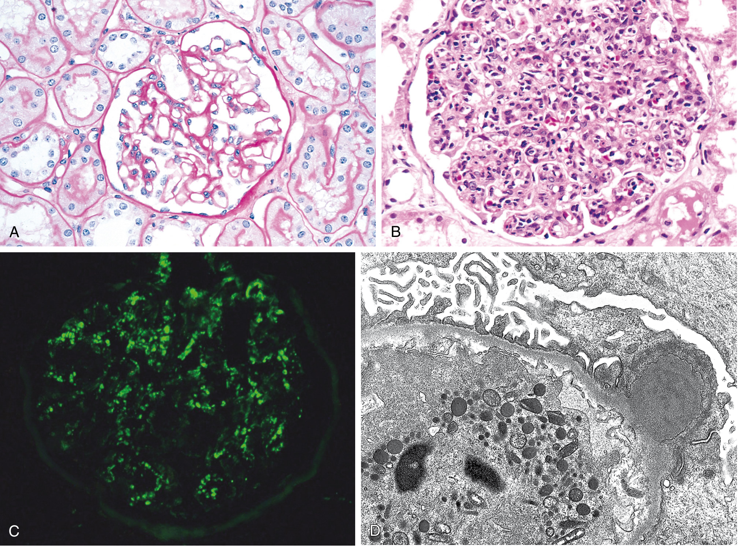
- Diffuse proliferative GN - all glomeruli affected
- Glomerular hypercellularity due to:
- Proliferation of mesangial and endothelial cells (intrinsic cells)
- Infiltration of neutrophils (most characteristic early on) and monocytes
- Glomeruli appear enlarged and hypercellular
- Capillary lumina occluded by swollen cells and infiltrating leukocytes
- May show subepithelial deposits as small bumps
- Tubules: contain RBC casts, protein casts
- In severe cases: crescent formation (indicates bad prognosis)
IMMUNOFLUORESCENCE:
- Granular ("starry sky") deposits of IgG and C3 in mesangium and along GBM
- Complement components predominate (C3 > IgG)
ELECTRON MICROSCOPY (MOST SPECIFIC):
- Subepithelial "humps" - discrete, electron-dense, dome-shaped deposits on epithelial side of GBM (PATHOGNOMONIC)
- Represent antigen-antibody-complement complexes
- Subendothelial and mesangial deposits also present
- Neutrophil in capillary lumen
Fig: Acute Proliferative GN - (A) Normal glomerulus (B) Glomerular hypercellularity - leukocyte infiltration + mesangial/endothelial proliferation (C) Immunofluorescence: coarsely granular C3 "starry sky" deposits (D) EM: Subepithelial electron-dense "HUMP" + neutrophil in lumen
CLINICAL COURSE
Presentation:
- Develops 1-4 weeks after streptococcal pharyngitis (or 3-6 weeks post-impetigo)
- Acute Nephritic Syndrome: sudden onset of:
- Hematuria (smoky/cola-colored urine - RBC casts)
- Oliguria
- Edema (periorbital and peripheral)
- Hypertension
- Azotemia (raised BUN, creatinine)
- Mild-moderate proteinuria (occasionally nephrotic range)
Laboratory:
- ↑ ASO titer (pharyngitis)
- ↑ Anti-DNAse B, Anti-hyaluronidase (impetigo/skin infection)
- ↓ Serum C3 (low complement - complement consumption)
- Urinalysis: hematuria, proteinuria, RBC casts, granular casts
Prognosis:
| Children | Adults |
|---|
| Excellent: >95% recover completely | More guarded |
| Latent period resolves in weeks | 15-50% develop chronic renal disease over years |
| Complement normalizes in 6-8 weeks | Especially if severe histologic injury |
| Some may develop RPGN (crescent formation) | |
QUICK COMPARISON TABLE (EXAM-READY)
| Feature | RPGN | PSGN | Membranous GN | Chronic Pyelo |
|---|
| Presentation | Nephritic + ARF | Nephritic | Nephrotic | CRF + Recurrent UTI |
| Gross Kidney | Enlarged, pale, petechiae | Enlarged, "flea-bitten" | Normal early | Irregular scars, asymmetric |
| LM | Crescents (>50% glomeruli) | Diffuse hypercellularity, PMN infiltrate | Capillary wall thickening | Thyroidization, interstitial fibrosis |
| IF | Linear (I), Granular (II), Negative (III) | Granular (IgG + C3) "starry sky" | Granular IgG + C3 along capillaries | Not glomerular |
| EM | GBM breaks, crescents | Subepithelial humps | Subepithelial electron-dense deposits + spikes | - |
| Key protein | Anti-GBM Ab / ANCA | Streptococcal SpeB antigen | Anti-PLA2R (IgG4) | - |
| Complement | Low (type I, II); Normal (III) | ↓↓ C3 | Normal (idiopathic) | Normal |
IMPORTANT MNEMONICS
RPGN Types: "Anti-GBM, Immune complex, Pauci-immune" = A-I-P (Anti-Immune-Pauci)
Nephrotic Syndrome: "Protein Heaps Exiting Losing" = Proteinuria, Hypoalbuminemia, Edema, hyperLipidemia
PSGN nephritogenic strains: "1, 4, 12 - Pharyngitis strains"
Chronic Pyelonephritis microscopy: "TICS" = Thyroidization, Interstitial fibrosis, Chronic inflammatory infiltrate, Scarring with calyceal blunting
RCC triad: "Pain, Hematuria, Mass" - only 10% have all three
Sources: Robbins Pathologic Basis of Disease (Robbins, Cotran & Kumar) - Chapter 20: Kidney; Robbins & Kumar Basic Pathology - Chapter 12 & 14. These closely align with Harsh Mohan's Textbook of Pathology used for MUHS exams.