I now have sufficient content to compile thorough BHMS-style notes. Let me write them up.
BHMS Practice of Medicine - PNEUMONIA
Complete Structured Notes
1. DEFINITION
Pneumonia is an acute infection of the lung parenchyma associated with:
- At least one respiratory symptom (cough, dyspnea, chest pain)
- Abnormal opacities on lung imaging
- With or without fever
- In the absence of an alternative diagnosis
It leads to consolidation of the usually air-filled alveoli and can be caused by bacteria, viruses, mycobacteria, mycoplasma, and fungi.
(Goldman-Cecil Medicine, p. 990)
2. CLASSIFICATION
| Type | Definition |
|---|
| Community-Acquired Pneumonia (CAP) | Acquired outside hospital/healthcare settings |
| Hospital-Acquired Pneumonia (HAP) | Develops ≥48 hrs after hospital admission |
| Ventilator-Associated Pneumonia (VAP) | In intubated ICU patients |
| Aspiration Pneumonia | Due to aspiration of oropharyngeal/gastric contents |
| Opportunistic Pneumonia | In immunocompromised hosts (PCP, fungal) |
3. ETIOLOGY / CAUSATIVE ORGANISMS
Typical Bacteria (Lobar/Consolidative Pattern)
- Streptococcus pneumoniae - most common cause of CAP
- Haemophilus influenzae - common in smokers and COPD
- Klebsiella pneumoniae - alcoholics, upper lobe with bulging fissure
- Staphylococcus aureus - post-influenza, IV drug users, MRSA
- Gram-negative bacilli - nosocomial/HAP settings
Atypical Bacteria (Interstitial/Walking Pneumonia Pattern)
- Mycoplasma pneumoniae - young adults, gradual onset
- Chlamydophila pneumoniae - community-acquired, mild illness
- Legionella pneumophila - legionnaires' disease, water exposure, smokers
Viruses
- Influenza A and B (most common viral cause)
- SARS-CoV-2 (COVID-19)
- Human metapneumovirus
- Respiratory syncytial virus (RSV)
- Measles, varicella (in children - can lead to bacterial secondary pneumonia)
Note: Respiratory viruses typically cause 20-30% of cases; bacterial causes account for up to 30%; in over 50% of cases no clear pathogen is confirmed.
(Goldman-Cecil Medicine, p. 993)
4. RISK FACTORS
Host Risk Factors:
- Old age (strongest risk factor - exponential increase after 65 years)
- Male sex
- Smoking
- Poor dental hygiene
- Crowded living / institutional settings (prisons, nursing homes)
- Malnutrition
- Alcohol use disorder
Underlying Diseases:
- Chronic lung disease (COPD, bronchiectasis, cystic fibrosis)
- Neurological disease / dementia / impaired gag reflex
- HIV infection (low CD4 count)
- Diabetes mellitus
- Immunosuppression (organ transplants, chemotherapy)
Medications:
- Opioids
- Proton pump inhibitors
- Corticosteroids / immunosuppressants
5. PATHOPHYSIOLOGY
The lung continuously receives microbes from inhaled air and subclinical microaspiration from the oropharynx. Pneumonia occurs when one or more pathogens become dominant in the lung microbiome and stimulate a host inflammatory response.
Routes of infection:
- Inhalation - most common (droplets, aerosols)
- Microaspiration - oropharyngeal secretions (commonest in elderly, alcoholics)
- Hematogenous spread - bacteremia from distant site
Defense mechanisms that fail:
- Decreased cough reflex
- Impaired mucociliary clearance
- Destruction of surfactant / alveolar macrophage function
- Immunosuppression
Stages of Lobar Pneumonia (classical)
| Stage | Timing | Pathology |
|---|
| Congestion | Day 1-2 | Vascular engorgement, serous exudate |
| Red hepatization | Day 3-4 | RBCs, fibrin, PMNs fill alveoli - lung looks like liver |
| Grey hepatization | Day 5-7 | RBCs lyse, fibrin persists, PMNs dominate |
| Resolution | Day 8+ | Enzymatic digestion, macrophage clearance |
6. CLINICAL FEATURES
Typical (Bacterial/Lobar) Presentation
- Rapid onset of high fever (>38.5°C), rigors
- Productive cough - rust-colored sputum (Pneumococcal pneumonia)
- Pleuritic chest pain - sharp, worsens with breathing
- Dyspnea - tachypnea
- Herpes labialis (cold sores around mouth) - in pneumococcal pneumonia
Atypical Presentation (Mycoplasma, Chlamydia, Viruses)
- Gradual onset over days
- Low-grade or absent fever
- Dry, non-productive cough ("walking pneumonia")
- Headache, myalgia, sore throat
- Minimal chest signs despite significant radiological changes
Signs on Physical Examination
| Finding | Significance |
|---|
| Tachypnea (>25 breaths/min) | Respiratory compromise |
| O2 saturation <92% | Significant hypoxia |
| Crackles (crepitations) | Over affected area - bacterial pneumonia |
| Bronchial breath sounds | Lobar consolidation |
| Egophony ("E-to-A" change) | Consolidation - patient says "eee", heard as "aaa" |
| Dullness to percussion | Consolidation or pleural effusion |
| Increased tactile fremitus | Pulmonary infiltrate (decreased over effusion) |
| Tachycardia | Systemic response |
| Hypotension | Suggests sepsis |
(Goldman-Cecil Medicine, p. 994)
7. INVESTIGATIONS
A. Blood Tests
- CBC: Leukocytosis (WBC >15,000/mm³) with neutrophilia - bacterial; normal WBC or lymphocytosis - viral/atypical
- CRP, ESR: Elevated in infection
- Serum creatinine / BUN: For CURB-65 scoring and renal function
- Blood cultures: 2 sets before antibiotics - positive in ~10-15% cases
- ABG / SpO2: To assess respiratory compromise
B. Sputum
- Gram stain and culture - before antibiotics
- Sputum must have <10 squamous epithelial cells per low-power field to be valid
- Pneumococcal pneumonia: Gram-positive diplococci
- Klebsiella: Gram-negative rods, "currant jelly" sputum
C. Urinary Antigen Tests
- Pneumococcal urinary antigen - rapid, specific
- Legionella urinary antigen - highly sensitive and specific for serogroup 1
D. Chest Radiograph (X-Ray) - KEY
- Lobar/segmental consolidation - Pneumococcal, Klebsiella, Legionella
- Interstitial/diffuse infiltrates - Viral, Mycoplasma
- Cavitation - Staph, Klebsiella, Anaerobes, TB
- Parapneumonic effusion - up to 60% of CAP cases
- Upper lobe cavitation - consider TB
- "Bulging fissure" sign - Klebsiella pneumoniae
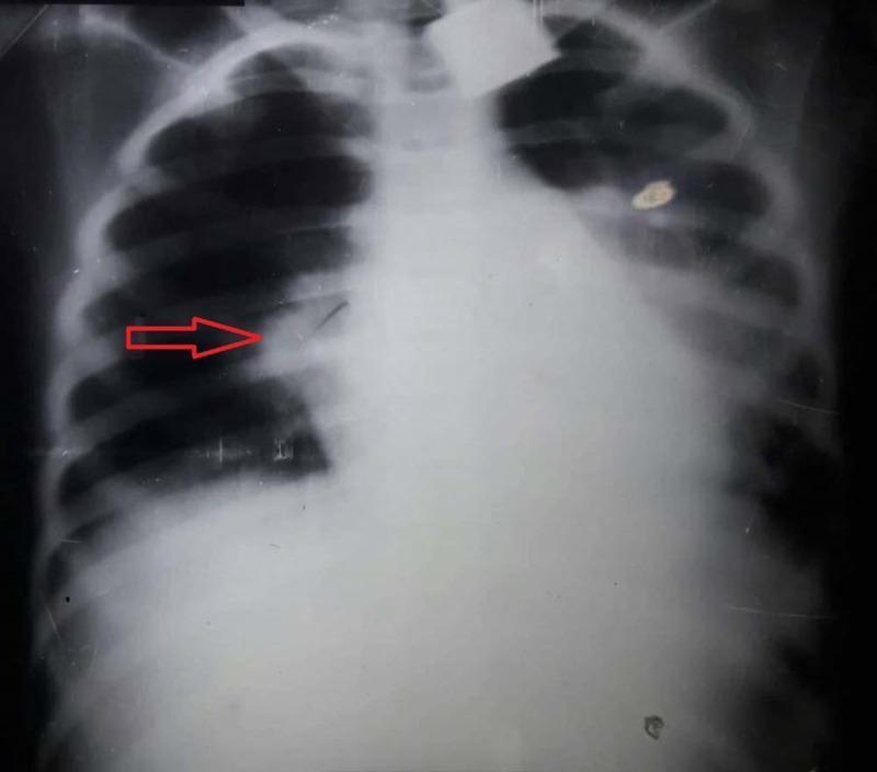
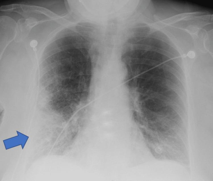
Chest X-ray showing right upper lobe consolidation with well-defined inferior border - classic lobar pneumonia
E. CT Chest
- Higher sensitivity than X-ray
- Used when X-ray is inconclusive or atypical
- Differentiates from pulmonary embolism, malignancy, interstitial lung disease
8. SEVERITY ASSESSMENT SCORES
CURB-65 Score
| Parameter | Points |
|---|
| C - Confusion (new onset) | 1 |
| U - Urea (BUN ≥20 mg/dL) | 1 |
| R - Respiratory rate ≥30/min | 1 |
| B - Blood pressure: systolic <90 or diastolic ≤60 mmHg | 1 |
| 65 - Age ≥65 years | 1 |
Management by Score:
- 0-1: Outpatient treatment
- 2: Short hospital admission or close outpatient follow-up
- 3-5: Hospitalization; score 4-5 = consider ICU
PSI (Pneumonia Severity Index)
| Score | Class | 30-Day Mortality |
|---|
| ≤70 | II | <1-3% |
| 71-90 | III | 3-4% |
| 91-130 | IV | 8% |
| >130 | V | 25% |
(Goldman-Cecil Medicine)
9. COMPLICATIONS
| Complication | Notes |
|---|
| Pleural effusion | Parapneumonic - most common complication |
| Empyema | Infected pleural effusion - 3-5% of CAP |
| Lung abscess | Cavitation - anaerobes, Staph, Klebsiella |
| Respiratory failure | Requires ventilatory support |
| Sepsis / Septic shock | Bacteremia |
| ARDS | Acute respiratory distress syndrome |
| Atrial fibrillation | 20-25% of hospitalized patients |
| Heart failure / Myocardial ischemia | Cardiovascular complications post-pneumonia |
| Hepatitis | Rare - Mycoplasma, Legionella |
| Meningitis | Pneumococcal dissemination |
10. TREATMENT / MANAGEMENT
A. General Supportive Measures
- Rest - bed rest, active as tolerated
- Hydration - adequate oral/IV fluids
- Oxygen therapy - if SpO2 <94%; target SpO2 94-98%
- Monitoring - vital signs, oxygen saturation frequently
- Antipyretics / Analgesics - Paracetamol for fever and pleuritic pain
- Positioning - semi-recumbent to minimize aspiration
B. Outpatient Antibiotic Therapy (Mild CAP)
Healthy adults with no comorbidities (CURB-65 = 0-1):
- Amoxicillin 500 mg TDS x 5 days, OR
- Doxycycline 100 mg BD x 5 days, OR
- Azithromycin 500 mg OD x 5 days
Adults with comorbidities (DM, heart disease, COPD):
- Respiratory fluoroquinolone monotherapy (Levofloxacin 750 mg OD x 5 days), OR
- Amoxicillin-clavulanate + Azithromycin/Doxycycline combination
C. Inpatient Antibiotic Therapy (Moderate CAP, CURB-65 = 2-3)
Without MRSA/Pseudomonas risk:
- Respiratory fluoroquinolone monotherapy (Levofloxacin/Moxifloxacin), OR
- Beta-lactam + Macrolide (Cefuroxime + Azithromycin)
Duration: Minimum 5 days; continue until clinical stability
D. ICU / Severe CAP (CURB-65 = 4-5)
- Dual therapy: Beta-lactam (Ceftriaxone/Piperacillin-tazobactam) + Macrolide or Fluoroquinolone
- If MRSA risk: Add Vancomycin or Linezolid
- If Pseudomonas risk: Antipseudomonal beta-lactam (Piperacillin-tazobactam, Meropenem)
E. Adjunctive Therapy
- Corticosteroids (e.g., prednisolone, dexamethasone) - benefit in severe CAP; also in severe COVID-19 pneumonia with supplemental oxygen need
- Anaerobic coverage (Clindamycin/Metronidazole) - only if lung abscess or empyema suspected
(Goldman-Cecil Medicine, p. 997-998)
11. SPECIAL SITUATIONS
Aspiration Pneumonia
- Common in elderly, neurological patients, alcoholics
- Organisms: Anaerobes, Gram-negative rods
- Treatment: Amoxicillin-clavulanate or Piperacillin-tazobactam + Metronidazole
Legionella Pneumonia (Legionnaires' Disease)
- Water-cooling towers, air conditioners
- Severe pneumonia with hyponatremia, elevated LFTs, diarrhoea
- Treatment: Fluoroquinolone or Azithromycin
Klebsiella Pneumonia
- Alcoholics, diabetics
- "Currant jelly" sputum (blood + mucus)
- Upper lobe consolidation with "bulging fissure"
- Treatment: Cefotaxime/Ceftriaxone + Aminoglycoside
Staphylococcal Pneumonia
- Post-influenza, IV drug users, neonates
- Rapid cavitation, pneumatoceles
- Treatment: Vancomycin (if MRSA) or Flucloxacillin (MSSA)
12. DIFFERENTIAL DIAGNOSIS
- Pulmonary tuberculosis
- Pulmonary embolism
- Lung malignancy
- Pulmonary edema (cardiac failure)
- Pleuritis / pleural effusion
- Bronchitis / acute exacerbation of COPD
- Asthma
- Interstitial lung disease
13. PREVENTION / PROPHYLAXIS
| Vaccine | Target Group |
|---|
| Pneumococcal vaccine (PCV15/PCV20/PPSV23) | All adults >65 yrs; chronic disease patients; immunocompromised |
| Influenza vaccine | Annual - all >50 yrs; chronic lung disease, DM, cardiac disease |
| COVID-19 vaccine | All eligible adults |
Other preventive measures:
- Smoking cessation
- Good dental hygiene
- Treat underlying comorbidities
- Revaccinate with pneumococcal vaccine after 10 years if first dose given before age 65
(Textbook of Family Medicine)
14. FOLLOW-UP
- Clinical review at 48-72 hrs - reassess if no improvement
- Chest X-ray clearance may require weeks - not routinely needed at discharge
- Warn patients about post-pneumonia cardiovascular complications (arrhythmias, heart failure, ACS)
- Supplemental oxygen may be required at discharge but usually temporary
- ICU-level monitoring for deterioration: worsening hypoxia, hemodynamic instability
15. QUICK REVISION TABLE - SPUTUM PATTERNS
| Organism | Sputum Character |
|---|
| Streptococcus pneumoniae | Rusty/rust-colored |
| Klebsiella pneumoniae | Currant jelly (blood + mucus) |
| Haemophilus influenzae | Purulent, greenish |
| Pseudomonas | Greenish-blue (pyocyanin) |
| Anaerobes | Foul-smelling, putrid |
| Viral/Mycoplasma | Scanty, mucoid |
16. MNEMONICS
CURB-65 = Confusion + Urea + Respiratory rate + Blood pressure + age 65
Signs of consolidation = DBLE
- D - Dullness to percussion
- B - Bronchial breathing
- L - Loud / increased vocal resonance
- E - Egophony
Atypical organisms = MCLL
- Mycoplasma
- Chlamydia
- Legionella
- Lungworm (Q fever / Coxiella)
Sources: Goldman-Cecil Medicine International Edition | Textbook of Family Medicine 9e | Murray & Nadel's Textbook of Respiratory Medicine | Fishman's Pulmonary Diseases and Disorders