

PXL_20260529_030839431~2.jpg
I am an ENT resident posted in OT. This is the diagnosis and procedure planned for today. Walk me through the diagnosis including findings, what it is and whatever a consultant might ask me. Then the procedure, begin with anatomy, physiology instruments investigation and step by step brief on the procedure
Reading File
The OT list shows two cases:
- Patient 75365 - Adenoid Hypertrophy + Bilateral Grommets insertion + Myringotomy
- Patient 584005 - Benign Follicular Nodule of Right Lobe of Thyroid - Right Hemithyroidectomy
I'll now do parallel searches across both cases simultaneously.
Finding Sources
Finding Sources
Finding Sources
Finding Sources
Let me broaden the searches with different terms:
Finding Sources
Finding Sources
Finding Sources
Finding Sources
Excellent - found rich sources. Let me now read the most relevant sections in parallel:
Reading File
Reading File
Reading File
Reading File
Reading File
Reading File
Finding Sources
Finding Sources
Reading File
Reading File
Reading File
Reading File
Reading File
Reading File
Reading File
Reading File
I have enough material from the textbooks. Now let me fetch the key images and read the Sabiston thyroidectomy surgical steps, then compile the final answer.
Loading Image
Loading Image
Reading File
Reading File
I now have comprehensive material from the textbooks. Let me compile the full answer.Here is your complete OT briefing for today's two cases:
OT BRIEFING - ENT
CASE 1 - Patient 75365
Diagnosis: Adenoid Hypertrophy
Procedure: Adenoidectomy + Bilateral Myringotomy + Grommet Insertion
THE DIAGNOSIS: ADENOID HYPERTROPHY
What It Is
There is only one nasopharyngeal adenoid - the term "adenoids" (plural) is technically incorrect but universally used. It is part of Waldeyer's lymphoid ring, sitting on the posterior wall of the nasopharynx. The most common cause of enlargement is physiological hypertrophy in childhood, peaking between ages 4-10 years, with spontaneous atrophy usually occurring at puberty.
Key point for consultants: Adenoid size alone does NOT correlate with disease severity. The role of the adenoid as a bacterial reservoir and its associated inflammation (adenoiditis) is more clinically relevant than pure size. Biofilms cover 88-99% of the surface area in adenoiditis/CRS cases vs only 0-6.5% in simple obstruction. (Scott-Brown's Otorhinolaryngology Vol 2)
Clinical Findings
Symptoms to know:
- Nasal obstruction, mouth breathing, hyponasal speech ("potato in mouth" voice)
- Snoring - often the chief complaint in children
- Obstructive sleep apnoea (OSA) - if concurrent tonsillar hypertrophy is present, the upper airway can be severely compromised during sleep
- Recurrent otitis media with effusion (OME/glue ear) - the eustachian tube orifice is near the adenoid; hypertrophy obstructs it
- Postnasal drip, chronic rhinosinusitis
- Recurrent acute rhinosinusitis
Examination findings:
- Mouth breathing child, adenoid facies (elongated face, high-arched palate, open mouth, crowded teeth)
- Hyponasal speech
- On anterior rhinoscopy: mucus, pale inferior turbinates
- Posterior rhinoscopy / flexible nasendoscopy: adenoid mass in postnasal space
Imaging:
Lateral soft tissue X-ray of the neck - the classic radiograph showing a large soft tissue pad in the postnasal space (arrow pointing to it, as below):
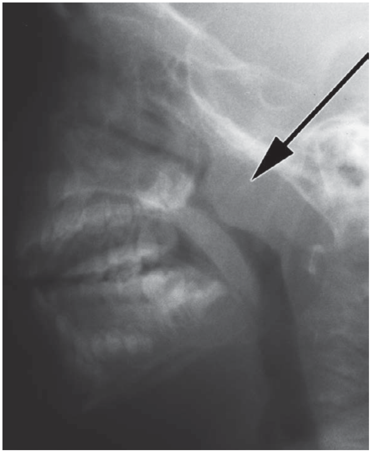
Plain lateral radiograph showing adenoid hypertrophy. The arrow points to the soft tissue mass in the postnasal space. - Bailey & Love, 28th ed.
And this is what it looks like on nasendoscopy - a smooth, lobulated, pinkish-red mass:
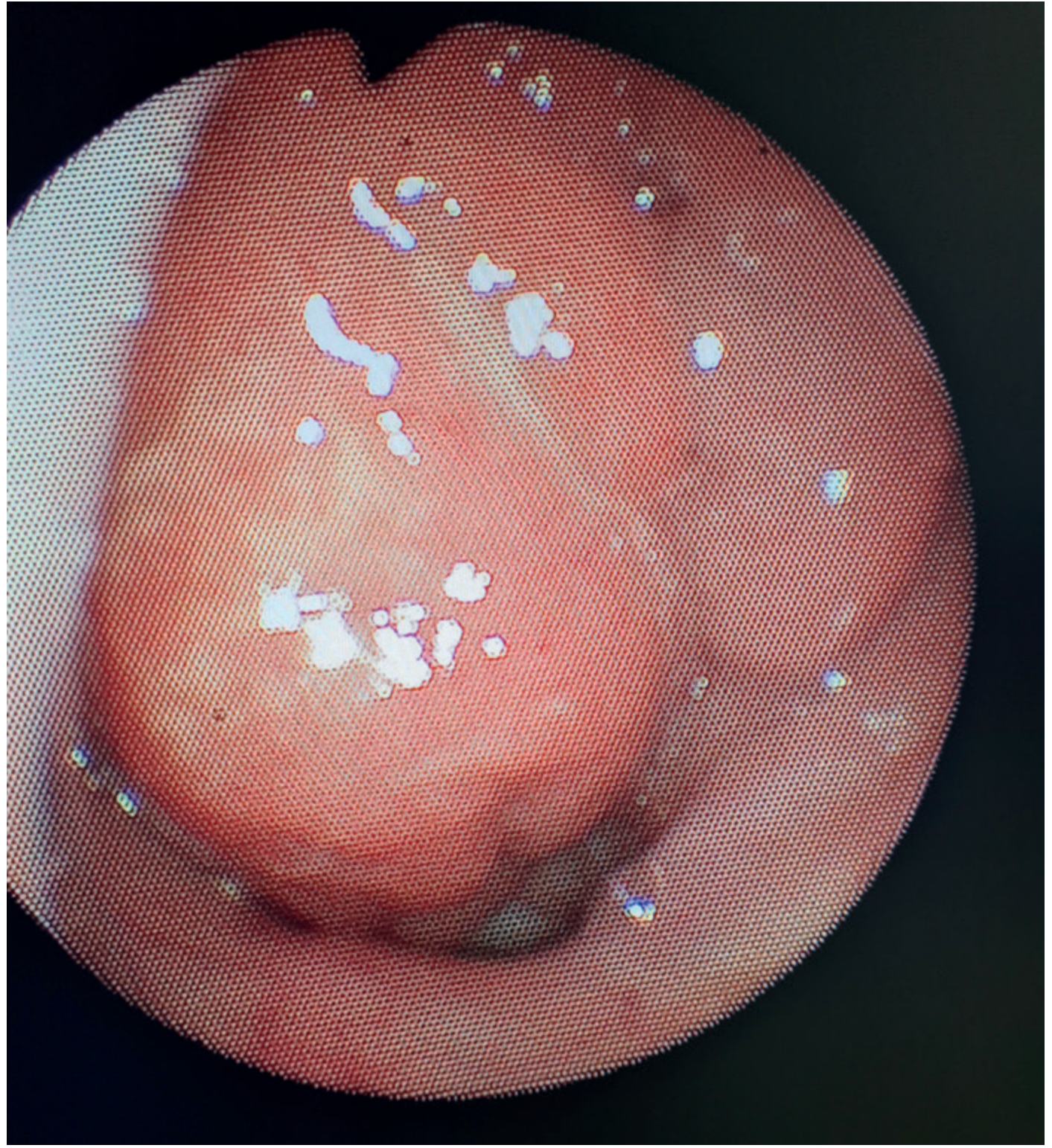
Nasendoscopic view of adenoid hypertrophy - Bailey & Love, 28th ed.
Investigations
- Pure tone audiometry (PTA) + tympanometry - to assess for conductive hearing loss and OME
- Lateral soft-tissue neck X-ray - adenoid/airway ratio
- Flexible nasendoscopy - direct visualisation
- Sleep study (polysomnography) if OSA is suspected
- Consider: allergy workup (skin prick testing, specific IgE)
Organisms in Adenoid Bacterial Reservoir
S. pneumoniae, Group A Streptococcus, H. influenzae, S. aureus, coagulase-negative staphylococci
What the Consultant Will Ask
-
What is Waldeyer's ring and what structures form it?
- Palatine tonsils (2), pharyngeal tonsil (adenoid), tubal tonsils (2), lingual tonsil (1)
-
Why does adenoid hypertrophy cause glue ear/OME?
- The eustachian tube orifice opens on the lateral nasopharyngeal wall near the adenoid. Physical obstruction of the tubal orifice + the adenoid acting as a bacterial reservoir seeding the middle ear via the eustachian tube - both mechanisms operate.
-
Size alone does not decide surgery - what does?
- Consequences: OSA, hearing loss from OME, recurrent rhinosinusitis, failure to thrive. Size is NOT an independent indication.
-
Adult with adenoid hypertrophy - what do you think?
- In an adult, lymphoproliferative disorder (lymphoma) or HIV infection must be excluded before attributing it to physiological hypertrophy. (Textbook of Family Medicine 9e)
-
What are the indications for adenoidectomy?
- OSA with postnasal obstruction
- Recurrent/prolonged OME (>3 months)
- Recurrent rhinosinusitis
- Postnasal discharge causing chronic cough
-
Relationship between adenoid hypertrophy and CRS?
- Adenoid is a bacterial reservoir; bacteria cultured from middle meatal swabs and adenoidal core cultures match in CRS (PPV 91.5%, NPV 84.3%). Adenoidectomy improves CRS symptoms in at least half of young children.
THE PROCEDURE: ADENOIDECTOMY + BILATERAL MYRINGOTOMY + GROMMET INSERTION
Anatomy
Nasopharynx:
- Posterior boundary: anterior arch of C1 vertebra
- Superior: sphenoid/base of skull (this is the danger zone - avoid curetting too superiorly)
- The adenoid sits on the posterior wall and roof, attached to the pharyngobasilar fascia
- Lateral walls contain the opening of the Eustachian tube (at the level of the inferior turbinate) and the fossa of Rosenmuller (pharyngeal recess) - injury here risks Eustachian tube damage
Tympanic membrane (for myringotomy):
The TM is divided into quadrants by the handle of malleus and the umbo:
- Anterior/posterior - by the long process of malleus
- Superior/inferior - by the umbo
- The safe quadrant for myringotomy and grommet insertion is the anteroinferior quadrant - away from ossicles, avoiding the round window niche (posteroinferior) and Shrapnell's membrane (pars flaccida, superiorly)
- Layers of TM: lateral epithelial, middle fibrous (radial + circular), medial mucosal
Physiology Relevant to OME
The Eustachian tube (ET) equates middle ear pressure with atmospheric pressure. Normal ET function: opens briefly on swallowing/yawning (tensor veli palatini muscle, innervated by CN V3). In adenoid hypertrophy, ET orifice obstruction + bacterial reservoir causes:
- Negative middle ear pressure
- Transudation of fluid from middle ear mucosa
- Glue ear (otitis media with effusion, OME)
- Conductive hearing loss (air-bone gap, typically 20-30 dB)
Tympanometry in OME: Type B tympanogram (flat, no peak - pathognomonic of middle ear fluid). Ipsilateral acoustic reflex absent.
Instruments for Adenoidectomy
| Instrument | Purpose |
|---|---|
| St Clair Thomson adenoid curette (guarded) | Classic instrument - scrapes adenoid off the posterior nasopharyngeal wall |
| Negus gag (or Boyle-Davis) | Mouth gag to hold mouth open and depress tongue |
| Draffin bipod rods | Support gag on chest |
| Postnasal mirror | Guide curette direction, check completeness |
| Yankauer / postnasal pack | Haemostasis post-curettage |
| Suction diathermy / Coblator | Alternatives to curette - better haemostasis |
Instruments for Myringotomy + Grommet Insertion
| Instrument | Purpose |
|---|---|
| Aural speculum | Exposure of TM |
| Operating microscope | Magnification |
| Myringotomy knife (Swann-Morton No 15 or dedicated myringotomy blade) | Make incision in TM |
| Alligator forceps / crocodile forceps | Pick up and insert grommet |
| Suction (fine bore Zoellner) | Aspirate middle ear fluid |
| Shah grommet (short-term) or Shepard / T-tube (long-term) | Ventilation tube |
Types of grommets:
- Short-term (Shah/Shepard): Stay 6-18 months, extrude spontaneously. Used in most children with OME.
- Long-term (T-tube): For recurrent OME post-short-term grommet extrusion; up to 55% of Shepard tubes extrude within 6 months.
Pre-operative Investigations
- PTA + tympanometry (confirm OME bilaterally)
- Bloods (FBC, coagulation) - bleeding risk
- Flexible nasendoscopy
- Lateral soft-tissue neck X-ray
Step-by-Step: The Procedure
Patient position: Supine, neck slightly extended (Rose position for adenoidectomy)
Part 1 - Adenoidectomy:
- GA + reinforced ETT / LMA, throat pack inserted
- Boyle-Davis/Negus gag placed to open mouth and depress tongue; Draffin rods support gag on chest
- Soft palate retracted with a rubber catheter passed through one nostril and out the mouth to give access to nasopharynx
- Postnasal mirror or 70-degree nasopharyngoscope used to visualise adenoid
- Guarded St Clair Thomson curette pressed firmly against the posterior nasopharyngeal roof and swept inferiorly in one firm stroke, delivering the adenoid mass into the oropharynx
- Repeat as needed; alternative - suction monopolar diathermy or Coblator (better haemostasis, preferred by many)
- Postnasal pack / ribbon gauze inserted and held for 5 minutes until haemostasis achieved
- Mirror check to confirm complete removal and haemostasis
- Throat pack removed and counted
Part 2 - Bilateral Myringotomy + Grommet Insertion (done under same GA):
- Head turned to one side; aural speculum inserted
- Operating microscope brought in
- TM examined under magnification - confirm opaque/amber TM, absent light reflex, possible air-fluid level
- Radial or circumferential incision made in the anteroinferior quadrant with myringotomy knife
- Middle ear fluid aspirated with fine Zoellner suction (though there is actually no strong evidence that aspiration improves hearing outcomes - aspiration can increase tympanosclerosis risk)
- Grommet loaded on alligator forceps and inserted into the myringotomy incision - the flanges grip the TM
- Confirm grommet is patent (suction through grommet lumen) and correctly seated
- Repeat on opposite side
- Ciprofloxacin-dexamethasone ear drops may be instilled post-insertion to reduce early infective otorrhoea
- Patient recovered and monitored
Complications to Know
Adenoidectomy:
- Haemorrhage (primary/reactionary/secondary) - most feared
- Velopharyngeal insufficiency (hypernasality) - rare, caused by excess removal
- Nasopharyngeal stenosis
- Atlanto-axial subluxation (Grisel's syndrome) - rare
- Eustachian tube injury (if too lateral)
Myringotomy/Grommets:
- Persistent TM perforation (1.8% anteroinferior vs 3.7% posteroinferior placement)
- Tympanosclerosis (whitish plaques on TM)
- Grommet blockage, premature extrusion
- Otorrhoea (infective)
- Hearing must be rechecked post-insertion (grommet restores hearing)
CASE 2 - Patient 584005
Diagnosis: Benign Follicular Nodule, Right Lobe of Thyroid
Procedure: Right Hemithyroidectomy
THE DIAGNOSIS: BENIGN FOLLICULAR NODULE OF THYROID
What It Is
A follicular nodule is a thyroid nodule composed of follicular epithelial cells. On FNAC, a benign follicular/colloid nodule (Bethesda II) shows macrofollicular architecture with abundant colloid - this is the most common FNAC result (60-90% of all aspirates return benign). The key cytological challenge is that a follicular neoplasm (Bethesda IV) CANNOT be distinguished from follicular carcinoma on cytology alone - capsular and vascular invasion can only be assessed on histology of the excised specimen. This is why the procedure is hemithyroidectomy.
FNAC and the Bethesda System
| Bethesda Category | Cytology | Risk of Malignancy | Management |
|---|---|---|---|
| I - Non-diagnostic | Inadequate | 1-4% | Repeat USS-guided FNA |
| II - Benign | Colloid nodule, thyroiditis | 0-3% | Clinical follow-up |
| III - AUS/FLUS | Atypia of undetermined significance | 5-15% | Repeat FNA / molecular testing |
| IV - Follicular Neoplasm | Cellular, microfollicular, scant colloid | 15-30% | Hemithyroidectomy |
| V - Suspicious for Malignancy | Features of PTC/MTC etc | 60-75% | Near-total or total thyroidectomy |
| VI - Malignant | Definitive malignancy | 97-99% | Total thyroidectomy |
This patient is likely Bethesda II or IV. A "benign follicular nodule" on the OT list likely means FNAC showed Bethesda II/IV with follicular features, and hemithyroidectomy is being done for definitive diagnosis + treatment.
(Cummings Otolaryngology; Sabiston Surgery 21e)
Clinical Findings/Presentation
History:
- Often incidental finding or patient notices a neck swelling
- Key red flags for malignancy: age <20 or >70, male sex, hoarseness (RLN involvement), dysphagia, rapid growth, hard/fixed nodule, cervical lymphadenopathy, family history of thyroid cancer, prior neck irradiation
- Symptoms of hyper/hypothyroidism should be sought but are usually absent with a single nodule
Examination:
- Neck: solitary nodule in right lobe of thyroid, moves with swallowing (attached to pre-tracheal fascia which moves with larynx/trachea on swallowing)
- Consistency, mobility, tenderness, size
- Check for cervical lymphadenopathy (levels II-VI)
- Check voice quality (RLN involvement)
BETHESDA IV findings on FNAC:
- Cellular smear, microfollicular pattern, rosette formation
- Scant colloid in background (contrast to benign colloid nodule with abundant colloid)
- Small uniform follicular cells with round nuclei, minimal atypia
Investigations
Essential:
- Serum TSH - screening; if suppressed, consider nuclear scan first (hot nodule rarely malignant)
- Neck Ultrasound (USS) - most important imaging; characterises nodule (TIRADS scoring)
- FNAC (USS-guided) - definitive workup step; Bethesda classification
- Indirect laryngoscopy - pre-op vocal cord assessment (baseline RLN function)
- Serum calcium - parathyroid baseline
USS features favouring malignancy (ACR TIRADS):
Solid, hypoechoic, irregular margins, taller-than-wide shape, microcalcifications, >3 cm size
Not routinely needed:
- Thyroglobulin - not useful pre-operatively (elevated in benign and malignant)
- Calcitonin - only if MTC/MEN-2 suspected
- CT neck/chest - for substernal goitre or suspected advanced disease
What the Consultant Will Ask
-
Why can't FNAC diagnose follicular carcinoma?
- Follicular carcinoma is defined by capsular invasion and/or vascular invasion - features that can ONLY be seen on histopathological examination of the whole excised specimen, not on cytology of cells alone.
-
What is NIFTP and why is it important?
- Non-invasive follicular thyroid neoplasm with papillary-like nuclear features. Previously classified as encapsulated follicular variant of PTC. Reclassified as NIFTP in 2016 because it has essentially zero malignant potential if completely excised. This reclassification changed the management of many Bethesda III-IV nodules.
-
What are the indications for hemithyroidectomy vs total thyroidectomy for a follicular nodule?
- Hemithyroidectomy (lobectomy) is appropriate for Bethesda IV solitary nodule <4 cm, no extrathyroidal extension, no cervical nodes. If final histology shows follicular carcinoma with capsular/vascular invasion, completion thyroidectomy may follow.
- Total thyroidectomy for bilateral disease, nodule >4 cm, high-risk USS features, or suspicious/malignant cytology.
-
What is the rate of malignancy in a Bethesda IV nodule?
- 15-30% (varies by institution and pathology practice)
-
What is Delphian node?
- The pre-cricoid/pre-tracheal lymph node (level VI), first node to be involved in thyroid carcinoma. If enlarged, suggests malignancy.
-
What are ATA risk categories for DTC?
- Low, intermediate, high - based on tumour size, histological variant, extrathyroidal extension, lymph node/distant metastasis, BRAF mutation status.
THE PROCEDURE: RIGHT HEMITHYROIDECTOMY
Anatomy
Gross anatomy of thyroid gland:
- Butterfly-shaped bilobed gland in the anterior neck
- Connected by isthmus overlying 2nd-4th tracheal rings
- Pyramidal lobe present in ~50% (remnant of thyroglossal duct), ascends from isthmus
- Lies deep to strap muscles (sternohyoid, sternothyroid) and within the pre-tracheal fascia
- Posterolateral to the gland: carotid sheath (common carotid artery, internal jugular vein, vagus nerve)
Arterial supply:
- Superior thyroid artery - first branch of external carotid artery; enters superior pole; the external branch of the superior laryngeal nerve (EBSLN) runs close to it - must be preserved (cricothyroid muscle innervation; injury causes inability to tense vocal cords, loss of high notes in voice)
- Inferior thyroid artery - from thyrocervical trunk of subclavian artery; runs posterior to carotid sheath; the recurrent laryngeal nerve (RLN) crosses at the inferior thyroid artery - perpendicular crossing is the landmark for RLN identification
- Thyroid ima artery - in ~2% of people; arises from aorta or innominate; midline; important to know pre-op
Venous drainage:
- Superior thyroid vein → internal jugular vein
- Middle thyroid vein → internal jugular vein (highly variable; must be ligated early in lateral dissection)
- Inferior thyroid veins → innominate veins
The Recurrent Laryngeal Nerve (RLN) - THE most important structure:
- Right RLN loops under subclavian artery; Left RLN loops under aortic arch - so right RLN has a more lateral course
- Ascends in the tracheo-oesophageal groove
- Crosses the inferior thyroid artery (usually passes posterior to it)
- Enters the larynx just posterior to the cricothyroid joint
- Supplies all intrinsic laryngeal muscles except cricothyroid
- Injury causes ipsilateral vocal cord paralysis - hoarseness (unilateral); bilateral injury - respiratory obstruction, may require tracheostomy
Parathyroid glands:
- Usually 4 in number (superior and inferior, bilateral)
- Superior parathyroids: posterior to midpole of thyroid, at level of cricoid cartilage - relatively constant position
- Inferior parathyroids: more variable; typically near inferior pole; embryologically derived from 3rd pharyngeal pouch (with thymus) - hence may descend into mediastinum
- Blood supply: inferior thyroid artery (predominantly)
- Must be identified and preserved; inadvertent removal → hypocalcaemia post-op
Lymphatic drainage:
- Level VI (central compartment - perithyroidal) nodes drain first
- Then lateral neck nodes (levels II, III, IV)
- Level VII = superior mediastinal nodes
Physiology
Thyroid hormone synthesis:
- Iodide trapping via Na/I symporter (NIS) on basolateral membrane
- Oxidation of I⁻ to I₂ by thyroperoxidase (TPO) in presence of H₂O₂
- Organification - iodination of tyrosyl residues on thyroglobulin → MIT, DIT
- Coupling: MIT+DIT = T3; DIT+DIT = T4 (on thyroglobulin scaffold)
- Pinocytosis of colloid into follicular cell
- Hydrolysis of thyroglobulin by lysosomal proteases → T3, T4 released into blood
- TSH (from anterior pituitary) drives all steps via cAMP pathway
- 80% of circulating T3 comes from peripheral conversion of T4 by deiodinases (D1/D2)
- T3 is the active hormone; T4 is prohormone
Preoperative check: Patient must be euthyroid before surgery - check TSH. If hypothyroid or hyperthyroid, treat first (hypothyroidism rare with a single nodule; for hyperthyroidism use thionamides + Lugol's iodine pre-op to reduce vascularity).
Instruments for Hemithyroidectomy
| Instrument | Purpose |
|---|---|
| Scalpel No 15, No 10 | Skin incision, strap muscle division |
| Langenbeck/Farabeuf retractors | Skin/soft tissue retraction |
| Self-retaining retractor (Joll's thyroid retractor) | Maintain exposure of thyroid |
| Lahey's dissecting forceps | Dissect along thyroid lobes, identify RLN |
| Kelly/Mosquito/Roberts artery forceps | Vessel ligation |
| Right-angle (Mixter) forceps | Pass ligatures around vessels |
| Ligaclips / vessel-sealing device (LigaSure/Harmonic scalpel) | Haemostasis of small vessels |
| Nerve Integrity Monitor (NIM) ETT | Intraoperative RLN monitoring |
| Lahey bag | Classic method - sweeping vessels forward |
| Bipolar diathermy | Haemostasis |
| Corrugated drain / suction drain (Redivac) | Post-op drainage |
Note for anaesthesia in your OT: A NIM (Neural Integrity Monitor) reinforced silicone ETT with surface electrodes must be placed in contact with the vocal cords. Muscle relaxants are contraindicated after intubation so RLN responses can be monitored. (Scott-Brown's Vol 1)
Preoperative Preparation
- Euthyroid state confirmed (TSH normal)
- Indirect laryngoscopy - bilateral vocal cord mobility documented
- USS + FNAC reports reviewed
- Consent: RLN injury (1-2% transient, 0.5-1% permanent), hypoparathyroidism (transient in 10-30%, permanent <2%), scar, bleeding, hypothyroidism (if total)
- Blood group and save
- Ca²⁺ baseline
Step-by-Step: Right Hemithyroidectomy
Position: Supine, neck extended over a shoulder roll, head ring. Arms tucked at sides.
-
Incision: Kocher's collar incision - 4-6 cm transverse incision in a skin crease approximately 2 cm above the sternal notch (over the thyroid isthmus level)
-
Raising flaps: Superior and inferior subplatysmal flaps raised in a plane deep to platysma. Superior flap to thyroid cartilage notch; inferior flap to sternal notch.
-
Midline division: Vertical incision through the linea alba between strap muscles (sternohyoid and sternothyroid). These are retracted laterally with Joll's retractor. (Strap muscles are cut only if tumour invades them.)
-
Isthmus division: The isthmus is clamped, divided, and ligated (or sealed with Harmonic/LigaSure) on the right side to mobilise the right lobe.
-
Superior pole dissection: Superior thyroid vessels skeletonised and ligated close to the gland (to protect the EBSLN). The superior thyroid artery and vein are clipped/ligated individually.
-
Middle thyroid vein ligation: Identified and ligated early to allow medial rotation of the lobe.
-
Lateral and posterior dissection - RLN identification: The right lobe is rotated medially. The inferior thyroid artery is identified. The RLN is identified at its crossing with the inferior thyroid artery - it runs in the tracheo-oesophageal groove. It is traced superiorly to its entry into the larynx and protected throughout. NIM confirms function.
-
Parathyroid identification: Superior and inferior parathyroids identified on the posterior thyroid capsule. Their blood supply preserved. If a parathyroid is inadvertently devascularised, it is minced and auto-transplanted into the sternocleidomastoid muscle.
-
Inferior pole dissection: Inferior thyroid vessels ligated close to gland. Lobe freed from trachea by dividing Berry's ligament (dense fibrous attachment of posterior thyroid capsule to trachea - the RLN is immediately lateral and vulnerable here).
-
Specimen delivery: Right lobe and isthmus removed. Sent for histopathology (frozen section if malignancy suspected intraoperatively).
-
Haemostasis and closure: Check for bleeding. Suction drain placed if needed. Strap muscles reapproximated in midline. Platysma closed with absorbable sutures. Skin closed with subcuticular absorbable suture.
-
Post-op: Voice check as soon as patient awake. Serum calcium at 6 hours, 24 hours. Watch for: haematoma (airway emergency - surgical evacuation), hypocalcaemia, hoarseness.
Complications to Know in Detail
| Complication | Cause | Rate | Management |
|---|---|---|---|
| RLN injury (unilateral) | Cutting, stretching, thermal injury | 0.5-1% permanent | Voice therapy, injection laryngoplasty |
| Bilateral RLN injury | Both nerves cut | Rare | Emergency tracheostomy |
| Hypoparathyroidism | Devascularisation/removal | Transient 10-30%; Permanent <2% | Calcium + Vit D supplementation |
| Post-op haematoma | Vessel slippage | 1-2% | Bedside wound opening, return to OT |
| Thyroid storm | Excess release in hyperthyroid | Rare | Beta-blockers, steroids, antithyroids, cooling |
| EBSLN injury | Ligating superior thyroid artery high | ~1-5% | Difficulty pitching voice high, subtle change |
| Hypothyroidism | After total thyroidectomy | 100% | Thyroxine replacement |
Berry's Ligament
This is the dense posterior suspensory ligament of the thyroid. The RLN is immediately lateral to it and sometimes passes through it. Division of Berry's ligament is the most dangerous step in the operation for RLN injury.
CONSULTANT VIVA - RAPID FIRE ROUND
Q: What forms Waldeyer's ring?
A: Paired palatine tonsils, single pharyngeal tonsil (adenoid), paired tubal tonsils, lingual tonsil.
Q: What nerve supplies tensor veli palatini?
A: CN V3 (mandibular division of trigeminal nerve). Opens eustachian tube.
Q: What is Type B tympanogram?
A: Flat curve, high compliance peak absent. Indicates middle ear fluid (OME) or TM perforation (with large volume).
Q: Why insert grommet anteroinferiorly?
A: Avoids ossicular chain (posterosuperior), reduces risk of permanent perforation, prolongs grommet survival (80% at 6 months vs 45% posterior placement).
Q: What is the safety triangle of tympanoscopy?
A: Anteroinferior quadrant - bounded by annulus fibrosus, anterior malleolar fold, inferior to umbo.
Q: Can FNAC diagnose follicular carcinoma?
A: No. Only histological capsular/vascular invasion diagnoses it.
Q: What is NIFTP?
A: Non-Invasive Follicular Thyroid neoplasm with Papillary-like nuclear features. Reclassified from encapsulated follicular variant PTC in 2016. Essentially benign behaviour if completely excised.
Q: Where does the RLN cross the inferior thyroid artery?
A: Usually posterior to it (passes behind the artery), but the relation is variable - may be anterior or between branches.
Q: What is Berry's ligament?
A: Posterior suspensory ligament of the thyroid attaching its posterior capsule to the trachea. The RLN passes immediately lateral to or through it - highest risk point during the operation.
Q: What is the Kocher incision?
A: Transverse skin crease incision 2 cm above the sternal notch, following Langer's lines, giving excellent cosmetic result and good access.
Sources: Bailey & Love's Short Practice of Surgery 28th Ed; Scott-Brown's Otorhinolaryngology Head & Neck Surgery Vols 1 & 2; Sabiston Textbook of Surgery 21e; Cummings Otolaryngology Head and Neck Surgery; KJ Lee's Essential OtolaryngologyThere's your complete OT briefing for both cases. A few things worth keeping at the front of your mind when you're scrubbed in today:
Case 1 (Adenoidectomy + Grommets):
- If the consultant asks why you're doing both procedures together - the grommet treats the OME caused by adenoid hypertrophy; single anaesthetic covers both
- Anteroinferior quadrant only for the myringotomy incision - mention this unprompted
- Know that aspiration of OME fluid is technically optional (evidence doesn't strongly support better hearing outcomes)
Case 2 (Right Hemithyroidectomy):
- The single most important structure in thyroid surgery is the RLN - identify it at the inferior thyroid artery crossing before doing anything irreversible
- Berry's ligament division is the highest-risk step for RLN injury - mention this
- Pre-op vocal cord check (indirect laryngoscopy) is non-negotiable - confirm it was done
- Post-op: first thing to check on waking = voice. Then serum calcium at 6 and 24 hours.
- Muscle relaxants are contraindicated intraoperatively if NIM is being used
Good luck in the OT today.
This is a shared conversation. Sign in to Orris to start your own chat.