
Do heart have m3 receptors
M3 muscarinic receptor heart cardiac
muscarinic receptor subtypes cardiac M2 M3
Do the Heart Have M3 Receptors?
Muscarinic Receptor Distribution in the Heart
| Receptor | Location in Heart | Role |
|---|---|---|
| M2 | Predominant subtype - SA node, AV node, atrial myocardium, ventricles | Main mediator of vagal/parasympathetic cardiac effects |
| M3 | Coronary circulation (endothelium & smooth muscle); also some non-M2 sites in the myocardium | Coronary vasodilation; minor cardiomyocyte effects |
M2 Receptors - The Dominant Cardiac Subtype
- Decrease heart rate (negative chronotropy) - slowing SA node pacemaker activity
- Slow AV conduction (negative dromotropy)
- Directly decrease atrial contractile force
- Exert inhibitory modulation of ventricular contractile force
- Inhibits adenylyl cyclase (reducing cAMP)
- Increases K+ conductance (via GIRK channels) → hyperpolarization
- Modulates Ca2+ channels and If (funny current)
M3 Receptors in the Heart - Where and Why
- Coronary vascular endothelium - stimulation causes release of nitric oxide (NO) → vasodilation
- Coronary vascular smooth muscle - direct relaxation
- Lung, bladder, exocrine glands, smooth muscle are the classic peripheral sites (Lippincott Pharmacology)
- Couples to Gq/11 protein
- Activates phospholipase C
- Generates IP3 (raises intracellular Ca2+) and DAG (activates PKC)
Clinical Relevance
- Anticholinergic drugs (e.g., atropine, glycopyrrolate) block both M2 and M3 receptors. At the heart, M2 blockade is the clinically relevant effect (causing tachycardia).
- Selective M3 antagonists (e.g., darifenacin, used in overactive bladder) are designed to spare M2-mediated cardiac effects. However, since M3 receptors are present in the coronary circulation, they can affect coronary tone.
- The reference to "loss of vagally mediated bradycardia in mice lacking M2 or M3 muscarinic receptors" (from Goodman & Gilman's bibliography) suggests M3 may contribute to vagal bradycardia in some contexts as well.
Summary
The heart has both M2 and M3 muscarinic receptors. M2 is the dominant, clinically important subtype responsible for all classic parasympathetic cardiac effects (bradycardia, reduced AV conduction). M3 receptors are found mainly in the coronary circulation where they mediate vasodilation via NO, and there is some evidence of non-M2 (including M3) receptors in the myocardium itself.
- Miller's Anesthesia (10th ed.), Neural Regulation of Cardiac Function
- Lippincott Illustrated Reviews: Pharmacology, Muscarinic Receptors section
- Goodman & Gilman's Pharmacological Basis of Therapeutics
Effect of parasympathetic
Effect of parasympathetic on which system/organ?
Effects of Parasympathetic Stimulation ("Rest and Digest")
Organ-by-Organ Effects
| Organ/System | Parasympathetic Effect |
|---|---|
| Eye | Pupillary constriction (miosis) via sphincter pupillae; increased lens curvature (accommodation for near vision) via ciliary muscle |
| Salivary Glands | Increased secretion - copious, watery saliva |
| Heart | Slowing of heart rate (bradycardia); decreased AV conduction; decreased atrial contractility |
| Lungs | Bronchoconstriction; increased bronchial secretions |
| GI Tract | Increased secretions; increased motility (peristalsis); relaxation of sphincters |
| Pancreas | Increased secretion (both exocrine and endocrine) |
| Male Sex Organs | Erection (point - parasympathetic = "P" for point) |
| Bladder | Detrusor contraction (micturition); internal urethral sphincter relaxation |
| Skin | No significant effect |
| Blood Vessels | Vasodilation in select beds (e.g., coronary via M3 on endothelium → NO release) |
Effects on the Heart - In Detail
- ACh acts on M2 receptors at the SA node
- Activates Gi → inhibits adenylyl cyclase (↓cAMP) + increases K+ conductance (GIRK channels) → hyperpolarization
- Slows diastolic depolarization → decreases firing rate
- Result: heart rate falls below 60 bpm (bradycardia)
- Extreme vagal stimulation can even stop the heart temporarily (e.g., carotid sinus syndrome causing syncope)
- ACh slows conduction through the AV node
- Increases PR interval on ECG
- Extreme stimulation can cause complete heart block
- Direct decrease in atrial contractile force
- Indirect inhibitory modulation of ventricular contractility
- Postganglionic fibers extend into coronary arteries
- Direct effect is mild vasoconstriction
- However, M3 receptor stimulation on endothelium releases NO causing vasodilation (as discussed in the previous response)
Mechanism of Action - M2 Receptor Signaling
ACh → M2 receptor → Gi/o protein →
  ├── Inhibits adenylyl cyclase → ↓cAMP → ↓PKA → ↓Ca²⁺ influx
  ├── Opens GIRK (K⁺) channels → hyperpolarization
  └── Modulates If (funny current) → slows pacemaker
Important Clinical Points
- Atropine (muscarinic antagonist) blocks all these effects - used to treat bradycardia and heart block
- Vagal tone at rest: the heart is under dominant parasympathetic influence at rest. Loss of vagal tone (e.g., cardiac vagotomy) increases resting heart rate from ~70 to ~160 bpm
- Vasovagal syncope: sudden, excessive vagal discharge causing profound bradycardia + vasodilation → loss of consciousness
- Carotid sinus massage: increases vagal tone deliberately - used to break certain supraventricular tachycardias
- THIEME Atlas of General Anatomy & Musculoskeletal System, Sympathetic/Parasympathetic Synopsis table
- Histology: A Text and Atlas (Pawlina), Systemic Regulation of Heart Function
- Guyton & Hall Textbook of Medical Physiology, Bradycardia & Vagal Stimulation sections
- Lippincott Illustrated Reviews: Pharmacology, Muscarinic Receptors
What is the function of ciliary muscle
Functions of the Ciliary Muscle
1. Accommodation (Primary Function)
| State | Ciliary Muscle | Zonular Fibers (Suspensory Ligaments) | Lens Shape | Vision |
|---|---|---|---|---|
| Near vision | Contracts (sphincter-like) | Relax (tension released) | Becomes more convex (rounder) | Near focus |
| Far/distant vision | Relaxes | Taut/tense | Becomes flatter | Far focus |
When the ciliary muscle contracts, it moves inward and forward, releasing tension on the zonular fibers attached to the lens. The elastic lens then bulges into a more convex shape, increasing its refractive power for near vision. When the muscle relaxes, zonular fibers pull the lens flat for distance vision.
- Innervation: Parasympathetic fibers via CN III (oculomotor nerve), relayed through the ciliary ganglion
- Receptor: M3 muscarinic receptors on ciliary smooth muscle
- Drugs that mimic this: pilocarpine, physostigmine
- Drugs that block this: atropine, cyclopentolate → cause cycloplegia (paralysis of accommodation)
2. Aqueous Humor Drainage - Reduces Intraocular Pressure
- This is the mechanism by which pilocarpine (a muscarinic agonist) lowers IOP in open-angle glaucoma
- Atropine has the opposite effect - blocks ciliary contraction, impairs drainage, can raise IOP (dangerous in glaucoma patients)
3. Aqueous Humor Production (Ciliary Body - Epithelium)
- Beta-adrenoceptors on ciliary epithelium stimulate aqueous humor secretion
- Beta-blockers (e.g., timolol eye drops) reduce secretion and lower IOP - another treatment for glaucoma
Innervation Summary
Parasympathetic (CN III → ciliary ganglion → short ciliary nerves)
  → M3 receptors on ciliary muscle
  → Contraction → accommodation + ↓IOP (trabecular meshwork opening)

Sympathetic
  → β receptors on ciliary epithelium
  → Aqueous humor secretion
Clinical Relevance
| Condition/Drug | Effect on Ciliary Muscle |
|---|---|
| Atropine (cycloplegic) | Blocks M3 → paralyzes accommodation (cycloplegia); used for refraction in children, uveitis |
| Pilocarpine | Stimulates M3 → contracts ciliary muscle → lowers IOP; treats glaucoma |
| Organophosphate poisoning | Excess ACh → sustained ciliary contraction (cyclospasm) → fixed near vision |
| Presbyopia | Lens hardens with age → accommodation lost despite normal muscle |
| CN III palsy | Loss of parasympathetic input → accommodation fails + pupil dilates + ptosis + eye turns "down and out" |
- Katzung's Basic & Clinical Pharmacology (16th ed.), Pharmacology of the Eye
- K.J. Lee's Essential Otolaryngology, The Globe
- Gray's Anatomy for Students, Cranial Nerves (CN III innervation table)
Does activation of parasympathetic causes passing stools
Parasympathetic Activation and Defecation
The Two Defecation Reflexes
Reflex 1: Intrinsic Myenteric Defecation Reflex (weak on its own)
- Feces distend the rectal wall
- Afferent signals spread through the myenteric (Auerbach's) plexus
- Triggers peristaltic waves in the descending colon, sigmoid, and rectum
- Relaxes the internal anal sphincter via inhibitory myenteric signals
- By itself this reflex is relatively weak and cannot effectively cause defecation
Reflex 2: Parasympathetic Defecation Reflex (the powerful amplifier)
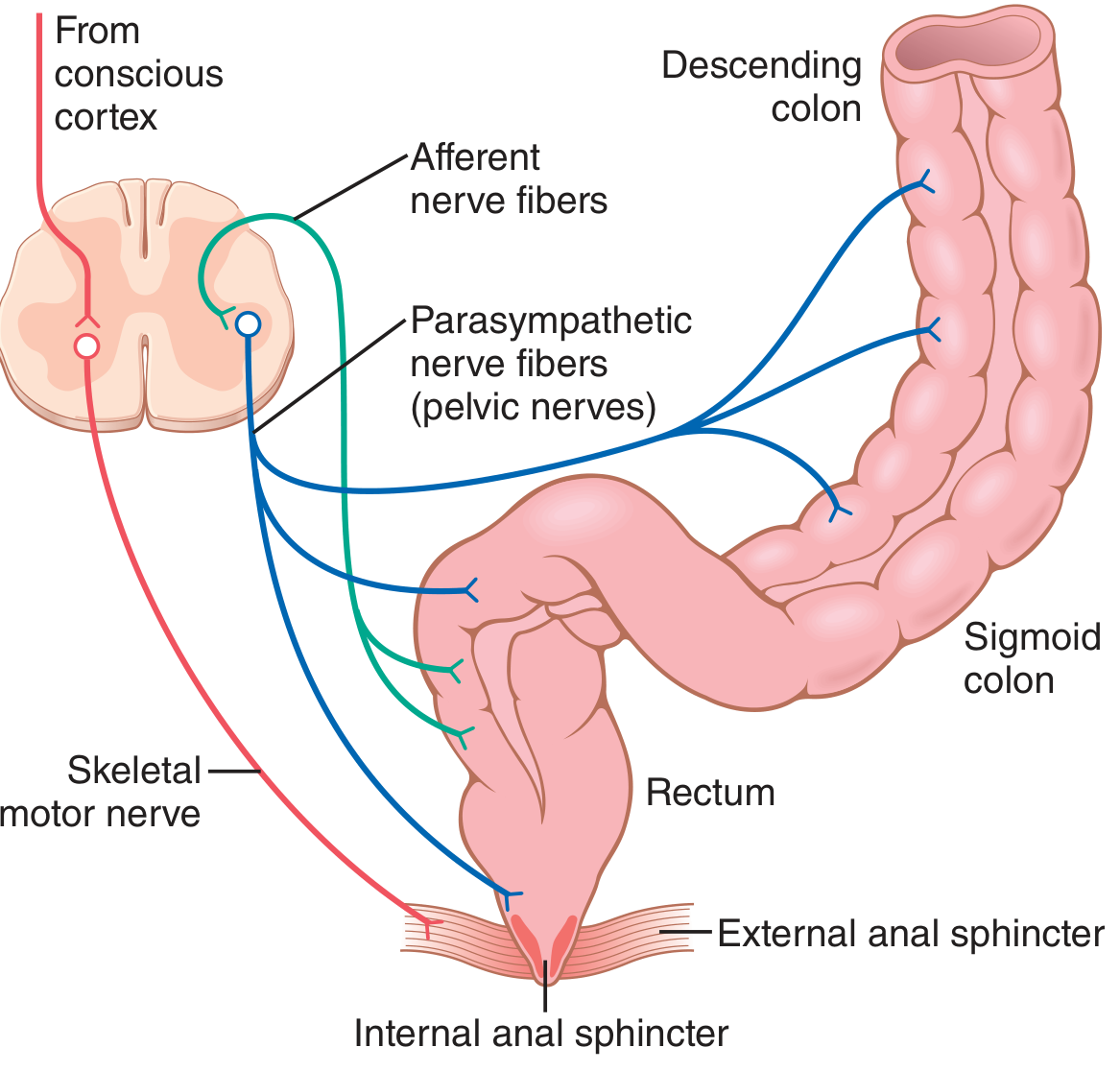
- Rectal distension signals travel afferently to the sacral spinal cord (S2-S4)
- A reflex signal returns via parasympathetic fibers in the pelvic nerves to the descending colon, sigmoid, rectum, and anus
- These parasympathetic signals:
- Greatly intensify peristaltic waves
- Relax the internal anal sphincter
- Convert the weak intrinsic reflex into a powerful, effective defecation reflex
What Happens Step-by-Step
1. Mass movement pushes feces into rectum
        ↓
2. Rectal wall distension → urge to defecate
        ↓
3. Afferent signals → sacral spinal cord (S2-S4)
        ↓
4. Parasympathetic efferents via pelvic nerves
        ↓
5. Intensified peristalsis in descending colon, sigmoid, rectum
   + Relaxation of INTERNAL anal sphincter (smooth muscle, involuntary)
        ↓
6. Additional signals cause:
   - Deep breath (diaphragm moves down)
   - Glottis closure (Valsalva)
   - Abdominal muscle contraction (↑intra-abdominal pressure)
   - Pelvic floor relaxation
        ↓
7. If EXTERNAL anal sphincter (voluntary, somatic/pudendal nerve) is
   consciously relaxed → DEFECATION occurs
The Two Sphincters - Key Distinction
| Sphincter | Type | Control | Parasympathetic Effect |
|---|---|---|---|
| Internal anal sphincter | Smooth muscle (involuntary) | Autonomic | Relaxed by parasympathetic → allows defecation |
| External anal sphincter | Skeletal muscle (voluntary) | Somatic (pudendal nerve, S2-S4) | Not directly controlled by parasympathetic - requires conscious voluntary relaxation |
Defecation requires both: parasympathetic relaxation of the internal sphincter AND voluntary relaxation of the external sphincter. This is why defecation can be consciously postponed even when the urge is strong.
Gastrocolic Reflex - Why You Feel the Urge After Eating
Clinical Relevance
| Condition | Mechanism |
|---|---|
| Spinal cord injury above sacral level | Lose voluntary control of external sphincter → automatic reflex emptying (reflex bowel) |
| Sacral cord injury (S2-S4) | Lose the parasympathetic defecation reflex → severe constipation, atonic bowel |
| Hirschsprung's disease | Absence of myenteric plexus in colon segment → no peristalsis, massive constipation |
| Anticholinergic drugs (atropine) | Block muscarinic receptors → decreased GI motility → constipation |
| Cholinomimetics (neostigmine, bethanechol) | Increase parasympathetic activity → increased motility → diarrhea/defecation |
| Habitual suppression of defecation urge | Weakens natural reflexes over time → chronic constipation |
- Guyton & Hall Textbook of Medical Physiology, Chapter 64 - Defecation
How does tetra benzine acts
Mechanism of Action of Tetrabenazine
Core Mechanism - VMAT2 Inhibition
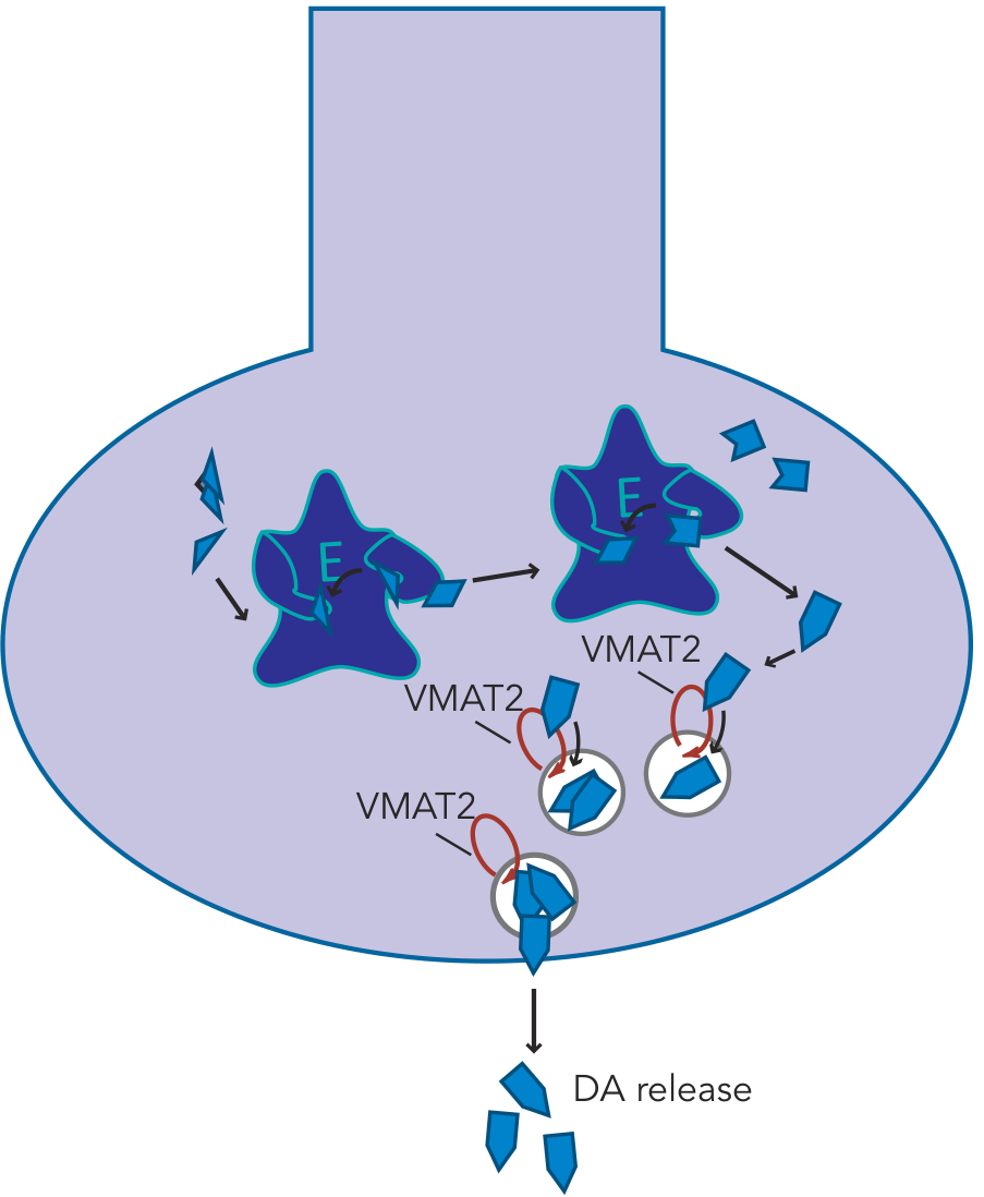
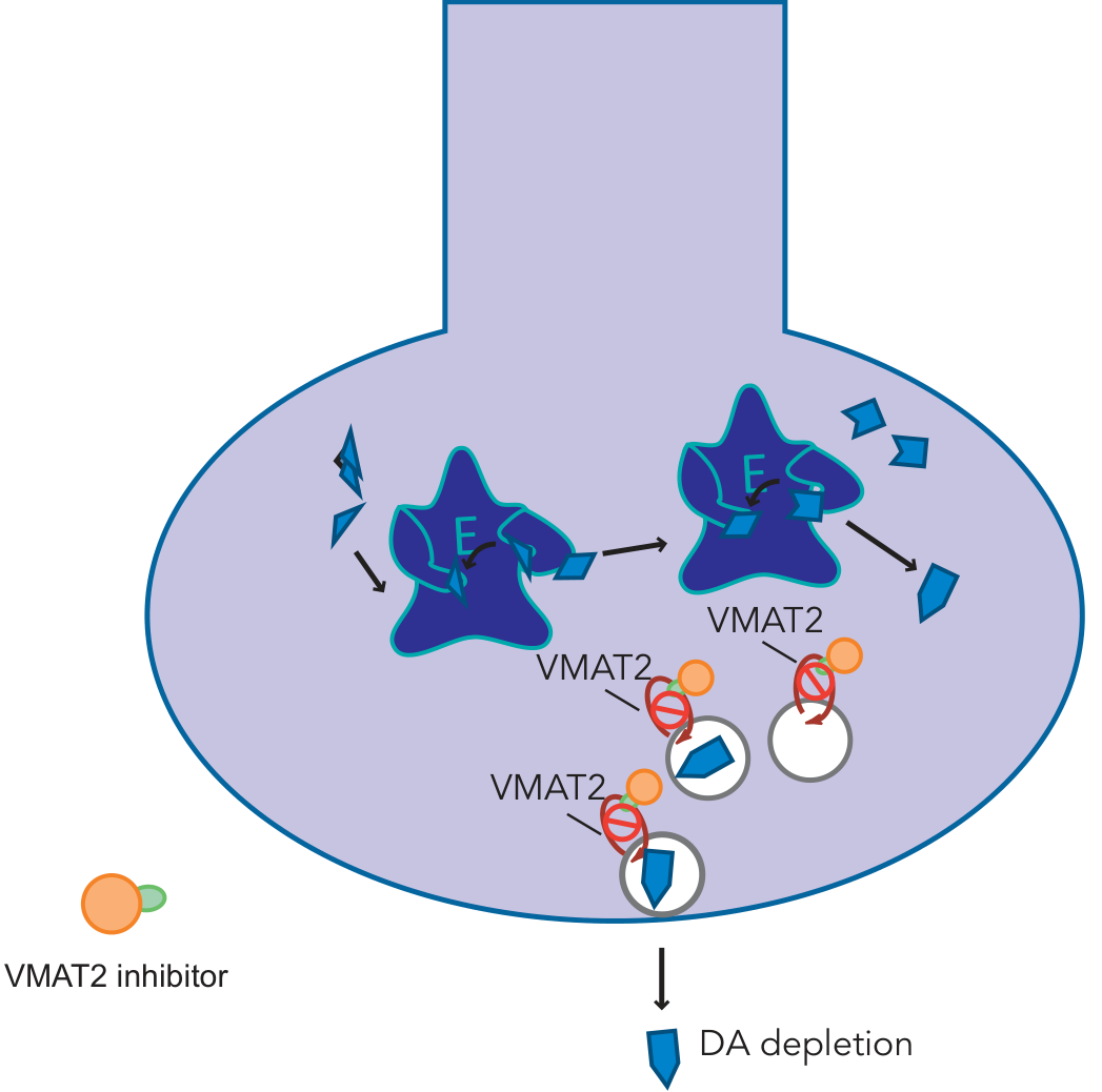
- Dopamine cannot be packaged into vesicles
- The unpackaged dopamine sitting in the cytoplasm is rapidly degraded by MAO (monoamine oxidase)
- This leads to progressive depletion of presynaptic dopamine stores
- Less dopamine is available for vesicular release into the synapse
- Result: reduced dopaminergic neurotransmission in the nigrostriatal pathway
Tetrabenazine is a Prodrug
| Active Metabolite | VMAT2 Potency | Additional Actions |
|---|---|---|
| +β-dihydro | Highest - responsible for most therapeutic effects | Main VMAT2 inhibitor |
| -α- and -β-dihydro | Moderate | Also antagonize 5-HT7 receptors and D2 receptors |
Selectivity: Tetrabenazine vs. Reserpine
| Drug | VMAT1 | VMAT2 | Effect |
|---|---|---|---|
| Reserpine | Inhibits both | Inhibits both | Peripheral side effects common (orthostatic hypotension, nasal congestion, GI effects) - because VMAT1 is in peripheral neurons |
| Tetrabenazine | No effect | Selective inhibitor | Fewer peripheral side effects; acts mainly in CNS |
Why It Works for Hyperkinetic Disorders
Tetrabenazine → Blocks VMAT2 → Dopamine not packaged
                              → Cytoplasmic dopamine degraded by MAO
                              → Depleted presynaptic dopamine stores
                              → Less DA released into synapse
                              → Reduced D2 receptor stimulation in striatum
                              → Less activation of "go" pathway
                              → Decreased involuntary movements
Approved Indications
| Drug | Approved Use | Key Features |
|---|---|---|
| Tetrabenazine | Chorea of Huntington's disease (FDA 2008) | TDS dosing; requires CYP2D6 genotyping at high doses; depression/suicide risk warning |
| Deutetrabenazine | Huntington's chorea + tardive dyskinesia | Deuterated form → slower CYP2D6 metabolism → longer half-life → less peak-dose side effects |
| Valbenazine | Tardive dyskinesia | Once-daily dosing; no genetic testing needed; no food requirements; no suicide warning |
Side Effects (from dopamine depletion)
- Drug-induced Parkinsonism (too much dopamine depletion in nigrostriatal pathway)
- Sedation
- Depression and suicidality - carries a black box warning (tetrabenazine reduces dopamine/serotonin which are important for mood)
- Akathisia (restlessness)
- QT prolongation (mild)
Reserpine
Reserpine - Complete Overview
Source
Mechanism of Action
Reserpine → Irreversible VMAT2 blockade
               ↓
  NE, DA, 5-HT cannot enter storage vesicles
               ↓
  Monoamines leak into cytoplasm
               ↓
  Degraded by MAO intraneuronally
               ↓
  Progressive depletion of NE, DA, serotonin
  (CNS + PNS + adrenal medulla)
               ↓
  No active transmitter released at nerve endings
  = "Pharmacological sympathectomy"
Reserpine vs. Tetrabenazine - Key Comparison
| Feature | Reserpine | Tetrabenazine |
|---|---|---|
| VMAT selectivity | Blocks VMAT1 + VMAT2 | Blocks VMAT2 only |
| Reversibility | Irreversible | Reversible |
| Peripheral effects | Significant (orthostatic hypotension, nasal stuffiness, GI effects) - because VMAT1 is in peripheral neurons | Fewer peripheral effects |
| CNS effects | Depletes CNS monoamines heavily | Mainly CNS dopamine |
| Duration of effect | Days to weeks after stopping | Hours (short half-life) |
| Adrenal medulla | Also depletes chromaffin granules | Less effect |
Pharmacological Effects
- Decreases both cardiac output and peripheral vascular resistance
- Lowers blood pressure gradually over several weeks
- At low doses: minimal postural hypotension
- Acts both centrally and peripherally
- Sedation and lassitude
- Mental depression (serious - can be severe and suicidal)
- Parkinsonism symptoms (due to dopamine depletion in corpus striatum)
- Nightmares
- Mild diarrhea and cramps
- Increases gastric acid secretion (exacerbates peptic ulcer)
Side Effects
| Side Effect | Mechanism |
|---|---|
| Depression / suicide | Depletion of brain NE and serotonin (the "monoamine hypothesis of depression" was originally based on reserpine-induced depression) |
| Parkinsonism | Dopamine depletion in striatum |
| Sedation | Central monoamine depletion |
| Nasal stuffiness | VMAT1 inhibition in peripheral neurons |
| Orthostatic hypotension | Peripheral NE depletion |
| Peptic ulcer exacerbation | Increased gastric acid secretion |
| Bradycardia | Sympathetic tone reduction + relative parasympathetic dominance |
Important: Depression may appear insidiously over weeks to months and can persist for months after stopping the drug (due to irreversible binding). The drug must be stopped at the first sign of depression and is absolutely contraindicated in patients with a history of depression.
Historical Significance
- It caused depression by depleting monoamines → led to the monoamine hypothesis of depression
- It was one of the first drugs used to treat psychosis (before chlorpromazine became dominant)
- Its antihypertensive use pioneered the concept of sympatholytic therapy
Therapeutic Use (Historical)
- Hypertension - used at low doses (0.05-0.25 mg/day), usually in combination with a diuretic, particularly in elderly patients with isolated systolic hypertension
- Now largely abandoned in favour of newer agents (ACE inhibitors, ARBs, CCBs, thiazides) which have better safety profiles and proven mortality benefits
- Goodman & Gilman's Pharmacological Basis of Therapeutics, Reserpine section
- Katzung's Basic & Clinical Pharmacology (16th ed.), Mechanism and Toxicity of Reserpine
- Stahl's Essential Psychopharmacology, Tetrabenazine/VMAT2 comparison
How does reserpine decreases ne but the tetra benzine decrease dopamine how if dopamine decreases ne also will decrease rat y both like how do they have significant effect
Why Reserpine Depletes NE (and Causes Hypotension) While Tetrabenazine Mainly Depletes Dopamine (and Treats Chorea)
The Two VMATs and Where They Live
| Transporter | Location | Neurotransmitters stored |
|---|---|---|
| VMAT1 | Peripheral nervous system neurons + CNS | NE, DA, 5-HT, histamine |
| VMAT2 | CNS neurons only | NE, DA, 5-HT, histamine |
RESERPINE  → blocks VMAT1 + VMAT2
                  ↓
         Acts in BOTH peripheral AND central neurons
                  ↓
         Depletes NE from peripheral sympathetic nerves
         (the ones that control blood vessels and heart)
                  ↓
         → Hypotension, bradycardia, nasal stuffiness
           (these are peripheral NE depletion effects)

TETRABENAZINE → blocks VMAT2 ONLY
                  ↓
         Acts ONLY in CNS neurons
                  ↓
         Cannot access peripheral sympathetic nerve vesicles
                  ↓
         → No significant peripheral NE depletion
         → No hypotension or peripheral side effects
But Why Does Tetrabenazine Preferentially Affect Dopamine if VMAT2 Also Stores NE and Serotonin?
"Although VMAT2 transports multiple neurotransmitters into synaptic vesicles (dopamine, norepinephrine, serotonin, and histamine), tetrabenazine preferentially affects dopamine transport at clinical doses."
Why Does This Matter Clinically?
| Effect | Reserpine | Tetrabenazine |
|---|---|---|
| Peripheral NE depletion | Yes (VMAT1 + VMAT2 in peripheral nerves) | No (VMAT2 is CNS only) |
| → Hypotension | Yes (therapeutic use!) | No |
| → Bradycardia, nasal stuffiness, GI effects | Yes | No |
| CNS dopamine depletion | Yes | Preferentially yes |
| → Treats chorea/TD | Yes (but too toxic) | Yes (approved use) |
| CNS NE + serotonin depletion | Yes (heavily) | Yes (but less than DA) |
| → Depression risk | High (depletes all three monoamines) | Present but relatively less |
| Drug-induced Parkinsonism | Yes | Yes (dopamine depletion in striatum) |
Simple Way to Remember It
RESERPINE
= Shotgun approach
= Kills ALL monoamines everywhere (PNS + CNS)
= Useful for BP (peripheral NE gone)
= Terrible for mood (CNS NE + 5-HT gone)
= Causes Parkinsonism (CNS DA gone)

TETRABENAZINE
= Sniper approach (CNS only, DA-preferential)
= No peripheral NE effect → no hypotension
= Mainly depletes DA in striatum → treats chorea
= Still causes some depression + Parkinsonism
  (because CNS DA and to lesser extent 5-HT/NE are still depleted)
- Stahl's Essential Psychopharmacology, Dopamine Depletion by VMAT2 Inhibition
- Katzung's Basic & Clinical Pharmacology (16th ed.), Reserpine Mechanism
But the dopamine is the main for ne synthesis ryt if u block in the cns then there should be no ne available for periphery even where does the ne come from to the periphery
Where Does Peripheral NE Come From? Is It Dependent on CNS Dopamine?
The Core Concept: Peripheral Sympathetic Neurons Are Autonomous Factories
The Catecholamine Synthesis Pathway
Blood tyrosine (amino acid from diet)
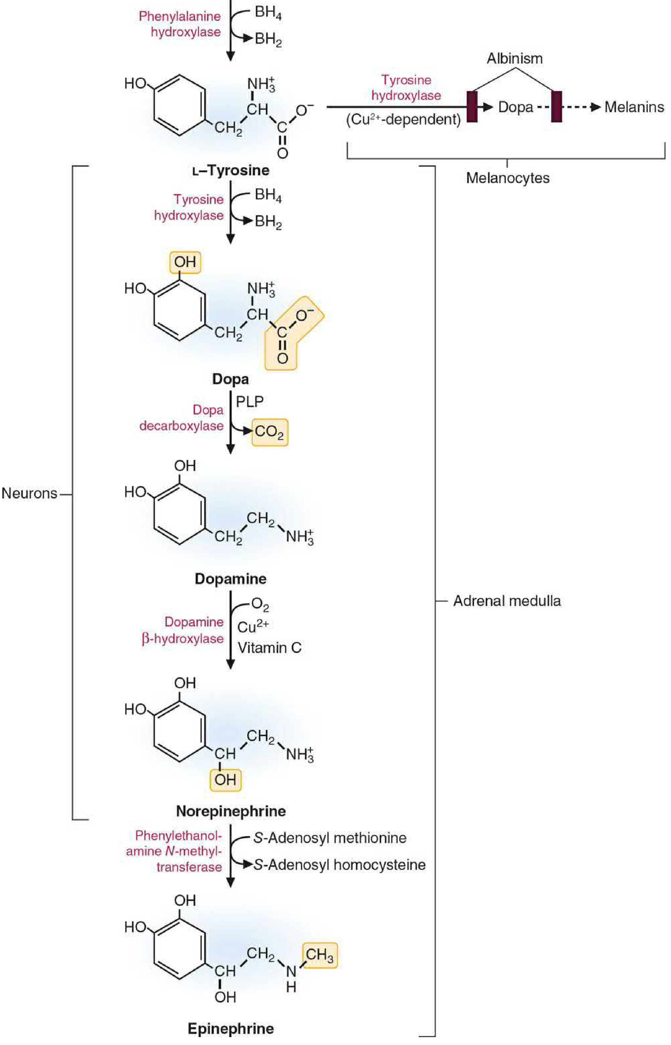
        ↓  [Tyrosine hydroxylase - rate-limiting step, in cytosol]
      L-DOPA
        ↓  [DOPA decarboxylase + pyridoxal phosphate, in cytosol]
     Dopamine  ← made right here in the nerve terminal cytosol
        ↓  [enters storage vesicle]
        ↓  [Dopamine β-hydroxylase (DBH) - inside the vesicle, needs Vit C + Cu2+]
  Norepinephrine  ← stored in vesicle, ready for release
So What Actually Happens in Each Neuron Type?
| Neuron Type | Enzymes Present | Final Product | Dopamine Role |
|---|---|---|---|
| Dopaminergic neuron (e.g. substantia nigra) | Tyrosine hydroxylase + DOPA decarboxylase | Dopamine (end product, released as NT) | Final neurotransmitter |
| Noradrenergic neuron (e.g. sympathetic nerve terminal, locus coeruleus) | Above + Dopamine β-hydroxylase inside vesicle | NE (dopamine is just a stepping stone) | Intermediate only |
| Adrenergic neuron / adrenal medulla | Above + PNMT | Epinephrine | Intermediate only |
Why CNS Dopamine Depletion Does NOT Affect Peripheral NE
Tetrabenazine blocks VMAT2 (CNS only)
        ↓
CNS dopamine neurons: dopamine cannot be packaged
                      → DA degraded by MAO → DA depletion
        ↓
Does this affect peripheral sympathetic NE?

NO - because:
1. Peripheral sympathetic neurons have THEIR OWN tyrosine hydroxylase
   → They make their own dopamine from tyrosine
2. That dopamine is converted to NE inside vesicles by DBH
3. VMAT2 in peripheral neurons is NOT blocked by tetrabenazine
   (VMAT2 in peripheral nerves? Actually, peripheral neurons use VMAT1 + VMAT2)
   → But tetrabenazine does not cross into peripheral nerve terminals effectively
4. The peripheral sympathetic neuron does not depend on the brain
   for its dopamine supply AT ALL
Why Reserpine DOES Deplete Peripheral NE
Peripheral sympathetic terminal:
- Tyrosine → DOPA → Dopamine (still synthesised normally in cytosol)
- But VMAT1 is blocked → Dopamine CANNOT enter the vesicle
- Dopamine stays in cytosol → degraded by MAO
- No dopamine in vesicle → No DBH conversion to NE
- No NE stored → No NE released
→ Peripheral sympathetic failure → Hypotension, bradycardia
Summary
| Question | Answer |
|---|---|
| Does brain dopamine supply peripheral NE? | No - each peripheral neuron makes its own |
| Where does peripheral NE synthesis start? | From dietary tyrosine in blood, taken up locally by each nerve terminal |
| Why doesn't tetrabenazine affect peripheral NE? | It only blocks CNS VMAT2; peripheral nerve terminals are not significantly affected |
| How does reserpine deplete peripheral NE? | By blocking VMAT1 in peripheral vesicles → dopamine cannot enter vesicles → DBH cannot convert it to NE → NE stores are depleted |
| Is dopamine just a precursor in NE neurons? | Yes - it is never released as a neurotransmitter there; it is immediately taken into vesicles and converted to NE |
- Basic Medical Biochemistry (6th ed.), Catecholamine Biosynthesis
- Stahl's Essential Psychopharmacology, VMAT2 and Dopamine Depletion
So epinephrine is significantly acting on alpha nor epinephrine also alpha but less than epinephrine but on beta 1 both have equal action then y non epinephrine consider as potential vaso constrictor though epinephrine has more action on alpha
Why NE is a More Potent Vasoconstrictor Than Epinephrine Despite Epi Having Stronger Alpha Action
Receptor Profiles - The Critical Difference
| Receptor | Epinephrine | Norepinephrine | Effect on Blood Vessels |
|---|---|---|---|
| Alpha-1 | +++ (strong) | ++ (moderate) | Vasoconstriction |
| Beta-1 | +++ | +++ (equal) | Heart (not vessels) |
| Beta-2 | +++ (strong) | + (very weak/negligible) | Vasodilation (in skeletal muscle, coronary, liver) |
What Beta-2 Does to Blood Vessels
- Skeletal muscle (the largest vascular bed in the body - ~40% of body mass)
- Coronary arteries
- Liver
The Net Effect - Why This Changes Everything
NOREPINEPHRINE:
  Alpha-1 → vasoconstriction in skin, viscera, kidneys
  Beta-2  → negligible (NE barely touches beta-2)
  ─────────────────────────────────────────────
  Net: PURE vasoconstriction
  → Total peripheral resistance ↑↑↑
  → Both systolic AND diastolic BP rise
  → Reflex bradycardia (baroreceptors fire)

EPINEPHRINE:
  Alpha-1 → vasoconstriction in skin, viscera, kidneys (stronger than NE)
  Beta-2  → VASODILATION in skeletal muscle (large vascular bed)
  ─────────────────────────────────────────────
  Net: Mixed effect
  → Vasoconstriction in some beds, vasodilation in others
  → Total peripheral resistance: ↓ or variable
  → Systolic BP rises (from cardiac output), but
  → Diastolic BP actually FALLS (beta-2 vasodilation dominates in periphery)
  → Pulse pressure widens
"Norepinephrine causes greater vasoconstriction than epinephrine, because it does NOT induce compensatory vasodilation via β2 receptors on blood vessels supplying skeletal muscles."
Katzung's Table - Net Vascular Resistance (This Makes it Crystal Clear)
| Drug | Skeletal Muscle Resistance | Total Peripheral Resistance | Diastolic BP |
|---|---|---|---|
| Phenylephrine (pure alpha) | ↑ | ↑↑↑ | ↑↑ |
| Epinephrine (alpha + beta-2) | ↓ or ↑ (variable - beta-2 fights alpha) | ↓ or variable | ↓ or variable |
| Isoproterenol (pure beta) | ↓↓ | ↓↓ | ↓↓ |
The "Epinephrine Reversal" Experiment - Proof of This Concept
- Alpha is blocked → only beta-2 acts on vessels
- BP actually FALLS (vasodilation dominates)
- This is called "epinephrine reversal"
- NE has no beta-2 → BP just decreases mildly (no reversal, no vasodilation)
- It cannot flip to vasodilation because there is nothing to flip with
"The actions of norepinephrine are not reversed but are diminished because norepinephrine lacks significant β2 agonist action on the vasculature."
Summary in One Sentence
Epinephrine has stronger alpha than NE, but its powerful beta-2 action vasodilates the massive skeletal muscle vascular bed, cancelling out much of the alpha vasoconstriction. NE has negligible beta-2, so its alpha action is unopposed, making the NET vasoconstriction much greater.
Quick Memory Aid
NE = Alpha warrior with no beta-2 opposition
   → Net: Pure ↑↑ peripheral resistance
   → BP ↑ (both systolic + diastolic)
   → Reflex bradycardia

Epi = Alpha warrior BUT beta-2 fights back
    → Net: ↑ heart + mixed vasculature
    → Systolic BP ↑, Diastolic BP ↓ or same
    → Pulse pressure widens
    → No reflex bradycardia (or even tachycardia)
- Lippincott Illustrated Reviews: Pharmacology, Cardiovascular actions of NE
- Katzung's Basic & Clinical Pharmacology (16th ed.), Cardiovascular responses table