
Angiotensin 1 and 2 receptors and their action
I'll start by reading the medical library skill, then search for information on angiotensin receptors in parallel.
Reading File
Now let me search the library and web in parallel:
Finding Sources
Finding Sources
Searching Images
angiotensin receptor AT1 AT2 signaling pathway diagram
Excellent results. Let me now read the most relevant sections in parallel - from Ganong's physiology (AT1/AT2 receptor section), Braunwald's (RAS activation), and Goodman & Gilman's (pharmacology of ARBs):
Reading File
Reading File
Reading File
Loading Image
Now let me get the detailed AT1/AT2 actions from Braunwald's and Campbell-Walsh, plus the Katzung pharmacology section:
Reading File
Reading File
Reading File
Loading Image
Excellent - I now have comprehensive information from multiple authoritative sources. Let me also get the Miller's Anesthesia section on AT1/AT2 in the heart:
Reading File
I now have rich, comprehensive data from five authoritative textbooks plus two excellent diagrams. Here is the complete answer:
Angiotensin Receptors: AT1 and AT2 - Structure and Actions
Background: The Renin-Angiotensin System (RAS)
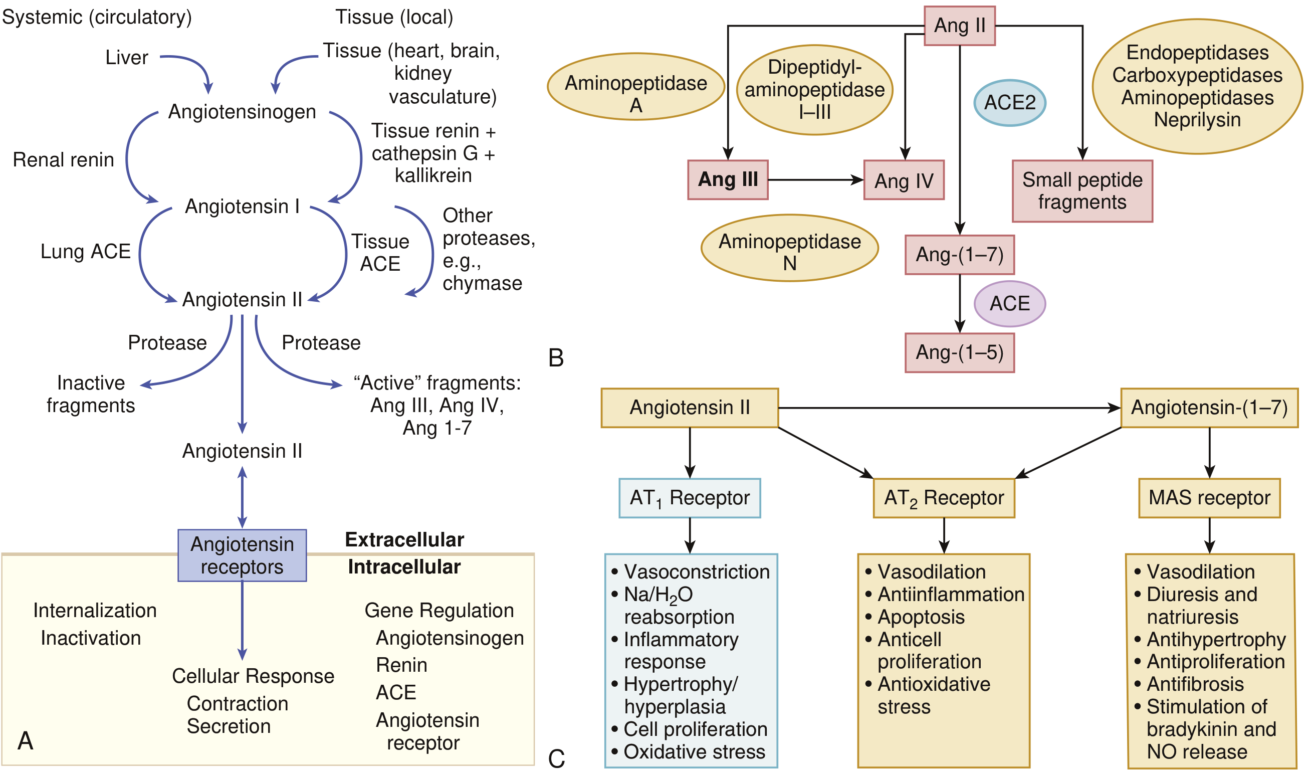
Before covering the receptors, a brief orientation. Renin (released from juxtaglomerular cells of the kidney) cleaves angiotensinogen (synthesized in the liver) to form Angiotensin I - a biologically inactive decapeptide. Angiotensin-converting enzyme (ACE), found predominantly (~90%) in tissue and ~10% in soluble interstitial form, removes two amino acids to form the active octapeptide Angiotensin II. ACE-independent pathways via chymase can also generate Ang II, especially in myocardium.
The diagram below (from Braunwald's Heart Disease) shows the full RAS cascade and both receptor pathways:
Angiotensin II Receptors: Overview
Angiotensin II exerts its biological effects by binding to two G protein-coupled receptors (GPCRs): AT1 and AT2. Both have seven transmembrane domains. Their downstream signaling and physiological effects are largely opposite to each other.
- Ganong's Review of Medical Physiology, 26e
- Braunwald's Heart Disease, 2-Vol Set
AT1 Receptor
Structure and Location
- G protein-coupled receptor; coupled to Gq protein → activates phospholipase C → increases cytosolic free Ca²⁺
- Also activates numerous tyrosine kinases
- Located in vascular smooth muscle (within caveolae), adrenal cortex, kidney, heart (predominantly in myocardial nerves), liver, brain, lungs
- In rodents, two subtypes exist: AT1A (blood vessels, brain, most organs - mediates the majority of Ang II effects) and AT1B (anterior pituitary and adrenal cortex); in humans, only one AT1 gene on chromosome 3 has been confirmed
- Predominant receptor in vasculature and in the normal adult human heart
Key Actions of AT1 Receptor
| System | Effect |
|---|---|
| Vasculature | Vasoconstriction (primary pressor mechanism) |
| Kidney | Na⁺/H₂O reabsorption; promotes sodium retention |
| Adrenal cortex | Stimulates aldosterone secretion from zona glomerulosa |
| Sympathetic NS | Enhances norepinephrine (NE) release from sympathetic nerve endings |
| Heart | Positive chronotropic and inotropic effects; cardiac hypertrophy and fibrosis |
| Cell biology | Promotes cell growth, proliferation, hypertrophy/hyperplasia |
| Inflammation | Pro-inflammatory response |
| Oxidative stress | Increases reactive oxygen species production |
AT1 Receptor Regulation
-
Vascular AT1 receptors: down-regulated by excess Ang II (desensitization)
-
Adrenocortical AT1 receptors: up-regulated by excess Ang II - making the adrenal gland more sensitive to aldosterone-stimulating effects
-
In heart failure: AT1 receptor mRNA and density are downregulated in failing human hearts
-
Ganong's Review of Medical Physiology, 26e
-
Miller's Anesthesia, 10e
-
Braunwald's Heart Disease
AT2 Receptor
Structure and Location
- Also a GPCR with seven transmembrane domains
- Gene located on the X chromosome in humans
- Acts via a G protein to activate phosphatases, which antagonize growth effects and open K⁺ channels
- Also increases nitric oxide (NO) production → raises intracellular cGMP
- More abundant in fetal and neonatal life; expression declines with development
- Persists in the brain and selected adult organs (fibroblasts and interstitium of the heart)
- In failing hearts and after injury/ischemia: AT2 receptors are upregulated
- In human myocardium: AT2 receptor predominates over AT1 in a 2:1 molar ratio
Key Actions of AT2 Receptor
| System | Effect |
|---|---|
| Vasculature | Vasodilation (via NO/cGMP) |
| Cell biology | Anti-proliferative, promotes apoptosis |
| Kidney | Natriuresis |
| Inflammation | Anti-inflammatory |
| Oxidative stress | Antioxidative |
| Heart | Counter-regulatory to AT1; generally antiproliferative |
| Fetal development | Vasodilation, cellular proliferation and apoptosis |
- Ganong's Review of Medical Physiology, 26e
- Braunwald's Heart Disease
- Campbell-Walsh-Wein Urology
AT1 vs. AT2: Head-to-Head Comparison
| Feature | AT1 Receptor | AT2 Receptor |
|---|---|---|
| G protein coupling | Gq → PLC → ↑Ca²⁺; also tyrosine kinases | G protein → phosphatases; ↑NO → ↑cGMP |
| Gene location | Chromosome 3 (humans) | X chromosome |
| Predominance | Adult tissues | Fetal/neonatal tissues |
| Vascular effect | Vasoconstriction | Vasodilation |
| Cell growth | Promotes (hypertrophy, proliferation) | Inhibits (anti-proliferative, pro-apoptotic) |
| Aldosterone | Stimulates secretion | - |
| Inflammation | Pro-inflammatory | Anti-inflammatory |
| Renal Na⁺ | Promotes retention | Natriuresis |
| In heart failure | Downregulated | Upregulated |
| Cardiac localization | Myocardial nerves | Fibroblasts and interstitium |
The MAS Receptor (Bonus: Third Arm of RAS)
Angiotensin-(1-7), formed from Ang II via ACE2, acts on the MAS receptor (another GPCR, expressed in brain, testes, and heart). Its effects mirror AT2:
- Vasodilation
- Diuresis and natriuresis
- Anti-hypertrophy, anti-proliferation, anti-fibrosis
- Stimulates bradykinin and NO release
This represents the counter-regulatory "protective" axis of the RAS, as opposed to the Ang II - AT1 "harmful" axis.
- Braunwald's Heart Disease
Clinical Relevance
-
Angiotensin Receptor Blockers (ARBs) - e.g., losartan, valsartan, candesartan - selectively and competitively block the AT1 receptor, preventing vasoconstriction, aldosterone release, and fibrosis. This leaves AT2 receptors unopposed (their activation may theoretically be beneficial, though this has not proven clinically superior to ACE inhibitors).
-
ACE inhibitors - block conversion of Ang I → Ang II, reducing stimulation of both AT1 and AT2. Benefit in heart failure is primarily attributed to reduced AT1 receptor activation.
-
Combination of ARB + ACEI: No additive prognostic benefit in heart failure; increases hypotension, hyperkalemia, and renal dysfunction - not recommended routinely.
-
Tissue RAS: Independent local RAS exists in heart, vasculature, kidney, brain, adrenal, and other organs. Angiotensin II generated locally acts as a growth factor - blockade of this (by ACE inhibitors or ARBs) contributes significantly to clinical benefit.
- Goodman & Gilman's Pharmacological Basis of Therapeutics
- Katzung's Basic & Clinical Pharmacology, 16e
This is a shared conversation. Sign in to Orris to start your own chat.